

 PDF
PDF ePub
ePub Citation
 Print
Print


Unilateral or bilateral superior oblique palsy (SOP) is commonly associated with horizontal deviation, especially with exotropia [1-4]. Minimal to moderate horizontal deviation may be resolved spontaneously after vertical muscle surgery in SOP. However, a large angle amount of associated exotropia frequently requires surgical treatment. Even though the exact cause of lateral incomitancy remains uncertain, the risk of consecutive esotropia has been shown to increase by a factor of eight in patients with lateral incomitancy [5]. Theoretically, the oblique muscles are abductors, so in exotropia associated with superior oblique palsy, lateral incomitancy could be more commonly observed because the abducting power of superior oblique (SO) is also reduced. Further, concerning additional reduction of abducting power following inferior oblique weakening surgery, it is not easy to determine the recession amount of lateral rectus muscles needs to prevent overcorrection and avoid excessive undercorrection. As far as we know, these concerns have not been mentioned in the abundant literature dealing with surgical procedures for SOP.
The purpose of this study is to suggest a surgical normogram for lateral rectus recession when performing inferior oblique recession in exotropia with SOP based on our experience. We evaluate the difference in incidence of lateral incomitancy and the effect of lateral rectus recession with reduced surgical requirements in patients who showed surgical success, dividing them into unilateral SOP and bilateral SOP groups.
Materials and Methods
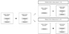
A retrospective review of medical records was performed on 71 patients who had undergone surgery for unilateral or bilateral superior oblique palsy with exotropia between February 2005 and July 2009 and who were successfully corrected more than 1 year after surgery. The criteria of successful correction were defined as follows: when the patients had no suppression and a deviation angle ranging from 5 prism diopters (PD) esodeviation to 10 PD exodeviation one year postoperative. We divided the patients into two groups; 34 patients who had undergone lateral rectus recession with unilateral inferior oblique (UIO) recession group and the remaining 37 patients who had undergone LR recession with bilateral inferior oblique (BIO) recession group (Fig. 1).
Patients who had undergone strabismus surgery or any intraocular surgery, had amblyopia, or follow-up of less than 12 months after surgery were excluded. Cyclopegic refraction, correction of refractive error, and stereopsis test were performed prior to surgical intervention. In all patients, we performed ocular motility testing, measurement of the deviation angle in all gazes by alternate prism and cover testing, including Bielschowsky head tilt test and fundus examination to diagnose exotropia associated with SOP.
To diagnose SOP, especially distinguishing from primary inferior oblique overaction, we carefully observed the presence of superior oblique underaction, fundoscopic foveal extorsion, positive Bielschowsky head tilt test, anomalous head posture of head tilting past or present, and facial asymmetry [6]. To measure lateral exo-angle to evaluate lateral incomitancy in our patients, we measured the angle with a 30° lateral head turn [5,7]. Lateral incomitancy was defined when the exoangle was reduced by more than 20% relative to the primary gaze.
All patients underwent simultaneous unilateral or bilateral inferior oblique (IO) recession and unilateral lateral rectus or bilateral lateral rectus (BLR) recession. All surgeries were performed under general anesthesia. To correct SOP, we used the same surgical method of 14 mm IO recession in all patients. To correct exotropia, we reduced the recession amount by 1 or 2 mm compared to the previous normogram [8], considering lateral incomitancy and laterality of IO recession. For example, in the case of 20 PD exodeviation, the previous normogram suggested 5 mm BLR recession; we performed 4 mm BLR recession (overall 2 mm reduction) if lateral incomitancy was associated, and 5/4 mm recession of each lateral rectus (1 mm reduction) if there was no accompanying lateral incomitancy. We compared the differences of total surgical amount between the two groups with a t-test. The surgical outcome was evaluated at one month, three months, six months, and one year postoperative.
The effect of lateral rectus (LR) recession (PD/mm) was analyzed. For each millimeter of amount of recession as the absolute value of the angle of preoperative deviation minus postoperative deviation on postoperative day one divided by total amount of recession [9,10]. The results were compared between the two groups using a previous surgical normogram as a reference (Parks' normogram) [8].
Results
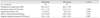
The mean age was 11.36 ± 5.86 years in the UIO group and 7.16 ± 3.94 years in the BIO group. The mean preoperative exodeviation was 20.4 PD in the UIO group and 26.4 PD in the BIO group. The recession amount of the LR muscle ranged from 4 to 8.5 mm in the UIO group and 5 to 9 mm in the BIO group. Lateral incomitancy was noted in 12 patients (36.4%) and 26 patients (70.3%) in each group, respectively (p = 0.02). The effect of LR recession was calculated as 3.23 ± 0.84 PD/mm in group A and 2.98 ± 0.62 PD/mm in group B and there was no statistically significant difference between two groups (p = 0.15) (Table 1).
Discussion
There have been many reports that SOP is associated with concomitant exotropia. The incidence of SOP with horizontal deviation has been reported to range from 36% to 55.6% [1-4]. Helveston et al. [1] reported that concomitant horizontal deviation was present in 36% of 190 patients with SOP. Of the unilateral palsies with a horizontal deviation, 75% were exotropias. Seventy-five percent of exotropias and 45% of esotropias resolved "spontaneously," in the sense that the patients underwent only cyclovertical muscle surgery, and horizontal deviation was not present postoperatively. Therefore, in the case of minimal or moderate horizontal deviation, it may be reasonable to perform only cyclovertical muscle surgery because the function of the inferior oblique muscle includes depression, incyclotorsion and abduction. However, large angle deviation as noted in our cases (20.4 to 26.4 PD) should be corrected with combined horizontal muscle surgery.
The usual amount of recession for 20 to 25 PD exotropia is 10 to 12 mm of lateral rectus if summing the recession amount of both eyes [8]. This amount is approximately a 10 PD overcorrection. In our cases, considering simultaneous weakening of abduction from the recession of the inferior oblique muscle, we reduced the recession amount by 1 or 2 mm lateral recti depending on the presence of lateral incomitancy and laterality of IO recession. It is expected that lateral incomitancy can be highly prevalent in exodeviation associated with SOP. The oblique muscles are abductors, so in exotropia associated with superior oblique palsy, lateral incomitancy could be more commonly observed because the abducting power of SO is also reduced. As a result, the medial rectus muscles became tighter than in the case of exotropia alone. However, this is a presumption and more research should be performed to determine the prevalence of lateral incomitancy in exotropia associated with SOP.
In this study, participants had good postoperative results for horizontal eye alignment due to the reduction of the amount of recession without significant risk for undercorrection. Even though our study lacked a control group that underwent standard full complete recession, our result suggests the reduction of LR recession, while considering lateral incomitancy and simultaneous IO recession, could be a safe regimen to prevent overcorrection during combination surgery. Moreover, we suggest the influence of horizontal weakening effect from IO recession alone. However, the influence of IO weakening on horizontal deviation remains controversial. Stager and Parks [11] reported that, in 50 patients with primary bilateral inferior oblique overaction without SOP, bilateral IO weakening did not influence the horizontal deviation and caused only 0.5 PD of exoshift on average. We think IO weakening for SOP may increase the already weakened abduction of the superior oblique. This also may cause increased incidence of lateral incomitancy and surgical overcorrection with the conventional amount of recession for exodeviation in SOP.
In addition, the results showed no significant difference between the two groups. The effect of LR recession was 3.23 ± 0.84 PD/mm in the UIO group and 2.98 ± 0.62 PD/mm in the BIO group. In Parks and Mitchell' normogram [8], the effect of LR recession can be calculated as about 3 mm/PD if its intended average postoperative results are 10 PD overcorrection. Comparing with this, the results of this study showed a more weakening effect horizontally. In conclusion, reducing the amount of LR recession by 1 to 2 mm was successful and a safe method to prevent overcorrection when combined with IO weakening, irrespective of the laterality of SOP.
 XML Download