

 PDF
PDF ePub
ePub Citation
 Print
Print


Various glaucoma drainage devices (GDDs) have been used for the treatment of refractory glaucomas, such as neovascular glaucoma (NVG), and the performance of GDDs can be significantly different among each individual product [1-3]. There are two types of GDDs, depending on the flow of aqueous humor being regulated. For example, the Molteno implant (IOP Inc., Costa Mesa, CA, USA) and Baerveldt implant (Pharmacia/UpJohn, Brdigewater, NJ, USA) do not regulate the flow of aqueous humor; however, the Krupin implant (Hood Laboratories, St. Pembroke, MA, USA) and Ahmed Glaucoma Valves (New World Medical Inc, Rancho Cucamonga, CA, USA) regulate the flow of aqueous humor. Of those, the Ahmed Glaucoma Valve (AGV) is one of the more commonly used flow-restricted implants in refractory glaucomas. The valve mechanism consists of two thin silicone elastomer membranes, 8 mm long and 7 mm wide, which allow one-way regulation of the flow with a goal of keeping the intraocular pressure (IOP) between 8 and 10 mmHg in the early postoperative period [4]. There are two surgical methods for tube coverage used in AGV implantation. In the first method, the drainage tube is covered with preserved donor sclera or Tutoplast (pericardium, dura, etc.), after it is inserted into the anterior chamber. The second method requires covering the drainage tube with an autologous scleral flap, after the tube is inserted into the anterior chamber under the partial-thickness scleral flap. Such differences may yield the cost-cutting and high dependence on donor sclera's availability in the graft group, but it will not be an issue for the flap counterpart, which only demands a mastery of surgical skill.
To the best of our knowledge, there have been few studies that compared the outcomes of two different surgical techniques used for AGV implantation. The purpose of this study is to compare the surgical outcomes between the scleral graft method and partial thickness scleral flap method in AGV implantation.
Materials and Methods
We retrospectively reviewed the medical records of patients diagnosed as NVG who underwent AGV implantation at the Eulji General Hospital in Seoul, Korea between January 2001 and December 2007. The institutional review board of Eulji Medical Center approved the study protocol before patient enrollment.
Inclusion criteria were as follows: 1) age between 20 to 85 years, 2) inadequately controlled NVG due to proliferative diabetic retinopathy (PDR) or central retinal vein occlusion (CRVO), and 3) an IOP above 22 mmHg in spite of maximum tolerated medical therapy. Patients who had no light perception preoperatively or who underwent the AGV implantation for other secondary glaucomas were excluded from this study.
All of the participants had follow-up periods of six months or longer after AGV implantation. Preoperative data included age, sex, implanted AGV model, preoperative IOP, and the number of topical IOP depressants. The eyes were divided into two groups according to the surgical methods covering the silicone drainage tube of AGV. In the graft group (n = 18), the drainage tube was inserted into the anterior chamber, and then covered with preserved donor sclera. In the flap group (n = 10), the drainage tube was inserted under the partial-thickness scleral flap, and then covered with the flap.
The selection of the surgical method for each patient depended on the donor sclera's availability when the operation was planned. That is, when the donor sclera was available, we performed the AGV surgery using the donor sclera graft method; otherwise we performed the surgery using the scleral flap method. The donor sclera used in AGV implantation was obtained from patients diagnosed with traumatic eyeball rupture at our hospital. The remaining cornea, lens, and uvea (iris, ciliary body, and choroid) of the enucleated eyeballs were clearly removed with scissors and blades. Only the sclera was preserved in 99.9% ethyl alcohol at a refrigerator temperature at 4℃.
The donor had no abnormal results on laboratory tests for infectious diseases such as hepatitis, acquired immunodeficiency syndrome, and syphilis. All patients were operated on by one surgeon (JSP). The S2 model had been used before 2004, while the FP7 model replaced the S2 model since 2004.
All patients underwent AGV implantation under retrobulbar anesthesia. A fornix-based conjunctival-Tenon's capsule flap was created in the superotemporal or superonasal quadrant, and blunt dissection was performed between Tenon's and the episclera for AGV implantation beyond the equator of the globe. The tip of the drainage tube was then irrigated with a 26-gauge blunt cannula containing BSS (Alcon Laboratories Inc., Fort Worth, TX, USA) for priming. The valved plate body was then inserted posteriorly into the sub-Tenon's space and sutured to the sclera with 9-0 nylon sutures through the anterior positional holes of the valved plate body, 8 mm posterior to the limbus. The tip of the drainage tube was then cut and beveled up in order to extend by 2 to 3 mm into the anterior chamber. A paracentesis was then made at the limbus, and a small amount of viscoelastic material was injected into the anterior chamber. In the graft group, the anterior chamber was then entered through the limbal area, approximately 0.5 to 1 mm posterior to the limbus, using a 23-gauge needle. The drainage tube was then inserted into the anterior chamber parallel to the iris plane and was secured to the sclera with 10-0 nylon sutures. The preserved donor sclera was then rehydrated in a BSS bath and trimmed to approximately 4 × 7 mm in size. The drainage tube was then covered with preserved donor sclera to avoid potential complications such as tube erosion, and the donor scleral graft was sutured to the underlying sclera at each corner with 10-0 nylon sutures. The conjunctiva and Tenon's capsule were then sutured back to the original position using 8-0 vicryl sutures.
In the flap group, a half-thickness, rectangular, 4 × 4 mm and limbal-based scleral flap was created. The valved plate body was then inserted posteriorly into the sub-Tenon's space and sutured to the sclera with 9-0 nylon sutures through the anterior positional holes of the valved plate body, 8 mm posterior to the limbus. The anterior chamber was then entered with a 23-gauge needle under the scleral flap, then the drainage tube was inserted into the anterior chamber and was secured to the sclera with 10-0 nylon sutures. The scleral flap over the drainage tube was re-attached to the sclera and sutured with 10-0 nylon sutures.
Complete surgical success is defined as 6 mmHg ≤ IOP ≤ 21 mmHg without IOP-lowering medication, additional glaucoma surgery, nor loss of light perception during the follow-up period. Qualified success is defined as 6 mmHg ≤ IOP ≤ 21 mmHg with supplemental medication, but without loss of light perception. Mean success periods are defined as a time to maintain "qualified success" or "complete success" from an initial visit, and those were compared to each other by log rank test. Failure is defined as follows: 1) constant hypotony below 6 mmHg, 2) IOP > 21 mmHg despite of supplemental medication at least three times consecutively, 3) further glaucoma surgical intervention or recommendation thereof, or 4) loss of light perception. Further surgical intervention for glaucoma is defined as additional glaucoma surgery that requires a return to the operating room, such as placement of another tube shunt, tube exchange, and tube removal, etc. Intracameral injection of air or viscoelastics for maintaining the anterior chamber was excluded from this category and would not be recorded as evidence of failure until three months postoperative.
We collected data such as IOP, and postoperative complications at one day, one week, two weeks, one month, three months, six months, and one year or more postoperatively until the last visit. IOP was measured with a Goldmann applanation tonometer. We prescribed topical IOP-depressants (maximally, three eye drops) for the patients with an IOP more than 21 mmHg during follow-up periods.
Statistical analysis was done by SPSS ver. 17.0 (SPSS Inc., Chicago, IL, USA). The preoperative data for the two groups were compared using the Mann-Whitney U-test or Fisher's exact test. The complete success rate, qualified success rate, and failure rate were analyzed by Fisher's exact test. The postoperative complications were compared using Pearson chi-square test or Fisher's exact test. The survival curves for the success rates in each group were calculated using the Kaplan-Meier method. A p-value of less than 0.05 was considered statistically significant.
Results
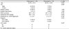
This study investigated 28 eyes (18 eyes in the graft group and ten eyes in the flap group). Table 1 summarizes the baseline characteristics for each group. The mean ages were 61.78 ± 10.11 years in the graft group and 64.10 ± 12.83 years in the flap group (p = 0.683). The preoperative mean IOP was 49.9 ± 9.8 mmHg in the graft group and 51.5 ± 12.1 mmHg in the flap group (p = 0.829). The male to female ratio, frequency of diabetes mellitus and systemic hypertension, and preoperative numbers of topical IOP-depressants were not significantly different between the two groups. The preoperative diagnoses in the two groups included NVG associated with PDR in 17 eyes (94.4%) in the graft group versus eight eyes (80.0%) in the flap group, and CRVO in one eye (5.6%) in the graft group versus two eyes (20.0%) in the flap group. All patients had angle-closure stage NVG. No eyes had undergone any glaucoma surgery before AGV implantation.
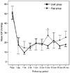
The mean IOP values in the graft group versus the flap group were as follows (Fig. 1): 17.4 ± 8.5 mmHg versus 17.3 ± 10.4 mmHg at postoperative one day (p = 0.530), 9.5 ± 3.1 mmHg versus 14.5 ± 7.2 mmHg at postoperative one week (p = 0.468), 15.8 ± 5.4 mmHg versus 14.3 ± 3.6 mmHg at postoperative two weeks (p = 0.580), 19.4 ± 5.2 mmHg versus 18.8 ± 7.4 mmHg at postoperative one month (p = 0.501), and 18.8 ± 5.5 mmHg versus 17.7 ± 6.6 mmHg at postoperative three months (p = 0.486). However, the stastically significant (p = 0.006) mean IOP at six months postoperatively was 16.4 ± 5.4 mmHg in the graft group and 27.4 ± 10.5 mmHg in the flap group. No other significant differences were observed (p = 0.967, 0.287, and 0.495 at 12 months, 18 months, and 24 months, respectively by Mann-Whitney U-test). Moreover, there was no significant difference between the two groups in the mean number of postoperative topical IOP-depressants.
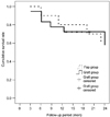
Six months postoperative, complete and qualified success rates were 22.2% (4 / 18) and 72.2% (13 / 18) in the graft group, and 10.0% (1 / 10) and 80.0% (8 / 10) in the flap group, respectively. At postoperative one year, the complete and qualified success rates were 22.2% (4 / 18) and 55.6% (10 / 18) in the graft group, and 10.0% (1 / 10) and 70.0% (7 / 10) in the flap group, respectively (Table 2). However, none of these were significantly different. On the other hand, the cumulative success rates were 94.4% versus 90.0% at six months and 77.8% versus 80.0% at one year in the graft and flap groups, respectively (Fig. 2). The mean success periods were 53.1 ± 10.1 months in the graft group and 50.9 ± 9.4 months in the flap group (p = 0.882 by log rank test).
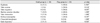
Table 3 summarizes postoperative complications for each group. We defined tube erosion as the exposure of the tube through conjunctival thinning or dehiscence, and tube migration as a dislocation of the tube posteriorly out of the anterior chamber or anteriorly contacting the corneal endothelium. Postoperative complications in the graft group included shallow anterior chamber in seven eyes (38.9%), hyphema in six eyes (33.3%), tube obstruction in five eyes (27.8%), early hypotony in two eyes (11.1%), tube erosion in one eye (5.6%), choroidal detachment in one eye (5.6%), and tube migration in no eye (0.0%). In the flap group, there were hyphema in five eyes (50.0%), shallow anterior chamber in four eyes (40.0%), tube erosion in three eyes (30.0%), tube migration in three eyes (30.0%), tube obstruction in three eyes (30.0%), and bullous keratopathy in one eye (10.0%). The frequency of tube migration was significantly higher in the flap group (p = 0.037). Despite being not statistically significant, the flap group had a higher frequency of tube erosion than the graft group (p = 0.116). A case of the tube erosion in the graft group underwent an additional patch graft of the preserved donor sclera. In the flap group, outward migration of the tube in addition to tube erosion occurred in two eyes. In these cases, we repositioned the tube and securing it again to the sclera. In one case in the flap group, the inward migration of the tube and severe tube erosion occurred, due to the dislocation of the valved plate body. Several trials to repair the exposure site with preserved donor sclera were made, but failed due to graft melting and further migration of the tube with tube-corneal touch, which caused the eye to develop bullous keratopathy.
Discussion
The aim of the present study was to compare the surgical outcomes of the two methods for AGV implantation. In our study, the graft group and flap group did not show significant differences in terms of postoperative IOP and the mean success period. At about six months postoperative, we observed three cases of tube obstruction and subsequent temporary elevation of IOP in the flap group. Such factors as a small sample size (n = 10) and an abrupt change of individual IOP might have affected the overall mean IOP, and thereby resulted in significant deviation from the normal. The overall success rate of AGV implantation varies among different types of glaucoma, ranging from 63% to 100% at one year of follow-up [5-9]. The one-year cumulative success rate (77.8% and 80.0%, respectively) after AGV implantation in this study was similar to that from previous studies.
All cases, which had shallow anterior chamber or early hypotony in the graft group, were spontaneously resolved in the postoperative one week. Two eyes, which had persistent shallow anterior chamber or hypotony in the flap group, were treated with intracameral injection of viscoelastics or air. We observed no tube migration in the graft group, but three eyes (30.0%) demonstrated tube migration in the flap group, which was statistically significant (p = 0.037). Due to the differences in the two surgical methods by nature, in the flap group, the tube penetrates half thickness of sclera, but in graft group, the tube penetrates the full thickness of sclera. As a result, the length of penetrance by the tube was shorter in the flap group. Consequently, the scleral support for the drainage tube can be weaker in the flap group than in the graft group, and is more likely to migrate from the original location. Furthermore, the scleral flap covering the drainage tube was thinner (half-thickness) and smaller (4 × 4 mm) in the flap group than the donor scleral graft (4 × 7 mm) in the graft group. As a result, we postulate that tube migration could be highly observable in the flap group relative to the graft group. In addition, in the flap group, making a partial-thickness scleral flap takes more time, requiring precise technique. Therefore, the surgeon's skill could affect the surgical complications more under the flap group.
However, we need to consider the potential problems when a preserved donor sclera is used for covering the drainage tube. Donor scleral grafts may transmit several infectious diseases. Seiff et al. [10] reported evidence of the human immunodeficiency virus (HIV) genome in sclera obtained from HIV-1 seropositive donors, despite treatment with heat, alcohol, or formalin. By contrast, there is no potential infectious disease transmission when the drainage tube is covered with an autologous scleral flap. Graft patch materials such as donor sclera and Tutoplast can also cause immune-mediated melting and tube erosion. According to Raviv et al. [11], no eyes had tube erosion when pericardial patch grafts were used in 44 eyes for 10.2 months, but graft thinning was noted in 5 of 44 cases. Smith et al. [12] followed up 64 glaucomatous eyes for at least 24 months after GDDs implantation using the Molteno implant, Baeveldt implant, or Krupin implant. They divided eyes into three groups according to graft material, (donor sclera, dura, or pericardium), and looked for signs of graft thinning and tube erosion. Their study shows that donor patch graft thinning occurred in 6 of 23 donor sclera eyes (26.1%), 4 of 18 donor dura eyes (22.2%), and 6 of 23 donor pericardium eyes (26.1%), none of which were statistically significant.
Although we used preserved donor sclera for covering the drainage tube, we were also able to use Tutoplast to cover the drainage tube. Tutoplast is more readily available and easier to handle because of its uniform quality and size. In addition, Tutoplast has been sterilized through gamma-irradiation and chemical treatment [13-15], but it is more expensive than donor sclera.
We concluded that the frequency of tube migration was significantly higher in the flap group than the graft group. However, postoperative IOP values and surgical success rates were not significantly different between the two methods. This study has some limitation due to the lack of long-term follow-up and small case number. Further studies with longer follow-up and larger case number will be needed to assure our conclusion.

 XML Download