

 PDF
PDF ePub
ePub Citation
 Print
Print


Normal-tension glaucoma (NTG) is a clinical entity defined as a chronic progressive optic neuropathy resulting in characteristic optic nerve head changes, retinal nerve fiber layer defects, and visual field defects. In NTG, the intraocular pressure (IOP) values are lower than 22 mmHg, which differentiates it from primary open-angle glaucoma (POAG).1,2 Despite many controversies about the origin and pathogenesis of NTG, many reports have suggested that IOP reduction by filtering surgery3-6 or medical treatment7,8 has favorable effects on visual field progression and optic disc change in NTG. Thus, the current major treatment modalities for NTG are directed toward IOP reduction.
Before the introduction of latanoprost, the proper lowering of IOP in NTG was not easy because the IOP-lowering effects of previous agents were not as potent as those of latanoprost. Latanoprost is a selective prostaglandin FP receptor agonist, and has been reported to lower IOP and to increase pulsatile ocular blood flow (POBF) significantly in NTG.9-11 Currently, latanoprost is being prescribed frequently for the management of NTG. However, few reports are available upon the long-term IOP-lowering efficacy of latanoprost in NTG.
In this study, we evaluated the IOP-lowering effect of latanoprost in NTG over a 12-month treatment period. The relationship between pre-treatment IOP levels and the efficacy of latanoprost was also assessed.
Materials and Methods
The medical records of 63 patients diagnosed with NTG who were treated with 0.005% latanoprost alone were reviewed retrospectively. The patients were all older than 20 years, and had unilateral or bilateral visual field loss as determined by at least two consecutive automated static threshold perimetry tests (a localized defect with a cluster of three adjacent non-edge points depressed 5 decibels [dB] from the average normal value for age, and a nucleus of at least one point depressed 10 dB from the normal value for age). In addition, optic nerve head cupping, notching of the neuroretinal rim, and/or retinal nerve fiber layer defects characteristic of glaucoma were observed. Open angles were also confirmed by gonioscopy. IOP values were less than 22 mmHg, as measured by Goldmann applanation tonometry in the absence of treatment or after a washout period. The minimum washout periods were: three days for cholinergic agonists, seven days for carbonic anhydrase inhibitors, 14 days for sympathomimetics, and 21 days for adrenergic antagonists.12 The IOP values of each patient were measured within the same time period one day before treatment and throughout the follow-up period after treatment. The patients had no history of ocular trauma, intraocular surgery, argon laser trabeculoplasty, use of contact lenses, infection and/or inflammation, conditions that could possibly affect the visual field (e.g., anterior ischemic optic neuropathy, optic neuritis, optic nerve head drusen, diabetic retinopathy, or macular diseases), use of systemic or ocular medications known to affect IOP, or cardiac or pulmonary diseases. The baseline untreated IOP and the IOPs after two weeks and after 1, 3, 6, 9, and 12 months of latanoprost treatment were obtained. We defined an insufficient IOP reduction as less than a 10% reduction in IOP from baseline at two subsequent visits, and switched to another medication if this condition was met.
Of 117 eyes in 63 patients, we randomly selected one eye per patient in the cases where both eyes were treated. Thus, 63 eyes in 63 patients were included in the statistical analyses. Of these, 47 eyes of 47 patients were treated for at least 12 months, and the other 16 eyes were not followed up for a full 12 months after treatment began because of follow-up loss or a change to other drugs.
Statistical analyses were performed using SAS/STAT software 8.1. A mixed model based on an autoregressive variance structure13 was used to evaluate the effect of latanoprost on IOP with time after the start of treatment and to evaluate the effect of the IOP pre-treatment level. Missing data for the 16 eyes that did not receive the full 12 months of treatment were estimated using the regression model based on a multiple imputation procedure.14,15 Mean IOP and mean IOP reduction from baseline were assessed and compared. The fixed effects Type 3 test was employed to identify potential variables that might affect the efficacy of latanoprost. A p-value of less than 0.05 was considered statistically significant.
To evaluate the differences in the efficacy of latanoprost according to the baseline IOP level, we divided the 63 patients into two groups: patients with pre-treatment IOP levels ≥15 mmHg and patients with pre-treatment levels <15 mmHg. The mean IOP reductions from baseline of these two groups were assessed and compared.
Results
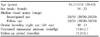
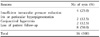
Patient demographics are shown in Table 1. The mean patient age was 59.2±12.8 years (range: 29 to 82 years) and the mean follow-up period was 15.2±9.1 months (range: 6 to 24 months). Eight patients had used ocular hypotensive medication before visiting our clinic, which was discontinued according to the washout schedule, and the other 55 patients were newly diagnosed with NTG. Among the 63 eyes, 47 eyes of 47 patients were treated for at least 12 months. The other 16 cases were not followed up for the full 12 months after treatment began; nine cases withdrew between 3 and 6 months, three cases withdrew between 6 and 9 months, and four cases withdrew between 9 and 12 months after treatment. The reasons for these withdrawals are presented in Table 2; the most common reason was spontaneous follow-up loss (50.0%).
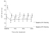
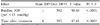
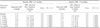
The mean IOP was significantly reduced after treatment, and the amount of IOP reduction was significantly greater in patients with a baseline IOP of ≥15 mmHg, compared to those with a baseline IOP of <15 mmHg (Table 3). The mean IOPs of the 63 patients versus time are shown in Table 4 and Figure 1. The mean baseline IOP before treatment was 15.0±2.73 mmHg. The missing IOP values of the 16 patients who were not followed up for 12 months after treatment began were imputed using the regression method, as described above. Significant reductions in IOP (p<0.05) from the baseline were obtained and maintained at two weeks and at 1, 3, 6, 9, and 12 months. The mean IOP reductions after administration of 0.005% latanoprost once daily were 2.6±0.2 (17.3%), 2.5±0.2 (16.7%), 2.3±0.2 (15.3%), 2.4±0.2 (16.0%), 2.3±0.2 (15.3%), and 2.4±0.2 (16.0%) at two weeks and 1, 3, 6, 9, and 12 months, respectively.
Patients with a baseline IOP of ≥15 mmHg achieved significantly higher IOP reductions than those with a baseline IOP of <15 mmHg at all follow-up periods (p<0.05). The groups versus time are shown in Figure 1 and Table 5. In patients with a baseline IOP of ≥15 mmHg, the mean IOP before treatment was 17.2 mmHg, and the IOP reduction from the baseline was 3.7 mmHg (21.5%) after 12 months of treatment. In those with a baseline IOP of <15 mmHg, the mean pretreatment IOP was 12.9 mmHg, and the IOP reduction was 1.1 mmHg (8.5%) after 12 months of treatment.
Age, gender, ocular laterality, lens status, and the time of day of IOP measurement were evaluated as potential confounding factors, but were not found to be significantly associated with IOP reduction.
Discussion
Treatment modalities for NTG are directed toward reducing IOP, which should prevent further deterioration of the visual field and further decay of the neuroretinal rim. It has been reported that reducing the IOP by 30% favorably influences NTG progression.7 Moreover, the findings that asymmetric NTG is often associated with asymmetric IOP,16-18 and that there is an inverse correlation between IOP and the neural rim area of the optic disc among NTG patients,19 suggest that the IOP, even when in the normal range, contributes to optic nerve damage.
Latanoprost is a prostaglandin analogue and reduces IOP by stimulating uveoscleral outflow.20 Long-term studies have shown the efficacy of latanoprost in patients with open-angle glaucoma and ocular hypertension, the reduction of IOP achieved by monotherapy was between 27.8 and 33%.21-25 In addition, latanoprost was reported to maintain IOP reduction, especially during the night, and to increase pulsatile ocular blood flow by 21%, indicating a remarkable increase in choroidal blood flow.26,27
Previously, the IOP-lowering effects of medication in NTG have been insufficient, because only modest IOP reductions could be achieved, but recently, several authors have suggested that latanoprost is effective in NTG patients. Tamada and colleagues28 performed a study on 31 patients, most of who had NTG, and were treated with once-daily latanoprost for eight weeks; they noted a 3.2 mmHg reduction in IOP (19.9%). In another study, Rulo and colleagues29 reported that in 29 patients with NTG, the IOP reduction was 3.6 mmHg (21.3%) at three weeks when treated with once-daily latanoprost 0.005%, and 2.4 mmHg (14.2%) when treated with twice-daily latanoprost 0.0015%. Both of these studies found that the patients with higher initial IOPs showed greater IOP reductions. The amount of IOP reduction in our study was smaller than those in previous reports by Tamada28 and Rulo.29 However, the baseline IOPs were 16.1 mmHg in the study by Tamada28 and 16.9 mmHg in the study by Rulo,29 while the baseline IOP in our study was 15.0 mmHg. Thus, the smaller IOP reduction in our study might be due to a lower baseline IOP compared to those in the previous reports by Tamada28 and Rulo.29 In our study, when the eyes with baseline IOP ≥15 mmHg (mean 17.2 mmHg) were considered, the IOP reduction after 12 months was 3.7 mmHg (21.5%), which was similar to the results of Rulo's study.29
In this study, latanoprost 0.005% administered once daily significantly reduced the IOP in NTG patients, and maintained this IOP reduction for up to 12 months. In 63 patients with NTG, the IOP reduction was 2.4 mmHg (16.0%) with 12 months of treatment. In most eyes, the full effect of latanoprost on IOP reduction was found to occur within two weeks from treatment initiation, and this reduction was maintained for at least 12 months with continued latanoprost treatment. Of the 63 eyes of 63 NTG patients treated with 0.005% latanoprost once a day, four eyes (6.3%) were withdrawn from latanoprost monotherapy due to insufficient IOP reduction during the 12-month follow-up. The risk of withdrawal from latanoprost during the 12-month treatment period due to an adverse event was 6.4%. Although conjunctival hyperemia was the most common side effect, most cases were mild, and this was not the main reason for discontinuance.
As in previous studies,28,29 we found that the higher the baseline IOP, the greater the IOP reduction, and that a statistically significant IOP reduction is more likely to occur at pre-treatment IOP levels of over 15 mmHg.
In conclusion, latanoprost was found to be well tolerated and to significantly reduce IOP in NTG patients. The initial IOP reductions by latanoprost were maintained throughout the 12-month treatment period, and latanoprost was found to be more effective at higher baseline IOP levels.

 XML Download