

 PDF
PDF ePub
ePub Citation
 Print
Print


Anatomical changes develop with time in the eyelid and orbit of anophthalmic patients who wear an ocular prosthesis following evisceration or enucleation. These changes consist primarily of enophthalmos, superior sulcus depression, upper lid ptosis, and lower lid laxity or retraction. These changes in the anophthalmic socket are defined as anophthalmic orbit syndrome.1 Lower lid laxity and retraction, which is relatively common in the anophthalmic sockets, was noted in 40% of 53 patients over a 5-year period,1 and caused cosmetic problems and inability to retain an ocular prosthesis safely.
The mechanisms suggested for lower lid laxity and retraction in the anophthalmic orbit are as follows. First, the orbital implant in an anophthalmic socket is predominantly supported by the lower lid, as is the prosthesis, and the weight and pressure effects of the orbital implant and prosthesis contribute to lower lid laxity and retraction.2 Second, frequent stretching of the canthal tendons by unnecessary and frequent removal of the prosthesis is another cause.3 Third, anatomical changes of the orbit such as the atrophy of structures supporting the lower lid and shrinkage of the posterior lamella are other causes of lower lid retraction.4
To correct lower lid laxity and retraction in anophthalmic patients, a fascia lata sling of the lower eyelid with or without horizontal lid shortening5,6 and a lateral tarsal strip procedure7,8 have been reported. Meanwhile, in such cases of lower lid retractions attributed to a contractured lower lid retractors such as thyroid associated ophthalmopathy,9 post-blepharoplasty,10 post orbital floor fracture repair,11 retractor recession and spacer insertion such as cartilage,12 or preserved sclera,13 or hard palate mucosa,14,15 have been widely used. However, retractor recession and posterior lamellar spacer insertion have rarely been reported for the correction of lower lid retraction (ptosis) in anophthalmic sockets.4,15
Lower lid entropion may develop in anophthalmic sockets because of cicatricial change (a deficiency of vertical lid height or fornix depth that results from chronic infection or inflammation in the socket) or involutional changes (lower lid laxity).2 In cases of mild cicatricial entropion of the lower lid, in which the inferior fornix is contractured minimally and deep enough, standard transverse blepharotomy (Wies operation) is sufficient to rotate the eyelid margin.2,16 When the inferior fornix is severely contractured and shallow, posterior lamellar lengthening procedures using various spacers such as fascia,17 sclera,18 cartilage,19 hard palate mucosa20 or PTFE (polytetrafluoroethylene)21 are preferred.
Moreover, when lower lid retraction is combined with entropion in cases of an anophthalmic socket wearing an ocular prosthesis, it seems rational that combinations of appropriate procedures could provide a solution. However, to our knowledge, no reports are available on the surgical results of simultaneous lower lid retraction correction combined with entropion in the anophthalmic orbit. Because severe cicatricial entropion needs posterior lamellar lengthening procedures, we considered that spacer insertions on the conjunctival side could correct the lower lid retraction and entropion, and that after spacer insertion, supplemental procedures of a less invasive and straightforward nature would be sufficient to correct residual entropion.
Thus we administered autogenous ear cartilage graft for posterior lamellar lengthening with selective and supplemental procedures, such as lateral tarsal strip and eyelasheverting procedure, to correct lower lid retraction combined with entropion in anophthalmic patients. We, here, report the surgical results of these combined procedures. The eyelasheverting procedure used was tarsal suturing of subciliary subcutaneous tissue, which is usually performed to correct epiblepharon.22-24
Materials and Methods
The medical records of 7 unilateral anophthalmic patients, who underwent posterior lamellar lengthening with autogenous ear cartilage graft and selective supplemental procedures, such as lateral tarsal strip or eyelash-everting procedure, for the simultaneous correction of lower lid retraction combined with entropion by one surgeon (SIK) from Mar 1998 to Mar 2003, were reviewed retrospectively. The ages of the seven patients ranged from 3 to 40 years (mean 16 years; sex ratio M:F = 2:5). The underlying causes of evisceration or enucleation are summarized in Table 1. The ocular prosthesis wearing time ranged from 29 months to 20 years (mean: 126 months) (Table 1). Preoperative status of anophthalmic sockets, including the amount of lower lid retraction and entropion, inferior fornix depth, surgical procedures, outcomes, and complications were investigated. In this study, the ear cartilage graft and supplemental procedures were indicated when an anophthalmic orbit was cosmetically unacceptable for lower lid retraction combined with entropion, and the inferior fornix was not severely contractured. Entropion was diagnosed if the shafts of the lower lid cilia were in an upright or inverted position and touched the ocular prosthesis. The degree of entropion was classified as (1) mild, when lower lid entropion disappeared after prosthesis removal, (2) moderate, when the shaft of the cilia remained in an upright position after taking off the prosthesis, or (3) severe, when the shaft of the cilia remained inverted after removing the prosthesis. The ear cartilage graft and supplemental procedures were performed for mild or moderate, not severe, entropion.
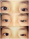
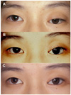
When the orbital volume was deficient, volume augmentation (implantation of a porous orbital implant) was performed about 6-12 months prior to the lid operations in order to reduce prosthesis thickness and weight. In five patients, primary or secondary implantation of a porous polyethylene orbital implant (Medpor®) was performed to correct for orbital volume deficiency such as enophthalmos, deep superior sulcus, and thick prosthesis, or to improve prosthetic motility, or remove an orbital cyst from 6 months to 1 year prior to the eyelid surgery. Prior surgical procedures are summarized in Table 1. Two patients (cases 1 and 2) with congenital anophthalmos, who had enophthalmos even after wearing a thick prosthesis over a primitive ocular remnant, underwent enucleation (removal of the primitive ocular remnant) and implantation of a 20-mm diameter, porous polyethylene orbital implant (Fig. 1). Another patient (case 4) who had undergone enucleation for retinoblastoma and implantation of a small Allen sphere for wearing a thick prosthesis, underwent removal of the small Allen sphere, secondary implantation of a 20-mm diameter, porous, polyethylene, orbital implant, and received an inferior fornix forming suture. Another patient (case 5) who was enucleated for retinoblastoma underwent excision of a large subconjunctival cyst, removal of a small Allen sphere and secondary implantation of a 20-mm diameter, porous, polyethylene, orbital implant (Fig. 2). Another patient (case 7), who was wearing a prosthesis over a severely phthisic eye and who showed deep superior sulcus deformity, wanted to improve the cosmesis and motility of his prosthesis, and therefore underwent enucleation and implantation of a 20-mm diameter, porous, polyethylene, orbital implant with later insertion of a motility coupling post.
Eyelid surgical procedures were performed after obtaining informed consent in all cases. General anesthesia was used in 5 patients (aged under 20 years), and local anesthesia in 2 patients (aged over 20 years).
Autogenous ear cartilage grafts were administered in all 7 cases. If lower lid retraction and entropion were not corrected sufficiently by ear cartilage grafting and horizontal lid laxity was present in the lower lid, a lateral tarsal strip procedure was performed supplementally to tighten the lower lid horizontally. When rotation of the cilia was insufficient after ear cartilage grafting and lateral tarsal strip procedure, tarsal suturing of subciliary subcutaneous tissue was added.22-24
1. Ear cartilage graft
After skin incision marking along the posterior surface of the helix, local anesthetic (2% lidocaine mixed with 1:100,000 epinephrine) was infiltrated subcutaneously on the anterior and posterior surface of the ear. By holding and everting the helix with a towel clamp, the posterior auricular surface was exposed. After skin incision along the previously marked line, dissection was performed between the posterior auricular surface skin and the ear cartilage to expose the ear cartilage. Cartilage of an adequate size and shape was then designed. To prevent ear deformity, ear cartilage with an auricular rim of at least 5 mm width was preserved.25 We attempted to harvest ear cartilage that was as flat as possible. The height of ear cartilage to be harvested was determined by the degree of lower lid retraction (approximately 1-2 mm of graft width for each millimeter of lid retraction)4 and the availability of flat cartilage.
The ear cartilage was incised as designed, dissected between the anterior auricular surface skin and cartilage, and excised at full-thickness. The skin incision was then closed. To prevent hematoma in the cartilage-deprived area, two or three sponges were then applied to the anterior and posterior surfaces of the helix, sutured through the full thickness, and removed one week later.
When the harvested cartilage had a curved contour, shallow incisions (scoring) were made on the concave surface to increase its flexibility and allow it to be flattened, and the margin of the harvested cartilage was trimmed to fit it into the recipient bed.
A horizontal conjunctival incision was made along the inferior margin of the lower lid tarsus to disinsert both the palpebral conjunctiva and the lower lid retractor from the lower tarsus. The lower lid retractor and conjunctiva were then recessed inferiorly. The harvested ear cartilage was placed in the space between the inferior border of the lower tarsus and the recessed lower retractor and conjunctiva, and sutured using 6-0 polyglactin (Fig. 3).
3. Eyelash-everting procedure
Tarsal suturing of subciliary subcutaneous tissue was performed as follows.22,23 A horizontal skin incision 2mm below the lower lid margin was followed by dissection between the lower tarsus and the pretarsal orbicularis oculi, fixation of subciliary subcutaneous tissue into the inferior margin of the lower tarsus with 8-0 Nylon (about 5 interrupted sutures), and finally skin repair. A Quickert suture was inserted as previously described.24
Results
The preoperative amount of lower lid retraction compared with the contralateral normal side ranged from 2 to 7 mm (mean: 4.6 mm) (Table 2). Entropion was mild in 5 patients and moderate in two.
During ear cartilage grafting, the height of the grafted ear cartilage, which was determined based on the available amount of flat cartilage in the patient's ear and on the degree of lower lid retraction, ranged from 4 mm to 7 mm.
In addition to ear cartilage grafting, supplemental procedures were performed in six patients (Table 2), while one patient (case 1) received only an ear cartilage graft without a lateral tarsal strip or eyelash-everting suture. Of the six patients that received supplementary procedures, one underwent the lateral tarsal strip procedure alone whereas five underwent both the lateral tarsal strip procedure and eyelash-everting suture insertion (tarsal suturing of subciliary subcutaneous tissue in four patients and Quickert suturing in one).
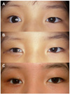
The postoperative follow-up period was 4-28 months (mean: 12.6 months). Lower lid retractions were corrected and there were no recurrences during follow-up (Fig. 1, 2, and 4). Lower lid entropions were corrected shortly after operation in all subjects, but recurred 4 to 6 months later in four (cases 1, 5, 6, and 7) (Fig. 2). Cilia of the lower lid were in the upright position, but did not touch the ocular prosthesis and did not require surgical corrections in any of the patients with recurred lower lid entropions. The epithelialization of ear cartilage from adjacent conjunctiva was complete 2-3 weeks after surgery in all patients. No cases of epithelialization failure occurred. All grafts showed good match with lower eyelid contours. All donor sites (posterior auricular surfaces) healed well without any complications including cosmetic deformity.
Discussion
To correct lower lid laxity (roughly, interchangeable with sagging or ptosis) in anophthalmic patients, various procedures have been suggested including lateral tarsal strip,7,26 fascia lata sling,5,6,27 autogenous ear cartilage graft into a preseptal or subcutaneous plane,28 or a lower lid prezygomatic flap with lateral canthoplasty.29
Spacer insertion on the conjunctival side of the lower eyelid is believed to be effective at correcting lower lid laxity or retraction in anophthalmos, but has rarely been investigated.4,12 Baylis et al4 reported on the successful surgical correction of lower lid retractions using an autogenous ear cartilage graft and a lateral tarsal strip, but their study subjects were mostly not anophthalmic patients and did not have combined entropion of the lower lid.
Lower lid entropions in anophthalmic patients may have cicatricial (chronic infection or inflammation in the socket, which leads to posterior lamellar shortening)30 or involutional causes.2 If entropion is caused by cicatricial changes of the posterior lamella, spacer insertions on the conjunctival side may correct the lower lid entropion, because severe cicatricial entropion requires posterior lamellar lengthening procedures. When lower lid laxity is present, entropion may develop due to the weight of the ocular prosthesis. When lower lid laxity is associated, the weight of the ocular prosthesis on the fornix imposes a constant downward force on the posterior lamella, which results in an inferior displacement of the fornix, while the anterior lamella remains in position. We believe that this discrepancy of force between the anterior and posterior lamella leads to lower lid entropion. We observed that in 5 of our 7 patients with sufficient inferior fornix, lower lid entropion disappeared when the prosthesis was removed, which supports this hypothesis.
Therefore, posterior lamellar lengthening by spacer insertion may resist the downward dragging of the posterior lamella and may ameliorate lower lid retraction and entropion simultaneously. In this study, we demonstrated that ear cartilage grafting alone corrected both lower lid retraction and entropion in one patient. For this reason, we believe that posterior lamellar lengthening by spacer insertion may represent a milestone for lower lid retraction in anophthalmic patients, especially if combined with entropion.
When lower lid entropions were not corrected sufficiently after ear cartilage grafting, we performed lateral tarsal strip as a supplementary procedure, because lower lid laxity has a greater tendency to lead to lower lid retraction31 and entropion.30 For the remaining cases of undercorrected lower lid entropion after ear cartilage grafting and the lateral tarsal strip procedure, tarsal suturing of subciliary subcutaneous tissue into the lower tarsus was undertaken. We believe that this stepwise approach effectively avoids unnecessary surgery, and achieves an appropriate amount of surgical correction whilst minimizing tissue injury.
Although the single lateral tarsal strip procedure without an ear cartilage graft might correct the lower lid laxity in the anophthalmos,7 lower lid entropion may require "more powerful" surgery such as the Wies operation16,32 or a mucous membrane graft.20 The Weis operation (transverse blepharotomy with marginal rotation) requires a full thickness eyelid incision, and, therefore, carries the risk of vascular injury with subsequent ischemic necrosis of the lid margin.33 We believe that spacer insertion provides strong lid elevation and that the lateral tarsal strip procedure provides additional elevation and eversion of the lower eyelid margin. Because of the fundamental effects of an ear cartilage graft and lateral tarsal strip, tarsal suturing of the subciliary subcutaneous tissue, which is performed using a half-thickness eyelid incision, and which is therefore less invasive and carries less risk than the Wies operation, could correct lower lid entropion.
It was found that lower lid entropion was resolved immediately after the eyelash-everting procedure in the present study, which suggests that the eyelash-everting procedure could be substituted for "more invasive" transverse blepharotomy with the help of spacer insertion and a lateral tarsal strip. The study also demonstrated that ear cartilage grafting could offer a major advance in the surgical correction of lower lid retraction combined with entropion in anophthalmic socket, and produce more durable and satisfactory results. A fascia lata sling may be effective at correcting lower lid retraction resulting from lower lid laxity, however, is not indicated for the correction of lower lid entropion, and cannot be performed with the Wies operation.
The delayed recurrence of lower lid entropions in 4 patients in the present study may have been due to progressing cicatricial contracture of the lower lid, but the exact mechanism of delayed recurrence remains to be elucidated. The loosening of suture adhesion of the tissue after the eyelash-everting procedure or primary marginal shortness of everting force may also be considered to be causes of the delayed recurrence of entropion. If this assumption about the limitation of the eyelash-everting procedure is true in anophthalmic socket, adding a procedure such as lid margin splitting or switching to more powerful entropion surgery may be considered to prevent recurrence of entropion.
We believe that ear cartilage is preferable to other spacers for posterior lamellar lengthening in anophthalmos. Although ear cartilage has disadvantages, such as an immobile lower eyelid during downgaze, an unnatural lid contour, being stiffer and thicker than tarsus, and ocular irritation due to the absence of a mucosal lining, it has the benefit of being resilient and thus is able to support the lower lid which must bear the weight of an ocular prosthesis and implant. Moreover, the delayed epithelialization of ear cartilage and the absence of mucosal lining, which may irritate ocular surfaces in other conditions, cause no discomfort in anophthalmos.
In conclusion, the results of this study suggest that lower lid retractions combined with entropion in anophthalmic patients can be effectively corrected by posterior lamellar lengthening with supplemental procedures such as lateral tarsal strip or an eyelash-everting procedure.
 XML Download