

 PDF
PDF ePub
ePub Citation
 Print
Print


Abstract
Purpose
To compare the efficacy and complications of scleral fixation of posterior chamber intraocular lens (IOL) and retropupillary fixation of iris claw IOL for dislocated IOL or aphakia without sufficient capsular support.
Methods
This retrospective study was comprised of 17 eyes of 16 patients undergoing scleral fixation and 14 eyes of 13 patients undergoing retropupillary fixation from August 2013 to June 2018. Uncorrected visual acuity (UCVA), best corrected visual acuity (BCVA), intraocular pressure (IOP), slit lamp examination, corneal topography, refractive indices, corneal curvatures, corneal endothelial cell density, and complications of both groups were examined preoperatively and 1 day, 1 week, 1 month, 2 months, and 6 months postoperatively.
Results
Six months after the operation, UCVA and BCVA improved in both groups; however, there were no significant differences between the two groups (UCVA, p = 0.162; BCVA, p = 0.418). IOP was temporarily higher in the scleral fixation group at one day postoperatively (p = 0.023). The mean absolute prediction error was smaller in the retropupillary iris fixation group at 6 months postoperatively (p = 0.034). Postoperative total astigmatism, corneal astigmatism, and corneal endothelial cell density were not significantly different between the two groups.
Conclusions
The retropupillary iris fixation group did not show significant improvement in visual acuity compared with the scleral fixation group. However, the retropupillary iris fixation group provided better mean absolute prediction error and a low risk of postoperative increase in IOP compared with the scleral fixation group. Retropupillary fixation of iris claw IOL is a promising option for scleral fixation of posterior chamber IOL for dislocated IOL or aphakia without sufficient capsular support.
Figures and Tables
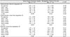
Figure 1
Changes of uncorrected visual acuity (UCVA), best corrected visual acuity (BCVA), and intraocular pressure (IOP) over time after scleral fixation of posterior chamber intraocular lens (IOL) or retropupillary fixation of iris claw IOL. (A) Six months after the operation, the UCVA in both groups significantly increased compared with that before the operation (the scleral fixation group, ‡p = 0.001; the retropupillary iris fixation group, ‡p = 0.001). In the sixth-month comparison, the UCVA in the retropupillary iris fixation group was not statistically better than the UCVA in the scleral fixation group (§p = 0.162). (B) Six months after the operation, the BCVA in both groups significantly increased compared with that before the operation (the scleral fixation group, ‡p = 0.001; the retropupillary iris fixation group, ‡p = 0.028). In the sixth-month comparison, the BCVA in the retropupillary iris fixation group was not statistically better than the BCVA in the scleral fixation group (§p = 0.418). (C) The IOP in the scleral fixation group was statistically higher than the IOP in the retropupillary iris fixation group in 1 day after the operation (†p = 0.023). Six months after the operation, the IOP in both groups did not significantly change compared with that before the operation, respectively (the scleral fixation group, ‡p = 0.481; the retropupillary iris fixation group, ‡p = 0.950). In the sixth-month comparison, the IOP in the scleral fixation group was not statistically higher than that in the retropupillary iris fixation group (§p = 0.058). logMAR = logarithm of minimal angle of resolution; Pre-op. = pre-operation; POD = post operative day; d = day; w = week; m = month(s). *Statistically significant differences (p < 0.05) among groups; †Mann Whitney U-test; ‡Wilcoxon signed rank test; §repeated-measures analysis of variance.
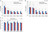
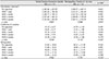
Figure 2
Changes of mean absolute prediction error over time after scleral fixation of posterior chamber intraocular lens (IOL) or retropupillary fixation of iris claw IOL. The mean absolute prediction error in the scleral fixation group was statistically higher than that in the retropupillary iris fixation group in 2 months, 6 months after the operation (*†p = 0.015, *†p = 0.034). Pre-op. = pre-operation; POD = post operative day; d = day; w = week; m = month(s). *Statistically significant differences (p < 0.05) among groups; †Mann Whitney U-test.
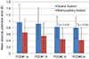
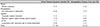
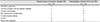
Table 2
Preoperative clinical characteristics of the scleral fixation group & the retropupillary iris fixation group
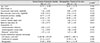
Table 3
Changes of autokeratometer refractive astigmatism, autokeratometer cornea front astigmatism, and Pentacam® cornea front astigmatism over time in the scleral fixation group & the retropupillary iris fixation group
References
1. Faria MY, Ferreira NP, Pinto JM, et al. Retropupillary iris claw intraocular lens implantation in aphakia for dislocated intraocular lens. Int Med Case Rep J. 2016; 9:261–265.

2. Gicquel JJ, Langman ME, Dua HS. Iris claw lenses in aphakia. Br J Ophthalmol. 2009; 93:1273–1275.
3. Jing W, Guanlu L, Qianyin Z, et al. Iris-claw intraocular lens and scleral-fixated posterior chamber intraocular lens implantations in correcting aphakia: a meta-analysis. Invest Ophthalmol Vis Sci. 2017; 58:3530–3536.
4. Zhang H, Zhao J, Zhang LJ, et al. Comparison of iris-fixated foldable lens and scleral-fixated foldable lens implantation in eyes with insufficient capsular support. Int J Ophthalmol. 2016; 9:1608–1613.
5. Wagoner MD, Cox TA, Ariyasu RG, et al. Intraocular lens implantation in the absence of capsular support: a report by the American Academy of Ophthalmology. Ophthalmology. 2003; 110:840–859.
6. Riazi M, Moghimi S, Najmi Z, Ghaffari R. Secondary artisan-verysise intraocular lens implantation for aphakic correction in post-traumatic vitrectomized eye. Eye (Lond). 2008; 22:1419–1424.
7. Teng H, Zhang H. Comparison of artisan iris-claw intraocular lens implantation and posterior chamber intraocular lens sulcus fixation for aphakic eyes. Int J Ophthalmol. 2014; 7:283–287.
8. Schallenberg M, Dekowski D, Hahn A, et al. Aphakia correction with retropupillary fixated iris-claw lens (Artisan) - long-term results. Clin Ophthalmol. 2014; 8:137–141.
9. Worst JG, Massaro RG, Ludwig HH. The introduction of an artificial lens into the eye using Binkhorst's technique. Ophthalmologica. 1972; 164:387–391.
10. Shenoy BH, Mittal V, Gupta A, et al. Refractive outcomes and prediction error following secondary intraocular lens implantation in children: a decade-long analysis. Br J Ophthalmol. 2013; 97:1516–1519.
11. Hoffer KJ, Savini G. IOL power calculation in short and long eyes. Asia Pac J Ophthalmol (Phila). 2017; 6:330–331.
12. Olsen T. Sources of error in intraocular lens power calculation. J Cataract Refract Surg. 1992; 18:125–129.
13. Kristianslund O, Østern AE, Drolsum L. Astigmatism and refractive outcome after late in-the-bag intraocular lens dislocation surgery: a randomized clinical trial. Invest Ophthalmol Vis Sci. 2017; 58:4747–4753.
14. Hayashi K, Hayashi H, Nakao F, Hayashi F. Intraocular lens tilt and decentration, anterior chamber depth, and refractive error after trans-scleral suture fixation surgery. Ophthalmology. 1999; 106:878–882.
15. Suto C, Hori S, Fukuyama E, Akura J. Adjusting intraocular lens power for sulcus fixation. J Cataract Refract Surg. 2003; 29:1913–1917.
16. Hara S, Borkenstein AF, Ehmer A, Auffarth GU. Retropupillary fixation of iris-claw intraocular lens versus transscleral suturing fixation for aphakic eyes without capsular support. J Refract Surg. 2011; 27:729–735.
17. Kaczmarek I, Prost M, Wasyluk J. Comparison of retropupillary iris-claw intraocular lens implantation and transscleral suture fixation of an intraocular lens for aphakic eyes. J Clin Diagn Res. 2018; 12:NC05–NC09.
18. Krause L, Bechrakis NE, Heimann H, et al. Implantation of scleral fixated sutured posterior chamber lenses: a retrospective analysis of 119 cases. Int Ophthalmol. 2009; 29:207–212.
19. Gonnermann J, Klamann MK, Maier AK, et al. Visual outcome and complications after posterior iris-claw aphakic intraocular lens implantation. J Cataract Refract Surg. 2012; 38:2139–2143.
20. Baykara M, Ozcetin H, Yilmaz S, Timuçin OB. Posterior iris fixation of the iris-claw intraocular lens implantation through a scleral tunnel incision. Am J Ophthalmol. 2007; 144:586–591.
21. Pavlin CJ, Rootman D, Arshinoff S, et al. Determination of haptic position of transsclerally fixated posterior chamber intraocular lenses by ultrasound biomicroscopy. J Cataract Refract Surg. 1993; 19:573–577.
22. Bellucci R, Marchini G, Morselli S, et al. Scleral fixation re-examined by ultrasound biomicroscopy. J Cataract Refract Surg. 1995; 7:326–330.
23. Stark W, Gottsch J, Goodman D, et al. Posterior chamber intraocular lens implantation in the absence of capsular support. Arch Ophthalmol. 1989; 107:1078–1083.
24. Uozato H, Okada Y, Hirai H, Saishin M. What is the tolerable limits of the IOL tilt and decentration? Jpn J Ophthalmol. 1988; 82:2308–2311.
25. Gabor SG, Pavlidis MM. Sutureless intrascleral posterior chamber intraocular lens fixation. J Cataract Refract Surg. 2007; 33:1851–1854.
26. Yamane S, Sato S, Maruyama-Inoue M, Kadonosono K. Flanged intrascleral intraocular lens fixation with double-needle technique. Ophthalmology. 2017; 124:1136–1142.
27. Alió JL, Mulet ME, Shalaby AM. Artisan phakic iris claw intraocular lens for high primary and secondary hyperopia. J Refract Surg. 2002; 18:697–707.
28. Kaynak S, Ozbek Z, Pasa E, et al. Transscleral fixation of foldable intraocular lenses. J Cataract Refract Surg. 2004; 30:854–857.
29. Dadeya S, Kumari Sodhi KP. Secondary intraocular lens (IOL) implantation: anterior chamber versus scleral fixation long-term comparative evaluation. Eur J Ophthalmol. 2003; 13:627–633.
30. Hazar L, Kara N, Bozkurt E, et al. Intraocular lens implantation procedures in aphakic eyes with insufficient capsular support associated with previous cataract surgery. J Refract Surg. 2013; 29:685–691.
 XML Download