

 PDF
PDF ePub
ePub Citation
 Print
Print


Abstract
Purpose
To report the pathological classification and incidence of conjunctival tumors in Korean patients.
Methods
A retrospective chart review was conducted for 234 patients who underwent excisional biopsy of conjunctival neoplasia between January 2007 and December 2016 at Soonchunhyang University Hospital. The clinical features, histological classification, and frequency of pigmented conjunctival masses were investigated.
Results
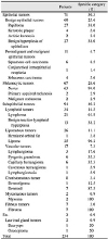
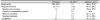
The mean age of the 234 patients who underwent excisional biopsy was 37.9 ± 21.6 years. The most common location of conjunctival lesions was the bulbar conjunctiva in 145 patients (62%), with pigmentary lesions in 68 patients (29.2%). Most tumors were benign (n = 201, 85.9%), while 34 malignant tumors were found as follows: 71 patients (30.3%) had epithelial tumors, 67 (28.6%) had melanocytic tumors, 33 (14.1%) had lymphoid tumors, and 26 (11.1%) had lipomatous tumors.
Conclusions
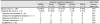
Most of the conjunctival tumors in Korean patients were benign. Epithelial tumors were the most common, and melanocytic tumors were less common then in Western studies. However, clinically malignant cases were not uncommon, occurring at a rate of 14.1%. Therefore, conjunctival tumors in elderly patients, lesions that are large, and the presence of a conjunctival sac are more likely to be malignant. Therefore, accurate histological diagnosis and treatment are needed.
Figures and Tables
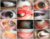
Figure 1
Representative pictures of conjunctival masses. (A) Nevus, (B) Conjunctival papilloma, (C) Lipoma, (D) Fibroma, (E) Pyogenic granuloma, (F) Capillary hemangioma, (G) Primary acquired melanosis, (H) Herniated orbital fat, (I) MALT lymphoma, (J) Squamous cell carcinoma, (K) Sebaceous cell carcinoma, (L) Malignant melanoma.
References
1. Shields JA, Shields CL. Atlas of eyelid and conjunctival tumors. Philadelphia: Lippincott Williams & Wilkins;1999. p. 98–99.
2. Shields CL, Shields JA. Tumors of the conjunctiva and cornea. Surv Ophthalmol. 2004; 49:3–24.


3. Othman IS. Ocular surface tumors. Oman J Ophthalmol. 2009; 2:3–14.

4. Young JL Jr. Surveillance, epidemiology, and end results: incidence and mortality data:1973–1977. Natl Cancer Inst Monogr. 1981; 1–1082.
5. Mahoney MC, Burnett WS, Majerovics A, Tanenbaum H. The epidemiology of ophthalmic malignancies in New York State. Ophthalmology. 1990; 97:1143–1147.
6. Seregard S, Koch E. Conjunctival malignant melanoma in Sweden 1969–91. Acta Ophthalmol (Copenh). 1992; 70:289–296.
7. Shields CL, Demirci H, Karatza E, Shields JA. Clinical survey of 1643 melanocytic and nonmelanocytic conjunctival tumors. Ophthalmology. 2004; 111:1747–1754.
8. Shields CL, Alset AE, Boal NS, et al. Conjunctival tumors in 5002 cases. comparative analysis of benign versus malignant counterparts. The 2016 James D. Allen lecture. Am J Ophthalmol. 2017; 106–133.
9. Grossniklaus HE, Green WR, Luckenbach M, Chan CC. Conjunctival lesions in adults. A clinical and histopathologic review. Cornea. 1987; 6:78–116.
10. Choi JH, Chi MJ, Baek SH. Clinical analysis of benign eyelid and conjunctival tumors. J Korean Ophthalmol Soc. 2003; 44:1268–1277.
11. Ash JE. Epibulbar tumors. Am J Ophthalmol. 1950; 33:1203–1219.
12. Wang Y, Zhao Y, Ma S. Racial differences in six major subtypes of melanoma: descriptive epidemiology. BMC Cancer. 2016; 16:691.
13. Nasiri AM, Al-Akeel ES, Rayes NH. Differences in survival by race/ethnicity among cutaneous melanoma patients in the United States over a period from 1982 to 2011. Int J Adv Med. 2018; 5:5–10.
14. Jang SG, Park BG, Park YM, Lee JS. Clinical manifestations of extruded conjunctival melanocytic mass. J Korean Ophthalmol Soc. 2016; 57:1691–1698.
15. Jakobiec FA, Folberg R, Iwamoto T. Clinicopathologic characteristics of premalignant and malignant melanocytic lesions of the conjunctiva. Ophthalmology. 1989; 96:147–166.
16. Reese AB. Tumors of the Eye. 2nd ed. New York: Harper & Row;1963. p. 161.
17. Brownstein S. Malignant melanoma of the conjunctiva. Cancer Control. 2004; 11:310–316.
18. Shields CL, Shields JA, Carvalho C, et al. Conjunctival lymphoid tumors: clinical analysis of 117 cases and relationship to systemic lymphoma. Ophthalmology. 2001; 108:979–984.
 XML Download