

 PDF
PDF ePub
ePub Citation
 Print
Print


Abstract
Purpose
We compared the anterior segment measurements using a Scheimpflug camera (Pentacam® [Oculus Inc., Wetzlar, Germany]) and a new anterior segment module of optical coherence tomography (Cirrus HD-OCT 5000® [Carl Zeiss Meditec, Dublin, CA, USA]).
Methods
Anterior segment measurements were evaluated in 47 eyes of 26 patients who visited for the purpose of cataract surgery. Measurements of the central corneal thickness (CCT), anterior chamber depth (ACD), anterior chamber angle (ACA; nasal and temporal side), and angle to angle distance (ATA) were compared between the Pentacam® and the new anterior seg-ment module of the Cirrus HD-OCT 5000®.
Results
The mean CCTs measured by the Pentacam® and the new anterior segment module of the Cirrus HD-OCT 5000® were 561.68 ± 31.35 μ m and 559.04 ± 36.43 μ m, respectively. There was no statistically significant difference (p = 0.387) and high correlations in the results using the two methods (r = 0.824, p < 0.001). The mean ACD measurements using the Pentacam® and Cirrus HD-OCT 5000® were 2.70 ± 0.44 mm and 2.68 ± 0.42 mm, respectively. The measurements were strongly correlated (r = 0.981, p < 0.001) without a statistically significant difference (p = 0.091). The measurements of ACA (nasal and temporal side) and ATA did not differ significantly different between the two methods (p = 0.109, p = 0.153, p = 0.322, respectively). High correlations were observed between the two methods (r = 0.944, r = 0.951, r = 0.955, respectively; p < 0.001 in all groups).
Conclusions
There was no significant difference between the CCT, ACD, ACA, and ATA measured by the Pentacam® and the new anterior segment module of the Cirrus HD-OCT 5000® in adult patients. However, there was a low degree of agreement for the CCT. The measurements of ACD, ACA, and ATA can therefore be considered interchangeable when used clinically.
References
1. Holladay JT. Standardizing constants for ultrasonic biometry, abdominal, and intraocular lens power calculations. J Cataract Refract Surg. 1997; 23:1356–70.
2. Alió JL, de la Hoz F, Pérez-Santonja JJ, et al. Phakic anterior abdominal lenses for correction of myopia; a 7-year cumulative analysis of complications in 263 cases. Ophthalmology. 1999; 106:458–66.
3. Olsen T. Sources of error in intraocular lens power calculation. J Cataract Refract Surg. 1992; 18:125–9.

4. Ou RJ, Shaw EL, Glasgow BJ. Keratectasia after laser in situ abdominal (LASIK): evaluation of the calculated residual stromal bed thickness. Am J Ophthalmol. 2002; 134:771–3.
5. Kashiwagi K, Tokunaga T, Iwase A, et al. Agreement between abdominal anterior chamber depth evaluation using the van Herick technique and angle width evaluation using the Shaffer system in Japanese. Jpn J Ophthalmol. 2005; 49:134–6.
6. Youn SM, Lim SH, Lee HY. Measurement comparison of anterior segment parameters between AL-Scan(R) and Pentacam(R). J Korean Ophthalmol Soc. 2014; 55:801–8.
7. Konstantopoulos A, Hossain P, Anderson DF. Recent advances in ophthalmic anterior segment imaging: a new era for ophthalmic abdominal? Br J Ophthalmol. 2007; 91:551–7.
8. Sakata LM, Wong TT, Wong HT, et al. Comparison of Visante and abdominal anterior segment optical coherence tomography in abdominal the anterior chamber angle. Eye (Lond). 2010; 24:578–87.
9. Chen D, Lam AK. Intrasession and intersession repeatability of the Pentacam system on posterior corneal assessment in the normal human eye. J Cataract Refract Surg. 2007; 33:448–54.
10. Miranda MA, Radhakrishnan H, O'Donnell C. Repeatability of corneal thickness measured using an Oculus Pentacam. Optom Vis Sci. 2009; 86:266–72.
11. Cheong YJ, Lee BR, Han KE, Jun RM. Corneal thickness abdominals using 2 kinds of spectral domain optical coherence abdominal, Pentacam, ultrasound pachymetry. J Korean Ophthalmol Soc. 2016; 57:1527–34.
12. Ha DY, Jung JW. Comparison of anterior segment parameters abdominal by anterior segment optical coherence tomography and dual rotating Scheimpflug camera. J Korean Ophthalmol Soc. 2017; 58:1341–8.
13. Jea SY, Jung SC, Oum BS. Quantified values of anterior chamber depth and angle measurements using ultrasound biomicroscopy and topography. J Korean Ophthalmol Soc. 2006; 47:97–104.
14. Park YS, Ahn H, Kim NR, et al. Comparison of measurement of abdominal segment parameters between Scheimpflug camera and abdominal biomicroscopy. J Korean Ophthalmol Soc. 2009; 50:1847–52.
15. Dada T, Sihota R, Gadia R, et al. Comparison of anterior segment optical coherence tomography and ultrasound biomicroscopy for assessment of the anterior segment. J Cataract Refract Surg. 2007; 33:837–40.
16. Kim DW, Yi KY, Choi DG, Shin YJ. Corneal thickness measured by dual Scheimpflug, anterior segment optical coherence abdominal, and ultrasound pachymetry. J Korean Ophthalmol Soc. 2012; 53:1412–8.
17. Borrego-Sanz L, Sáenz-Francés F, Bermudez-Vallecilla M, et al. Agreement between central corneal thickness measured using Pentacam, ultrasound pachymetry, specular microscopy and optic biometer Lenstar LS 900 and the influence of intraocular pressure. Ophthalmologica. 2014; 231:226–35.
18. O'Donnell C, Hartwig A, Radhakrishnan H. Comparison of Central Corneal Thickness and Anterior Chamber Depth Measured Using LenStar LS900, Pentacam, and Visante AS-OCT. Cornea. 2012; 31:983–8.
19. Fu J, Wang X, Li S, et al. Comparative study of anterior segment measurement with Pentacam and anterior segment optical abdominal tomography. Can J Ophthalmol. 2010; 45:627–31.
20. Doors M, Cruysberg LP, Berendschot TT, et al. Comparison of abdominal corneal thickness and anterior chamber depth measurements using three imaging technologies in normal eyes and after phakic intraocular lens implantation. Graefes Arch Clin Exp Ophthalmol. 2009; 247:1139–46.
21. Dinc UA, Gorgun E, Oncel B, et al. Assessment of anterior abdominal depth using Visante optical coherence tomography, slitlamp optical coherence tomography, IOL Master, Pentacam and Orbscan IIz. Ophthalmologica. 2010; 224:341–6.
22. Yazici AT, Bozkurt E, Alagoz C, et al. Central corneal thickness, anterior chamber depth, and pupil diameter measurements using Visante OCT, Orbscan, and Pentacam. J Cataract Refract Surg. 2010; 26:127–33.
23. Rabsilber TM, Khoramnia R, Auffarth GU. Anterior chamber measurements using Pentacam rotating Scheimpflug camera. J Cataract Refract Surg. 2006; 32:456–9.
24. Shin CJ, Lee JE, Kim JY, Tchah HW. Changes in anterior chamber depth and angle after phacoemulsification measured by anterior segment optical coherence tomography. J Korean Ophthalmol Soc. 2010; 51:353–8.
25. Lee DG, Choi SH. Measurement of anterior segment using visante OCT in Koreans. J Korean Ophthalmol Soc. 2009; 50:542–50.
26. Han JY, Kim YC. Relation between ocular biometry and abdominal parameters in adult Koreans with cataracts. J Korean Ophthalmol Soc. 2016; 57:1205–9.
27. Kim TG, Moon SW, Yang JH, Jin KH. Clinical usefulness of UBM in the sitting position in anterior chamber depth and angle measurements. J Korean Ophthalmol Soc. 2014; 55:1007–16.
28. Yi JH, Lee H, Hong S, et al. Anterior chamber measurements by Pentacam and AS-OCT in eyes with normal open angles. Korean J Ophthalmol. 2008; 22:242–5.
29. Mou D, Fu J, Li S, et al. Narrow- and open-angle measurements with anterior-segment optical coherence tomography and Pentacam(TM). Ophthalmic Surg Lasers Imaging. 2010; 41:622–8.
30. Lee JH, Park WC, Rho SH. The effects of pilocarpine on the anterior chamber depth and angle. J Korean Ophthalmol Soc. 1994; 35:572–9.
31. Kim SK, Kim HM, Song JS. Comparison of internal anterior abdominal diameter imaging modalities: 35-MHz ultrasound abdominal, Visante optical coherence tomography, and Pentacam. J Cataract Refract Surg. 2010; 26:120–6.
Figure 1.
Device picture. New anterior segment module that can be attached to Cirrus HD-OCT 5000® (Carl Zeiss Meditec, Dublin, CA, USA).
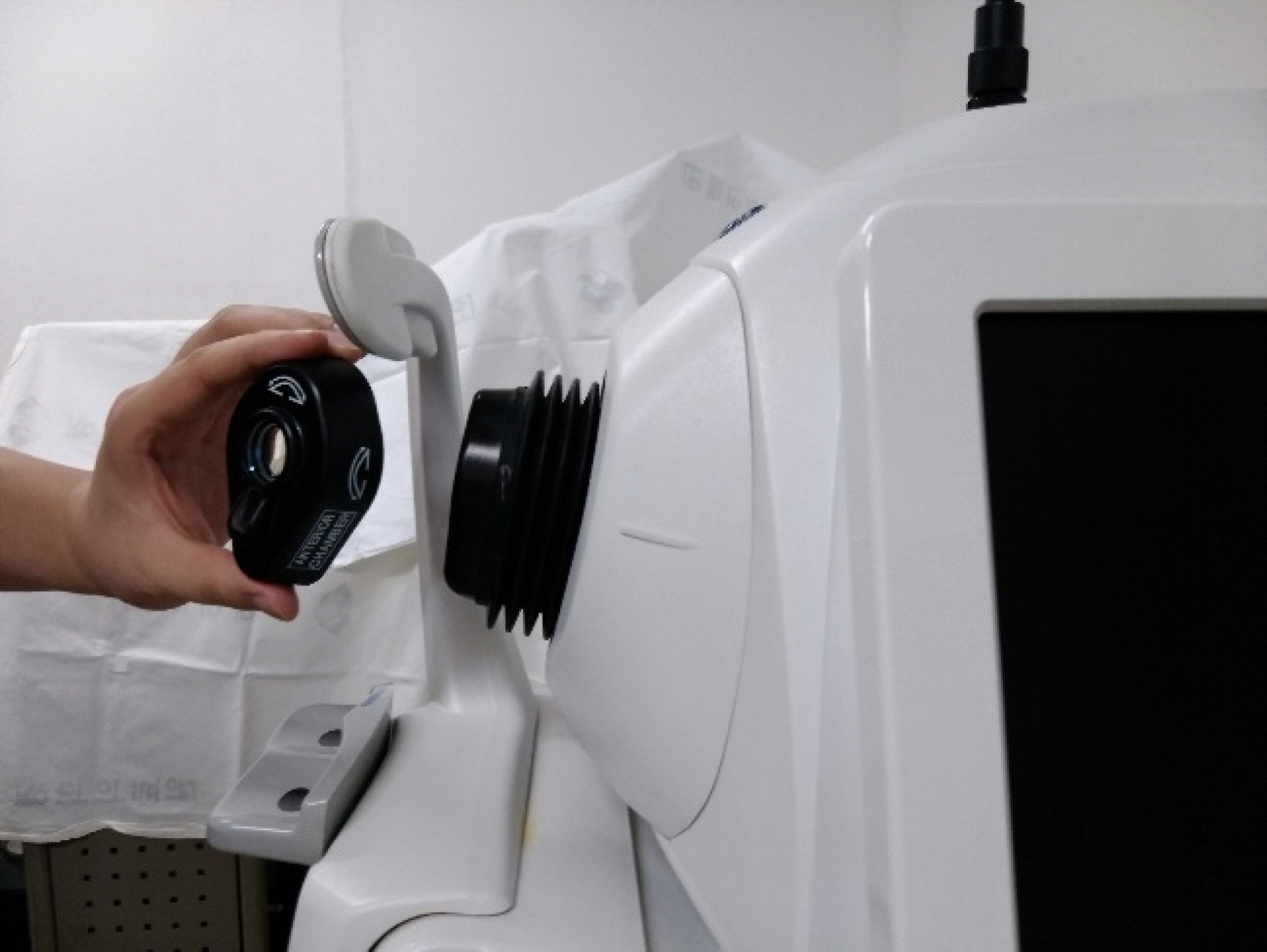
Figure 2.
An example of anterior chamber mode of the Cirrus HD-OCT 5000® (Carl Zeiss Meditec, Dublin, CA, USA). Anterior chamber analysis screen with linear measurement tool of the Cirrus HD-OCT 5000®.
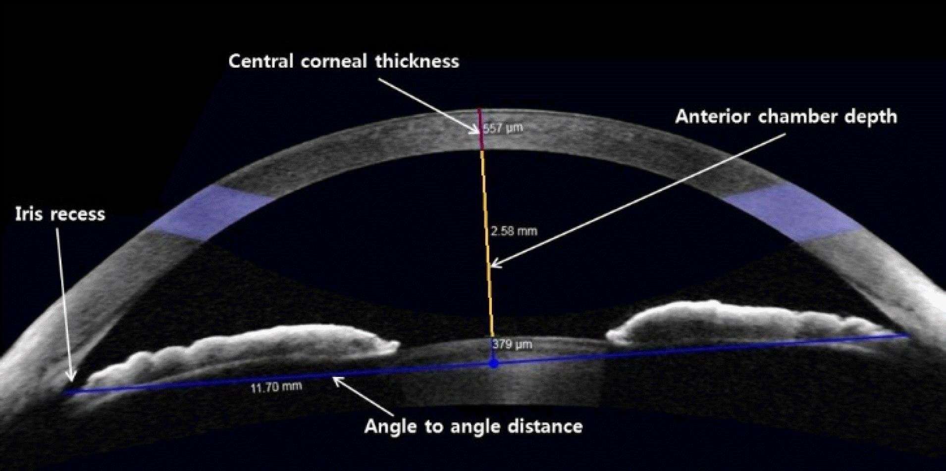
Figure 3.
An example of wide angle to angle mode of the Cirrus HD-OCT 5000® (Carl Zeiss Meditec, Dublin, CA, USA). The anterior chamber angle was measured on a line per-pendicular to the trabecular meshwork at points 500 μ m from the scleral spur.
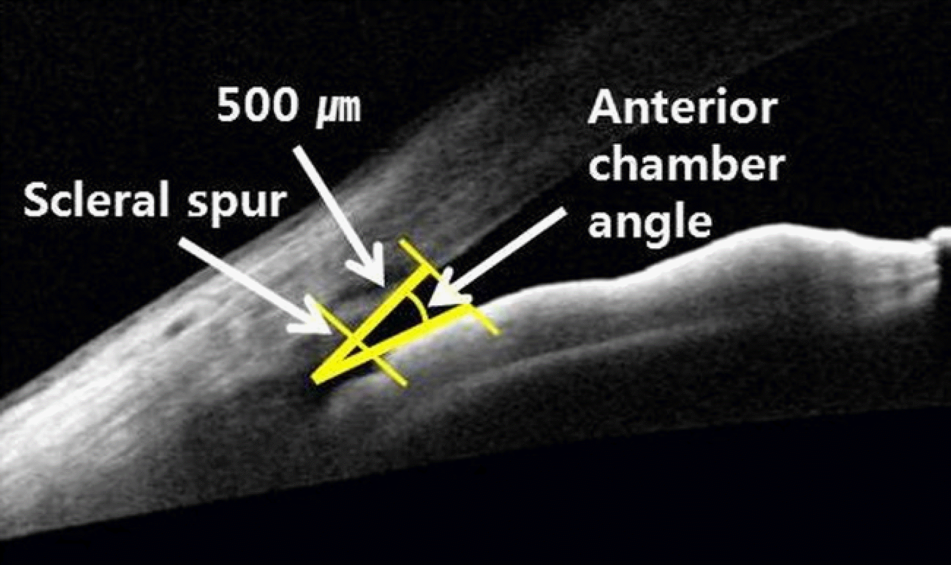
Figure 4.
Pearson correlation of Pentacam® (Oculus Inc., Wetzlar, Germany) and Cirrus OCT® (Carl Zeiss Meditec, Dublin, CA, USA) in anterior segment parameters. (A) Central corneal thickness (CCT). (B) Anterior chamber depth (ACD). (C) Anterior chamber angle (nasal) (ACA_N). (D) Anterior chamber angle (temporal) (ACA_T). (E) Angle to angle distance (ATA).

Figure 5.
Bland-Altman plots of Pentacam® (Oculus Inc., Wetzlar, Germany) and Cirrus OCT® (Carl Zeiss Meditec, Dublin, CA, USA) in anterior segment parameters. (A) Central corneal thickness (CCT). (B) Anterior chamber depth (ACD). (C) Anterior chamber angle (nasal) (ACA_N). (D) Anterior chamber angle (temporal) (ACA_T). (E) Angle to angle distance (ATA).
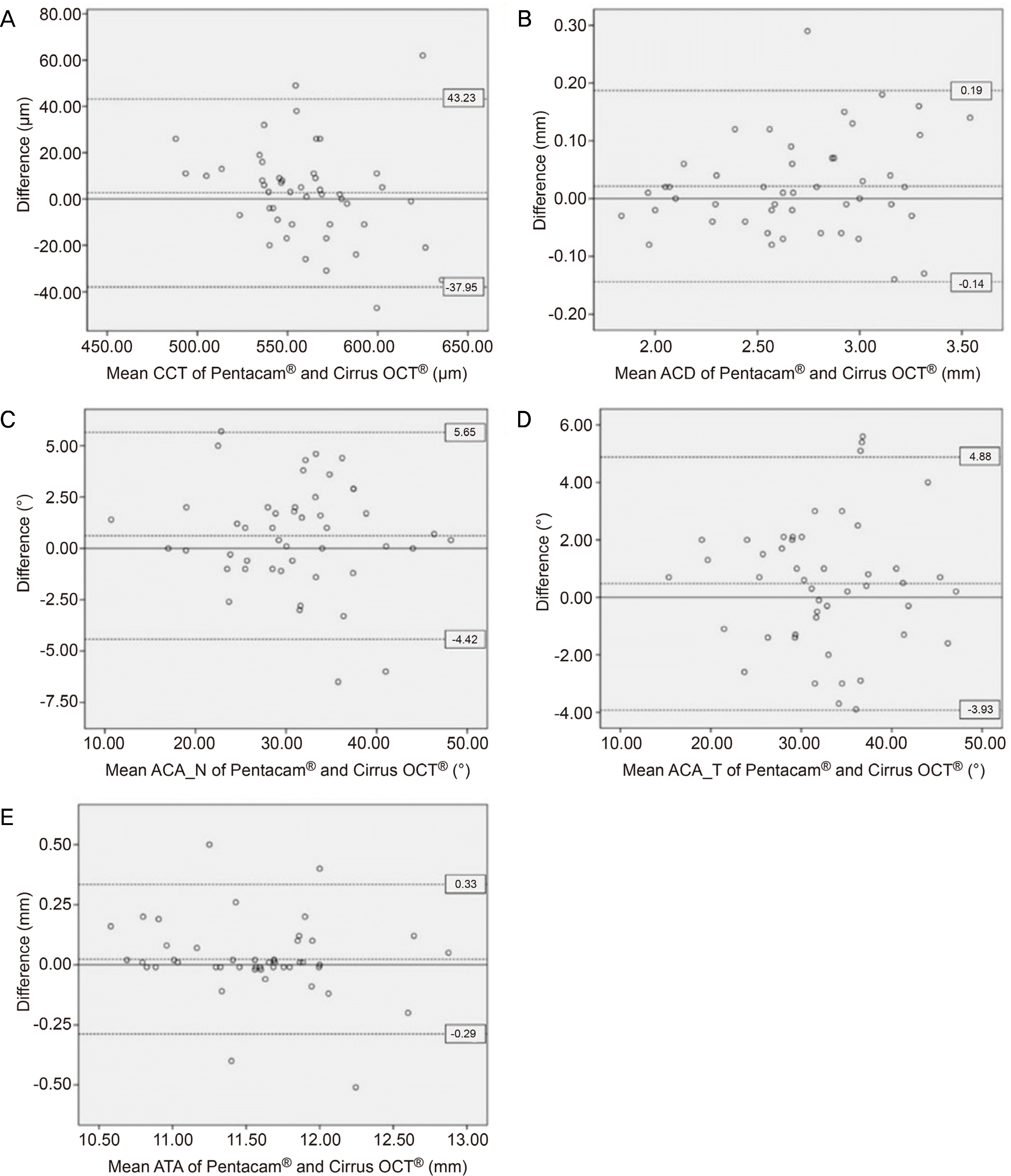
Table 1.
Comparison of anterior segment measurements using Pentacam® and Cirrus OCT®
| Pentacam® | Cirrus OCT® | p-value* | r | |
|---|---|---|---|---|
| CCT (μ m) | 561.68 ± 31.35 | 559.04 ± 36.43 | 3 0.387 | 0.824 |
| ACD (mm) | 2.70 ± 0.44 | 2.68 ± 0.42 | 0.091 | 0.981 |
| ACA_N (°) | 31.25 ± 7.46 | 30.64 ± 7.76 | 0.109 | 0.944 |
| ACA_T (°) | 32.80 ± 7.21 | 32.32 ± 7.18 | 0.153 | 0.951 |
| ATA (mm) | 11.58 ± 0.50 | 11.56 ± 0.53 | 0.322 | 0.955 |
Table 2.
Intercorrelation of each method for anterior segment measurements
| ICC (95% CI) (Pentacam®-Cirrus OCT®) | |
|---|---|
| CCT (μ m) | 0.898 (0.816–0.943) |
| ACD (mm) | 0.990 (0.982–0.995) |
| ACA_N (°) | 0.971 (0.947–0.984) |
| ACA_T (°) | 0.975 (0.952–0.986) |
| ATA (mm) | 0.976 (0.955–0.988) |
Table 3.
Intraexaminer repeatability of each method for ante rior segment measurements
 XML Download