

 PDF
PDF ePub
ePub Citation
 Print
Print


Abstract
Purpose
To investigate sexual differences and correlations among refractive error, axial length (AL), and corneal power.
Methods
A retrospective review of the medical records for 2,006 eyes of children aged 5–16 years was conducted. Cycloplegic refraction and AL measurements were performed on all eyes. Sexual differences in corneal power and AL were investigated in emmetropic eyes and after adjustment for the spherical equivalent (SE). The distribution of AL with every 1 diopter (D) interval was determined. Quantitative correlations among SE, corneal power, and AL were analyzed using multiple regression analyses.
Results
The mean age of the subjects was 7.62 years and the mean SE was −0.10 D. Males had a longer AL and lower corneal power than females both in emmetropic eyes and in all subjects after adjustment for the SE. The AL increased 0.40 mm for every −1 D change of the SE. When compared to a 1 D interval of the SE, the AL difference between the upper and lower values of a 95% confidence interval was 2.98 mm, which showed the variability of the AL distribution. Eyes with a long AL had lower corneal power (p < 0.001). Every 1 mm change of AL resulted in a −2.1 D change in the SE, and every 1 D change of corneal power resulted in a −0.8 D change in the SE (p < 0.001).
References
1. Lee MJ, Lee YH, Shyn KH. The progression of myopia with age. J Korean Ophthalmol Soc. 1987; 28:151–5.
2. Goss DA, Cox VD, Herrin-Lawson GA, et al. Refractive error, abdominal length, and height as a function of age in young myopes. Optom Vis Sci. 1990; 67:332–8.
3. Jung SI, Han J, Kwon JW, et al. Analysis of myopic progression in childhood using the Korea National Health and Nutrition Examination Survey. J Korean Ophthalmol Soc. 2016; 57:1430–4.

4. Zadnik K. The Glenn A. Fry Award Lecture (1995). Myopia abdominal in childhood. Optom Vis Sci. 1997; 74:603–8.
5. Chia A, Lu QS, Tan D. Five-year clinical trial on atropine for the treatment of myopia 2: myopia control with atropine 0.01% eyedrops. Ophthalmology. 2016; 123:391–9.
6. Cho P, Cheung SW. Retardation of myopia in orthokeratology (ROMIO) study: a 2-year randomized clinical trial. Invest Ophthalmol Vis Sci. 2012; 53:7077–85.
7. Ojaimi E, Rose KA, Morgan IG, et al. Distribution of ocular abdominal parameters and refraction in a population-based study of Australian children. Invest Ophthalmol Vis Sci. 2005; 46:2748–54.
8. Twelker JD, Mitchell GL, Messer DH, et al. Children's ocular abdominal and age, gender, and ethnicity. Optom Vis Sci. 2009; 86:918–35.
9. Li SM, Wang N, Zhou Y, et al. Paraxial schematic eye models for 7- and 14-year-old Chinese children. Invest Ophthalmol Vis Sci. 2015; 56:3577–83.
10. Li SM, Li SY, Kang MT, et al. Distribution of ocular biometry in 7- and 14-year-old Chinese children. Optom Vis Sci. 2015; 92:566–72.
11. Li SM, Iribarren R, Kang MT, et al. Corneal power, anterior abdominal length and lens power in 14-year-old Chinese children: the Anyang childhood eye study. Sci Rep. 2016; 6:20243.
12. Xiong S, Zhang B, Hong Y, et al. The associations of lens power with age and axial length in healthy Chinese children and adolescents aged 6 to 18 Years. Invest Opthalmol Vis Sci. 2017; 58:5849–55.
13. Olsen T. On the calculation of power from curvature of the cornea. Br J Ophthalmol. 1986; 70:152–4.
14. Lee EK, Lee DB, Jin KH, Kim JM. The study of the correlation abdominal axial length and refractive error in Korean children. J Korean Ophthalmol Soc. 1993; 34:654–60.
15. Jung JW, Kim YE, Paik HJ. Clinical comparison of autorefractor versus retinoscopic refraction in children according to the age. J Korean Ophthalmol Soc. 2005; 46:1931–5.
16. Choi MJ, Baek SH, Gong SM. Comparison of autorefraction and clinical refraction with or without in children. J Korean Ophthalmol Soc. 2005; 46:837–46.
Figure 1.
Box-and-Whiskers plot for the distribution of axial length according to spherical equivalent (inverted X-axis). Box denoted interquartile range (IQR, 25th–75th percentile) of axial length for a specific spherical equivalent value. Whiskers denoted 1.5 × IQR, and dots denoted outliers. A horizontal line and a diamond in a box denoted median and mean value of axial length, each. N = number of subjects; AL = axial length; CI = confidence interval.
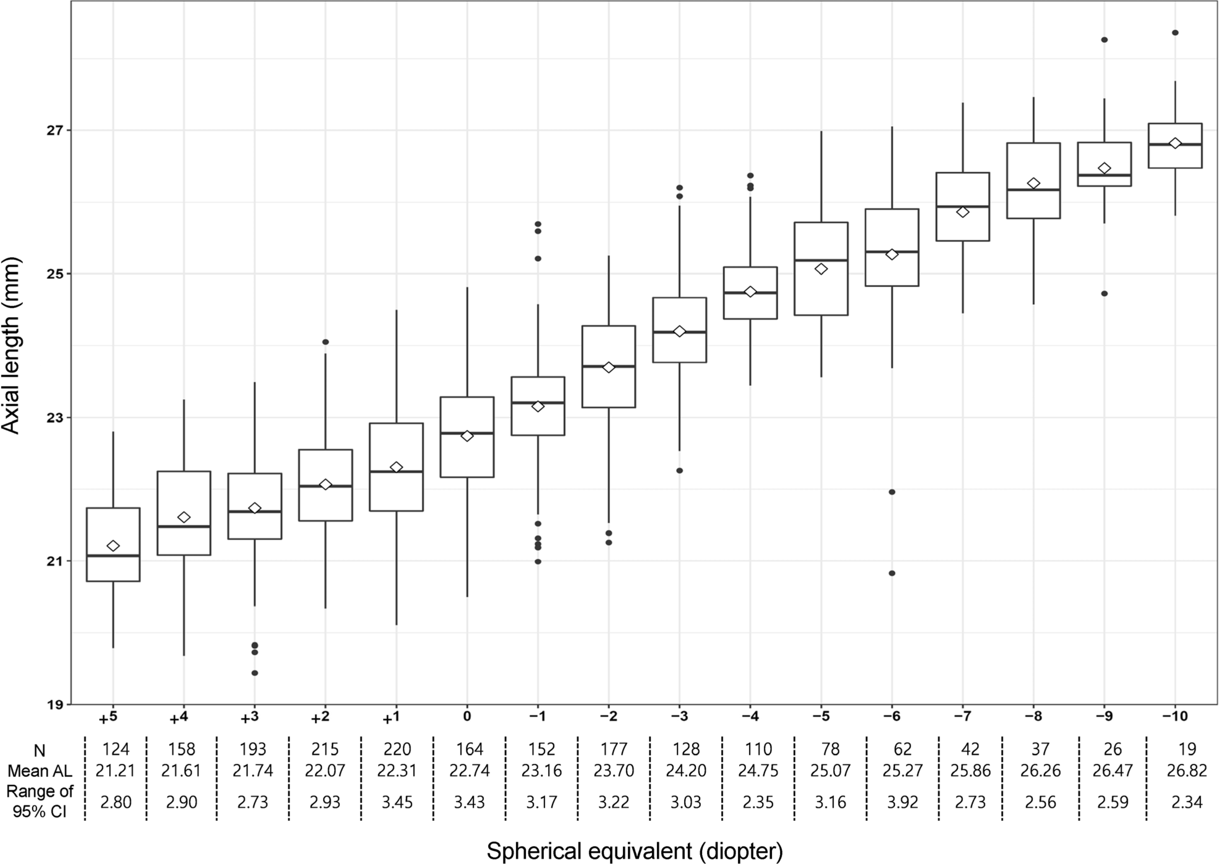
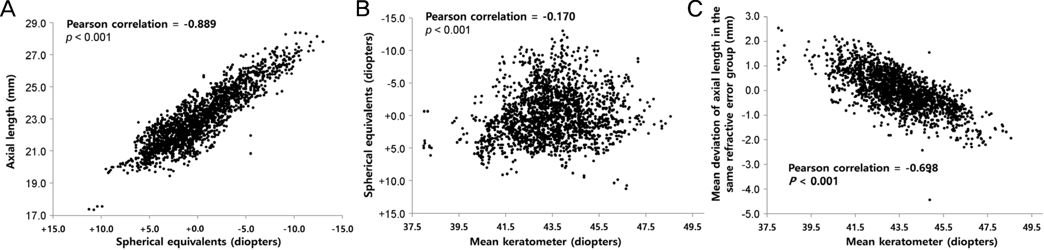
Figure 2.
Correlation among ocular parameters and refractive parameters. (A) Definite negative correlation between spherical equivalents (inverted X-axis) and axial length was noted. (B) Weak correlation between mean keratometer and spherical equivalent (inverted Y-axis) was noted. (C) Definite negative correlation between mean keratometer and mean deviation of axial length in the same refractive error group.
 XML Download