

 PDF
PDF ePub
ePub Citation
 Print
Print


Abstract
Purpose
To compare the new swept-source optical coherence tomography based IOL Master 700 to both the partial coherence interferometry based IOL Master 500 and ultrasound A-scan in terms of the ocular biometry and the prediction of postoperative refractive outcomes.
Methods
A total 67 eyes of 55 patients who received cataract surgery were included in our study. The axial length, anterior chamber depth, and keratometry were measured using IOL Master 700, IOL Master 500, and A-scan. The predictive errors, which are the differences between predictive refraction and post-operative refraction 1 month after surgery, were also compared.
Results
Axial length measurements were not successful in 5 eyes measured using IOL Master 700 and in 12 eyes measured using IOL Master 500. The mean absolute postoperative refraction predictive errors were 0.63 ± 0.50 diopters, 0.66 ± 0.51 diopters, and 0.62 ± 0.51 diopters for IOL Master 700, IOL Master 500, and A-scan, respectively, and these values exhibited no statistically significant differences. The mean axial lengths were 24.25 ± 2.41 mm, 24.24 ± 2.40 mm, and 24.22 ± 2.39 mm; the mean anterior chamber depths were 3.09 ± 0.39 mm, 3.17 ± 0.39 mm, and 3.15 ± 0.46 mm; and the mean keratometry values were 44.12 ± 1.82 diopters, 44.57 ± 2.10 diopters, and 43.98 ± 1.84 diopters for the IOL Master 700, IOL Master 500, and A-scan groups, respectively. None of these parameters showed statistically significant differences between the three groups. Regarding pair-wise comparison, there were significant differences between the IOL Master 700 and the other devices.
Conclusions
The ocular biometric measurements measured using IOL Master 700, IOL Master 500, and A-scan showed no significant differences. However, IOL Master 700 demonstrated a superior ability to successfully take biometric measurements compared to IOL Master 500. Therefore, IOL Master 700 is capable of measuring ocular biometry for cataract surgery in clinical practice.
Figures and Tables
Figure 1
Ocular biometry measurement success rate obtained with IOL Master 700, IOL Master 500, and Ultrasound A-scan. Axial length measurements were not successful in 5 eyes measured using IOL Master 700 and in 12 eyes measured using IOL Master 500.
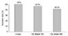
Figure 2
Histogram of mean absolute postoperative refractive error for A-scan, IOL Master 700, and IOL Master 500. The mean absolute postoperative refraction predictive errors exhibited no statistically significant differences between the three groups.
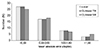
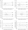
Figure 3
Bland-Altman plots comparing IOL Master 700 with IOL Master 500 and with A-scan. The middle lines indicate means and the dot lines above and below represent 95% limits of agreement. (A) Axial length with IOL Master 700 and IOL Master 500. (B) Axial length with IOL Master 700 and A-scan. (C) Anterior chamber depth with IOL Master 700 and IOL Master 500. (D) Anterior chamber depth with IOL Master 700 and A-scan. (E) Corneal power with IOL Master 700 and IOL Master 500. (F) Corneal power with IOL Master 700 and A-scan. SD = standard deviation. AXL = axial lengh; ACD = anterior chamber depth; K = corneal power from keratometry.

References
1. Mylonas G, Sacu S, Buehl W, et al. Performance of three biometry devices in patients with different grades of age-related cataract. Acta Ophthalmol. 2011; 89:e237–e241.
2. Rose LT, Moshegov CN. Comparison of the Zeiss IOLMaster and applanation A-scan ultrasound: biometry for intraocular lens calculation. Clin Exp Ophthalmol. 2003; 31:121–124.
3. Sheng H, Bottjer CA, Bullimore MA. Ocular component measurement using the Zeiss IOLMaster. Optom Vis Sci. 2004; 81:27–34.
4. Akman A, Asena L, Güngör SG. Evaluation and comparison of the new swept source OCT-based IOLMaster 700 with the IOLMaster 500. Br J Ophthalmol. 2016; 100:1201–1205.
5. Telenkov SA, Mandelis A. Fourier-domain biophotoacoustic subsurface depth selective amplitude and phase imaging of turbid phantoms and biological tissue. J Biomed Opt. 2006; 11:044006.
6. McAlinden C, Wang Q, Gao R, et al. Axial length measurement failure rates with biometers using swept-source optical coherence tomography compared to partial-coherence interferometry and optical low-coherence interferometry. Am J Ophthalmol. 2017; 173:64–69.
7. Shin DH, Lim DH, You JY, et al. Formula comparison for intraocular lens power calculation using IOL master and ultrasound for the ZCB00 IOL. J Korean Ophthalmol Soc. 2014; 55:527–533.
8. Ha DY, Lee KW, Jung JW. Comparison of ocular biometry measurements using a-scan ultrasound and two types of partial coherence interferometers. J Korean Ophthalmol Soc. 2016; 57:757–762.
9. Kaswin G, Rousseau A, Mgarrech M, et al. Biometry and intraocular lens power calculation results with a new optical biometry device: comparison with the gold standard. J Cataract Refract Surg. 2014; 40:593–600.
10. Arriola-Villalobos P, Almendral-Gómez J, Garzón N, et al. Agreement and clinical comparison between a new swept-source optical coherence tomography-based optical biometer and an optical low-coherence reflectometry biometer. Eye (Lond). 2017; 31:437–442.
11. Kurian M, Negalur N, Das S, et al. Biometry with a new swept-source optical coherence tomography biometer: Repeatability and agreement with an optical low-coherence reflectometry device. J Cataract Refract Surg. 2016; 42:577–581.
12. Srivannaboon S, Chirapapaisan C, Chonpimai P, Loket S. Clinical comparison of a new swept-source optical coherence tomography-based optical biometer and a time-domain optical coherence tomography-based optical biometer. J Cataract Refract Surg. 2015; 41:2224–2232.
13. Hirnschall N, Leisser C, Radda S, et al. Macular disease detection with a swept-source optical coherence tomography-based biometry device in patients scheduled for cataract surgery. J Cataract Refract Surg. 2016; 42:530–536.
14. Karunaratne N. Comparison of the Pentacam equivalent keratometry reading and IOL Master keratometry measurement in intraocular lens power calculations. Clin Exp Ophthalmol. 2013; 41:825–834.
15. Hoffer KJ, Hoffmann PC, Savini G. Comparison of a new optical biometer using swept-source optical coherence tomography and a biometer using optical low-coherence reflectometry. J Cataract Refract Surg. 2016; 42:1165–1172.
 XML Download