

 PDF
PDF ePub
ePub Citation
 Print
Print


Abstract
Case summary
A 47-year-old women presented with a one-month history of ocular pain and decreased visual acuity of the right eye. She had a family history of crystalline lens dislocation but showed no systemic abnormality or trauma history. Intraocular pressure was 45 mmHg in the right eye, which showed a myopic shift (-6.5 D). The crystalline lens of the right eye was subluxated to the anterior chamber, and the angle was closed. Phacoemulsification with scleral fixation of the posterior chamber intraocular lens of the right eye was performed. After that, best corrected visual acuity of the right eye was 1.0, and intraocular pressure was 15 mmHg. After 2 years, she presented with intermittent ocular pain and decreased visual acuity of the left eye. The crystalline lens of the left eye was subluxated to the anterior chamber. Phacoemulsification with scleral fixation of the posterior chamber intraocular lens of the left eye was performed. After that, intermittent ocular pain and visual acuity of the left eye were improved. Genetic testing confirmed an FBN1 gene mutation in the patient.
Conclusions
A bilateral ectopia lentis patient without history of definite trauma should undergo complete systemic and ophthalmic examination to rule out accompanying disease, and a detailed family history should be collected. If hereditary ectopia lentis is suspected, genetic testing of probands and their family should be performed and will be helpful for genetic counseling and ophthalmic surveillance.
Figures and Tables
Figure 1
Pedigree of proband with ectopia lentis and FBN1 gene analysis for the proband. Medical history taking of proband's family was performed. FBN1 genetic analysis was performed only on proband (arrow). Pedigree suggested that the mode of inheritance was autosomal dominant pattern. Among seven siblings of proband, three were affected with definite lens dislocation surgery after 5th decade, but the surgery history of proband's mother was uncertain because she was already deceased. Squares indicate males; circles indicate females; solid symbols denote that the member is affected; open symbols mean the member is unaffected; slashed symbols indicate that the member is deceased.
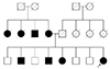
Figure 2
Anterior segment photography and anterior segment optical coherence tomography (OCT) of both eyes at initial examination. (A, B) Preoperative anterior segment photography and optical coherence tomography of the right eye demonstrated anteriorly subluxated crystalline lens with shallow anterior chamber and closed angle. (C, D) Crystalline lens of the left eye was not subluxated. Anterior chamber of the left eye was shallow but not closed. (E, F) One month after the right eye lens extraction surgery, anterior segment photography and OCT of right eye demonstrated well located intraocular lens, deep anterior chamber, and widely opened angle. OD = oculus dexter; OS = oculus sinister.
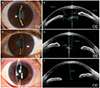
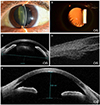
Figure 3
Anterior segment photography, anterior segment optical coherence tomography (OCT), and ultrabiomicroscopy (UBM) of the left eye at 2 years after the right eye surgery. (A) Preoperative anterior segment photography of the left eye demonstrated anteriorly subluxated lens with shallow anterior chamber. (B) Preoperative retroillumination view of the left eye demonstrated superotemporal displacement of the crystalline lens. (C) Preoperative anterior segment OCT of the left eye demonstrated anteriorly subluxated lens with shallow anterior chamber. (D) Preoperative UBM of left eye demonstrated no ciliary body abnormality and mass effect. (E) One month after the left eye lens extraction surgery, anterior segment optical coherence tomography of the left eye demonstrated well located intraocular lens, deep anterior chamber, and widely opened angle. OS = oculus sinister.
References
1. Nelson LB, Maumenee IH. Ectopia lentis. Surv Ophthalmol. 1982; 27:143–160.
2. Jarrett WH II. Dislocation of the lens. A study of 166 hospitalized cases. Arch Ophthalmol. 1967; 78:289–296.
3. Neely DE, Plager DA. Management of ectopia lentis in children. Ophthalmol Clin North Am. 2001; 14:493–499.
4. Sadiq MA, Vanderveen D. Genetics of ectopia lentis. Semin Ophthalmol. 2013; 28:313–320.
5. Chandra A, Aragon-Martin JA, Hughes K, et al. A genotype-phenotype comparison of ADAMTSL4 and FBN1 in isolated ectopia lentis. Invest Ophthalmol Vis Sci. 2012; 53:4889–4896.
6. Dureau P. Pathophysiology of zonular diseases. Curr Opin Ophthalmol. 2008; 19:27–30.
7. Loeys BL, Dietz HC, Braverman AC, et al. The revised Ghent nosology for the Marfan syndrome. J Med Genet. 2010; 47:476–485.
8. Mudd SH, Skovby F, Levy HL, et al. The natural history of homocystinuria due to cystathionine beta-synthase deficiency. Am J Hum Genet. 1985; 37:1–31.
9. Shafique M, Muzaffar W, Ishaq M. The eye as a window to a rare disease: ectopia lentis and homocystinuria, a Pakistani perspective. Int Ophthalmol. 2016; 36:79–83.
10. Yu YS, Awh JY, Lee JH. Management of ectopia lentis in children. J Korean Ophthalmol Soc. 1999; 40:1973–1978.
11. Faivre L, Collod-Beroud G, Loeys BL, et al. Effect of mutation type and location on clinical outcome in 1,013 probands with Marfan syndrome or related phenotypes and FBN1 mutations: an international study. Am J Hum Genet. 2007; 81:454–466.
12. Robinson PN, Godfrey M. The molecular genetics of Marfan syndrome and related microfibrillopathies. J Med Genet. 2000; 37:9–25.
13. Deng T, Dong B, Zhang X, et al. Late-onset bilateral lens dislocation and glaucoma associated with a novel mutation in FBN1. Mol Vis. 2008; 14:1229–1233.
14. Hubmacher D, Apte SS. ADAMTS proteins as modulators of microfibril formation and function. Matrix Biol. 2015; 47:34–43.
15. Collin GB, Hubmacher D, Charette JR, et al. Disruption of murine Adamtsl4 results in zonular fiber detachment from the lens and in retinal pigment epithelium dedifferentiation. Hum Mol Genet. 2015; 24:6958–6974.
 XML Download