

 PDF
PDF ePub
ePub Citation
 Print
Print


Abstract
Purpose
To compare the measurements between manifest refraction and cycloplegic refraction using retinoscopy or an autorefractor in children and to investigate factors affecting the difference.
Methods
A total of 388 children with a mean age of 7.4 ± 3.6 years were examined using retinoscopy and a Grand Seiko GR-3500KA autorefractor before and after cycloplegia. We compared the difference in spherical and cylindrical components between refractions and analyzed the results according to gender, age, type of refractive error, amblyopia, strabismus, and neu-ro-developmental disorder. A difference in refractions of ±0.50 D or more was considered as a significant discrepancy.
Results
Before cycloplegia, the spherical portion of the refractive error via autorefractor measurement was more myopic than for the retinoscopic measurement in 47.2% of patients, and the cylindrical portion was greater in 37.1%. The spherical discrepancies were more common in children aged < 7 years, with hyperopia, or amblyopia (respectively, p = 0.002, p < 0.001, and p = 0.033). After cycloplegia, the spherical component of the refractive error by autorefraction differed from retinoscopic measurement in 29.4% of patients, and the cylindrical portion differed in 30.7%. However, the difference was not significant and there was no difference according to clinical features. More than half of the children with discrepancies in the spherical component between retinoscopic refractions before and after cycloplegia had a discrepancy between autorefraction and retinoscopic refraction before cycloplegia, and the two discrepancies had a significant correlation.
Conclusions
Auto-refraction after cycloplegia can estimate retinoscopic values partially. Nevertheless, 30% of the children still showed a discrepancy. The discrepancy of manifest refraction or autorefraction compared to retinoscopic refraction with cycloplegia should be considered in younger children, cases with hyperopia or amblyopia, and cases with a difference in autorefraction and retinoscopic refraction before cycloplegia.
References
1. Lu Q, Zheng Y, Sun B, et al. A population-based study of visual abdominal among abdominal children in Beijing: the Beijing study of visual impairment in children. Am J Ophthalmol. 2009; 147:1075–81.
2. Kim MS, Chang HR. The evaluation of noncycloplegic and abdominal autorefraction in children according to the age. J Korean Ophthalmol Soc. 1998; 39:728–34.
3. Choi MJ, Baek SH, Gong SM. Comparison of autorefraction and clinical refraction with or without in children. J Korean Ophthalmol Soc. 2005; 46:837–46.
4. Jung JW, Kim YE, Paik HJ. Clinical comparison of autorefractor versus retinoscopic refraction in children according to the age. J Korean Ophthalmol Soc. 2005; 46:1931–5.
5. Choong YF, Chen AH, Goh PP. A comparison of autorefraction and subjective refraction with and without cycloplegia in primary school children. Am J Ophthalmol. 2006; 142:68–74.

6. Hashemi H, Khabazkhoob M, Asharlous A, et al. Cycloplegic abdominal versus subjective refraction: the Tehran Eye Study. Br J Ophthalmol. 2015; Nov 5:pii: bjophthalmol-2015–307871. [Epub ahead of print].
7. Fotouhi A, Morgan IG, Iribarren R, et al. Validity of noncycloplegic refraction in the assessment of refractive errors: the Tehran Eye Study. Acta Ophthalmol. 2012; 90:380–6.
8. Zhao J, Mao J, Luo R, et al. Accuracy of noncycloplegic abdominal in school-age children in China. Optom Vis Sci. 2004; 81:49–55.
9. Chua SY, Ikram MK, Tan CS, et al. Relative contribution of risk factors for early-onset myopia in young Asian children. Invest Ophthalmol Vis Sci. 2015; 56:8101–7.
10. Ma Y, He X, Zou H, et al. Myopia screening: combining visual acuity and noncycloplegic autorefraction. Optom Vis Sci. 2013; 90:1479–85.
11. Lai YH, Tseng HY, Hsu HT, et al. Uncorrected visual acuity and noncycloplegic autorefraction predict significant refractive errors in Taiwanese preschool children. Ophthalmology. 2013; 120:271–6.
12. Rajavi Z, Sabbaghi H, Baghini AS, et al. Accuracy and abdominal of refractive error measurements by photorefractometry. J Ophthalmic Vis Res. 2015; 10:221–8.
13. Charman WN, Jennings JA, Whitefoot H. The refraction of the eye in relation to spherical aberration and pupil size. Br J Physiol Opt. 1978; 32:78–93.
14. Kirschen D, Isenberg SJ. The effectiveness of an autorefractor with eye-tracking capability in pediatric patients. J AAPOS. 2014; 18:217–21.
15. Oral Y, Gunaydin N, Ozgur O, et al. A comparison of different abdominal with retinoscopy in children. J Pediatr Ophthalmol Strabismus. 2012; 49:370–7.
16. Trager MJ, Dirani M, Fan Q, et al. Testability of vision and abdominal in preschoolers: the strabismus, amblyopia, and refractive error study in Singaporean children. Am J Ophthalmol. 2009; 148:235–41.e6.
17. Pai AS, Rose KA, Samarawickrama C, et al. Testability of abdominal, stereopsis, and other ocular measures in preschool abdominal: the Sydney Paediatric Eye Disease Study. J AAPOS. 2012; 16:185–92.
18. McBrien NA, Millodot M. The relationship between tonic abdominal and refractive error. Invest Ophthalmol Vis Sci. 1987; 28:997–1004.
19. Kasthurirangan S, Glasser A. Age related changes in abdominal dynamics in humans. Vision Res. 2006; 46:1507–19.
20. Mordi JA, Ciuffreda KJ. Static aspects of accommodation: age and presbyopia. Vision Res. 1998; 38:1643–53.
21. Rosenfield M, Ciuffreda KJ, Ong E, Azimi A. Proximally induced accommodation and accommodative adaptation. Invest Ophthalmol Vis Sci. 1990; 31:1162–7.
22. Hiraoka T, Miyata K, Nakamura Y, et al. Influences of cycloplegia with topical atropine on ocular higher-order aberrations. Ophthalmology. 2013; 120:8–13.
23. Seo HJ, Kim SH, Suh YW, et al. Influence of watching 3D tele-vision on refractive error in children with exodeviation. J Korean Ophthalmol Soc. 2014; 55:1525–9.
Figure 1.
Graph showing the correlation of the difference between retinoscopic MR and auto-MR, and the difference between retinoscopic MR and CR (linear regression analysis; R2 = 0.063, p < 0.001). MR = manifest refraction; auto-MR = autorefraction before cycloplegia; CR = retinoscopic cycloplegic refraction.
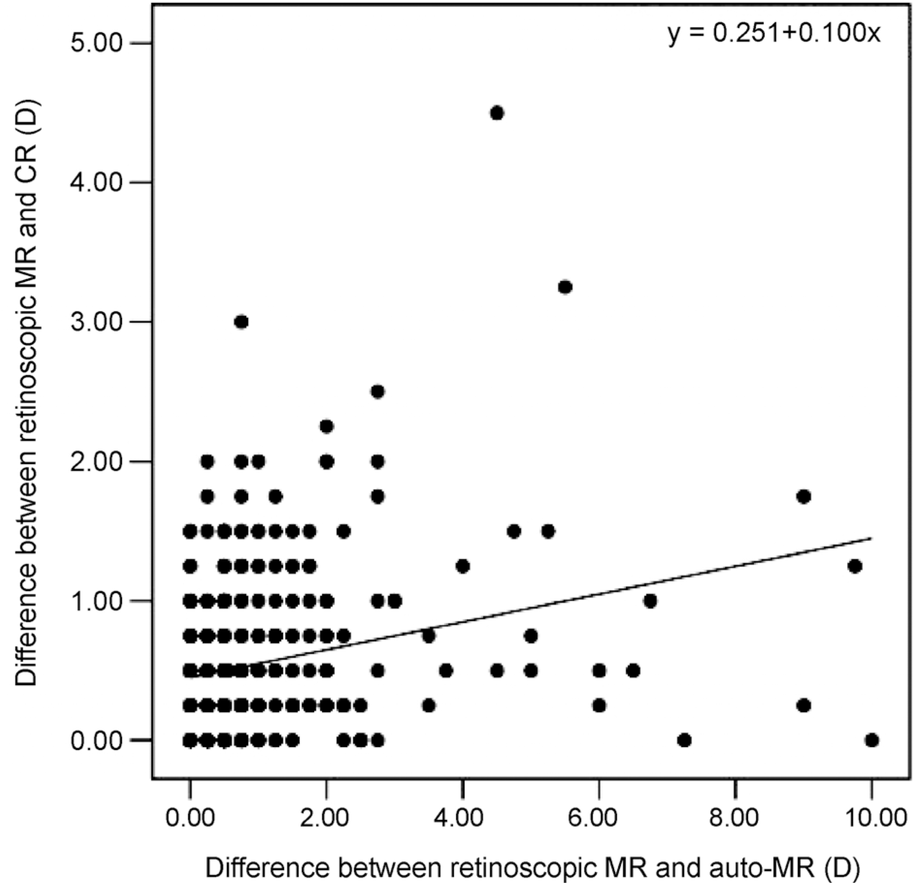
Table 1.
Proportion of eyes of disagreement between retinoscopic refractions before and after cycloplegia according to component
| Component | Same* | Different† |
|---|---|---|
| Sphere | 504 (64.9) | 272 (35.1) |
| Cylinder | 624 (80.4) | 152 (19.6) |
| Axis | 696 (89.7) | 80 (10.3) |
| Spherical equivalent | 494 (63.7) | 282 (36.3) |
Table 2.
Comparison of characteristics between same and different groups divided by the difference in sphere component between retinoscopic refractions before and after cycloplegia
| Overall (n = 388) | Same* (n = 252) | Different† (n = 136) | p-value | |
|---|---|---|---|---|
| Gender (male) | 180 (46.4) | 115 (45.6) | 65 (47.8) | 0.382‡ |
| Age (year) | 7.4 ± 3.6 | 7.9 ± 3.5 | 6.6 ± 3.5 | 0.001§ |
| <7 years | 210 (54.1) | 112 (44.4) | 98 (72.1) | <0.001‡ |
| ≥7 years | 178 (45.9) | 140 (55.6) | 38 (27.9) | |
| Type of refractive errors | <0.001‡ | |||
| Emmetropia | 85 (21.9) | 63 (25.0) | 22 (16.2) | |
| Hyperopia | 134 (34.5) | 60 (23.8) | 74 (54.4) | |
| Myopia | 138 (35.6) | 115 (45.6) | 23 (16.9) | |
| Astigmatism | 31 (8.0) | 14 (5.6) | 17 (12.5) | |
| Anisometropia | 77 (19.8) | 47 (18.7) | 30 (22.1) | 0.250‡ |
| Amblyopia | 29 (7.5) | 12 (4.8) | 17 (12.5) | 0.010‡ |
| Aniso:Strabismic:Combined | 17:6:6 | 8:2:2 | 9:4:4 | |
| Strabismus | 189 (48.7) | 129 (51.2) | 60 (44.1) | 0.214‡ |
| Exo:Eso:Vertical | 156:29:4 | 110:16:3 | 46:13:1 | |
| Neuro-developmental disorders | 38 (9.8) | 25 (9.9) | 13 (9.6) | 0.522‡ |
Table 3.
Proportion of eyes of disagreement between retinoscopic refraction and autorefraction before cycloplegia according to component
| Component | Same* | Different† |
|---|---|---|
| Sphere | 410 (52.8) | 366 (47.2) |
| Cylinder | 488 (62.9) | 288 (37.1) |
| Axis | 332 (42.8) | 444 (57.2) |
| Spherical equivalent | 368 (47.4) | 408 (52.6) |
Table 4.
Comparison of characteristics between same and different groups divided by the difference in sphere component between retinoscopic refraction and autorefraction before cycloplegia
| Overall (n = 388) | Same* (n = 205) | Different† (n = 183) | p-value | |
|---|---|---|---|---|
| Gender (male) | 180 (46.4) | 101 (49.3) | 79 (43.2) | 0.136‡ |
| Age (year) | 7.4 ± 3.6 | 7.9 ± 3.5 | 6.9 ± 3.6 | 0.005§ |
| <7 years | 210 (54.1) | 96 (46.8) | 114 (62.3) | 0.002‡ |
| ≥7 years | 178 (45.9) | 109 (53.2) | 69 (37.7) | |
| Type of refractive errors | <0.001‡ | |||
| Emmetropia | 85 (21.9) | 49 (23.9) | 36 (19.7) | |
| Hyperopia | 134 (34.5) | 53 (25.9) | 81 (44.3) | |
| Myopia | 138 (35.6) | 90 (43.9) | 48 (26.2) | |
| Astigmatism | 31 (8.0) | 13 (6.3) | 18 (9.8) | |
| Anisometropia | 77 (19.8) | 35 (17.1) | 42 (23.0) | 0.093‡ |
| Amblyopia | 29 (7.5) | 9 (4.4) | 20 (10.9) | 0.033‡ |
| Aniso:Strabismic:Combined | 17:6:6 | 4:2:3 | 13:4:3 | |
| Strabismus | 189 (48.7) | 100 (48.8) | 89 (48.6) | 0.095‡ |
| Exo:Eso:Vertical | 156:29:4 | 89:10:1 | 67:19:3 | |
| Neuro-developmental disorders | 38 (9.8) | 25 (12.2) | 13 (7.1) | 0.069‡ |
Table 5.
Proportion of eyes of disagreement between retinoscopic refraction and autorefraction after cycloplegia according to component
| Component | Same* | Different† |
|---|---|---|
| Sphere | 548 (70.6) | 228 (29.4) |
| Cylinder | 538 (69.3) | 238 (30.7) |
| Axis | 380 (49.0) | 396 (51.0) |
| Spherical equivalent | 524 (67.5) | 252 (32.5) |
Table 6.
Comparison of characteristics between same and different groups divided by the difference in sphere component between retinoscopic refraction and autorefraction after cycloplegia
| Overall (n = 388) | Same* (n = 274) | Different† (n = 114) | p-value | |
|---|---|---|---|---|
| Gender (male) | 180 (46.4) | 126 (46.0%) | 54 (47.4) | 0.445‡ |
| Age (year) | 7.4 ± 3.6 | 7.6 ± 3.6 | 7.1 ± 3.6 | 0.227§ |
| <7 years | 210 (54.1) | 139 (50.7) | 71 (62.3) | 0.069‡ |
| ≥7 years | 178 (45.9) | 135 (49.3) | 43 (37.7) | |
| Type of refractive errors | 0.075‡ | |||
| Emmetropia | 85 (21.9) | 62 (22.6) | 23 (20.2) | |
| Hyperopia | 134 (34.5) | 84 (30.7) | 50 (43.9) | |
| Myopia | 138 (35.6) | 105 (38.3) | 33 (28.9) | |
| Astigmatism | 31 (8.0) | 23 (8.4) | 8 (7.0) | |
| Anisometropia | 77 (19.8) | 57 (20.8) | 20 (17.5) | 0.250‡ |
| Amblyopia | 29 (7.5) | 20 (7.3) | 9 (7.9) | 0.299‡ |
| Aniso:Strabismic:Combined | 17:6:6 | 13:4:3 | 4:2:3 | |
| Strabismus | 189 (48.7) | 137 (50.0) | 52 (45.6) | 0.288‡ |
| Exo:Eso:Vertical | 156:29:4 | 117:17:3 | 39:12:1 | |
| Neuro-developmental disorders | 38 (9.8) | 27 (9.8) | 11 (9.6) | 0.564‡ |
Table 7.
Linear regression analysis for the spherical and cylindrical components of autorefraction before and after cycloplegia against those of retinoscopic refraction with cycloplegia
 XML Download