

 PDF
PDF ePub
ePub Citation
 Print
Print


Abstract
Purpose
We investigated the clinical manifestations of combination therapy with photodynamic therapy (PDT) and intravitreal bevacizumab in idiopathic macular telangiectasia type I patients.
Methods
The present study included 7 patients who visited our clinic from May 2008 to February 2011 complaining of decreasing visual acuity and diagnosed as idiopathic macular telangiectasia type I including visible aneurysms at juxtafoveal area and telangiectatic vessels leakage based on fluorescein angiography. Additionally, all patients were treated with combination therapy with PDT and intravitreal bevacizumab.
Results
Visual acuity improved from 0.48 ± 0.14 (log MAR) to 0.18 ± 0.17 (log MAR) after the combination therapy, however, there was no change in intraocular pressure between before (17.9 ± 3.1) and after (16.8 ± 2.3) therapy. After combination therapy, fluorescein angiography showed decreased leakage of telangiectatic vessels and optical coherence tomography showed only minimal intraretinal edema. Central subfield macular thickness decreased from 301.9 ± 50.7 μ m to 193.6 ± 58.8 μ m after the combination therapy.
References
1. Hutton WL, Snyder WB, Fuller D, Vaiser A. Focal parafoveal abdominall telangiectasis. Arch Ophthalmol. 1978; 96:1362–7.
2. Gass JD, Blodi BA. Idiopathic juxtafoveolar retinal telangiectasis. Update of classification and follow-up study. Ophthalmology. 1993; 100:1536–46.
3. Yannuzzi LA, Bardal AM, Freund KB, et al. Idiopathic macular telangiectasia. Arch Ophthalmol. 2006; 124:450–60.

4. Kovach JL, Rosenfeld PJ. Bevacizumab (avastin) therapy for abdominal macular telangiectasia type II. Retina. 2009; 29:27–32.
5. Charbel Issa P, Holz FG, Scholl HP. Findings influorescein abdominal and optical coherence tomography after intravitreal abdominal in type 2 idiopathic macular telangiectasia. Ophthalmology. 2007; 114:1736–42.
6. Gamulescu MA, Walter A, Sachs H, Helbig H. Bevacizumab in the treatment of idiopathic macular telangiectasia. Graefes Arch Clin Exp Ophthalmol. 2008; 246:1189–93.
7. Takayama K, Ooto S, Tamura H, et al. Intravitreal bevacizumab for type 1 idiopathic macular telangiectasia. Eye (Lond). 2010; 24:1492–7.
8. Chopdar A. Retinal telangiectasis in adults: fluorescein abdominal findings and treatment by argon laser. Br J Ophthalmol. 1978; 62:243–50.
9. Park DW, Schatz H, McDonald HR, Johnson RN. Grid laser photo-coagulation for macular edema in bilateral juxtafoveal telangiectasis. Ophthalmology. 1997; 104:1838–46.
10. Alldredge CD, Garretson BR. Intravitreal triamcinolone for the treatment of idiopathic juxtafoveal telangiectasis. Retina. 2003; 23:113–6.
11. Battaglia Parodi M, Da Pozzo S, Ravalico G. Photodynamic abdominal in chronic central serous chorioretinopathy. Retina. 2003; 23:235–7.
12. Khosla PK, Rana SS, Tewari HK, et al. Evaluation of visual abdominal following argon laser photocoagulation in central serous retinopathy. Ophthalmic Surg Lasers. 1997; 28:693–7.
13. Klais CM, Ober MD, Ciardella AP, et al. Retina. 4th ed.2. Los Angeles: Mosby;2006. p. 1153–9.
14. Kotoula MG, Chatzoulis DZ, Karabatsas CH, et al. Resolution of macular edema in idiopathic juxtafoveal telangiectasis using PDT. Ophthalmic Surg Lasers Imaging. 2009; 40:65–7.
15. Kurup S, Lew J, Byrnes G, et al. Therapeutic efficacy of intravitreal bevacizumab on posterior uveitis complicated by neovascularization. Acta Ophthalmol. 2000; 87:349–52.
16. Dhalla MS, Shah GK, Blinder KJ, et al. Combined photodynamic therapy with verteporfin and intravitreal bevacizumab for abdominal neovascularization in age-related macular degeneration. Retina. 2006; 26:988–93.
17. Rishi P, Shroff D, Rishi E. Combined photodynamic therapy and intravitreal ranibizumab as primary treatment for subretinal neo-vascular membrane (SRNVM) associated with type 2 idiopathic macular telangiectasia. Graefes Arch Clin Exp Ophthalmol. 2008; 246:619–21.
18. Bashshur ZF, Schakal A, Hamam RN, et al. Intravitreal abdominal vs verteporfin photodynamic therapy for neovascular age-related macular degeneration. Arch Ophthalmol. 2007; 125:1357–61.
Figure 1.
Fluorescein angiography and opti-cal coherence tomography before treatment. Fluorescein angiography of telangiectatic vessels at the early phase (A) and leakage at the late phase (B). (C) Optical coherence tomography of intraretinal edema before combination treatment.
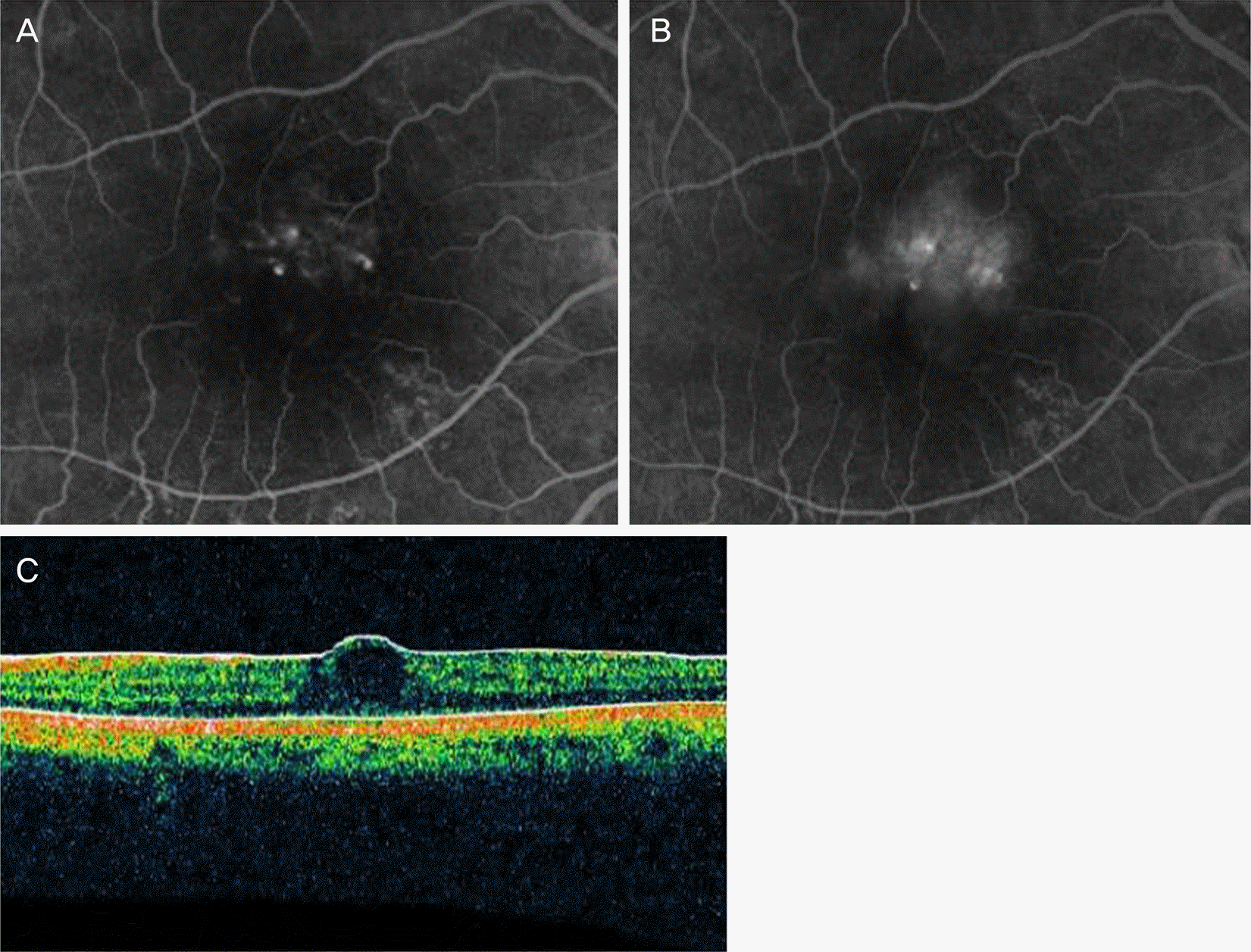
Figure 2.
Fluorescein angiography and optical coherence tomography after treatment. Fluorescein angiography of a regression of the telangiectatic vessels (A) and a decreased leakage of telangiectatic vessels at the late phase after combination therapy(B). (C) Optical coherence tomography of minimal intraretinal edema and restored foveal depression after treatment.

Table 1.
Clinical characteristics of the patient at presentation
Table 2.
Clinical outcomes after combined therapy for idiopathic macular telangiectasia type I
 XML Download