

 PDF
PDF ePub
ePub Citation
 Print
Print


Abstract
Purpose
To describe a case of sympathetic ophthalmia due to corneal perforation caused by exposure keratitis in a patient with recurrent sphenoid wing meningioma.
Case summary
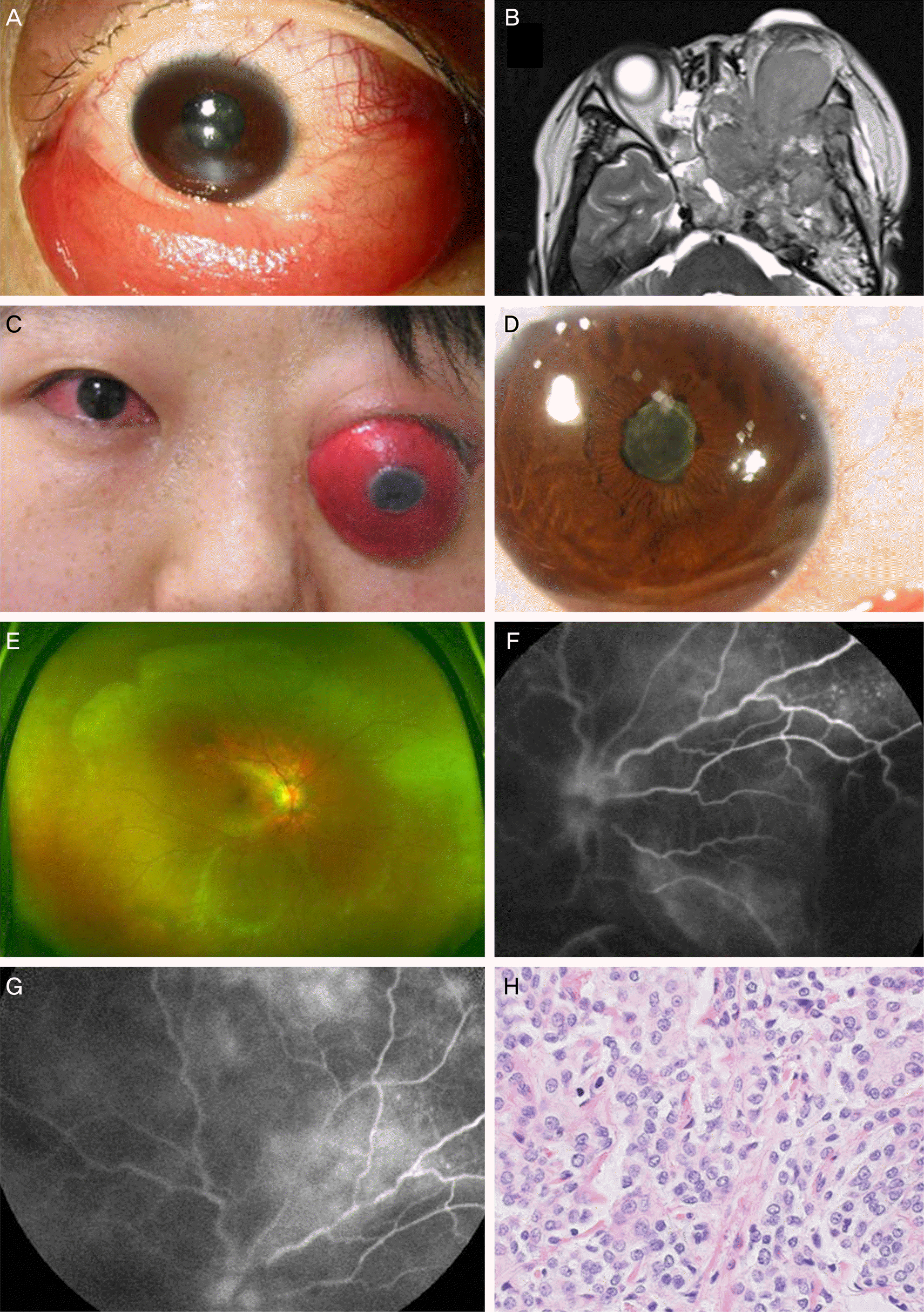
A 34-year-old female patient presented with proptosis in her left eye caused by left sphenoid greater wing meningioma despite tumor debulking surgery and radiation treatment. The cornea was perforated with prolapsed iris due to exposure keratitis, thus enucleation of the left eye was performed. After 2 weeks, an inflammatory reaction occurred in both eyes, keratic precipitates on corneal endothelium, exudative retinal detachment, and multiple granulomatous nodules on the right eye retina. The patient was diagnosed with sympathetic ophthalmia, thus enucleation of the left eye and debulking of the tumor were performed followed by a high-dose intravenous steroid therapy. At 5 months postoperatively, slit lamp biomicroscope showed no chamber reaction; improved disc swelling and exudative retinal detachment in the right eye were observed.
References
1. Albert DM, Diaz-Rohena R. A historical review of sympathetic ophthalmia and its epidemiology. Surv Ophthalmol. 1989; 34:1–14.

2. Mackenzie W. A practical treatise on the diseases of the eye. 3rd ed.London: Longmans;1840.
3. Makley TA Jr, Azar A. Sympathetic ophthalmia. A long-term fol-low-up. Arch Ophthalmol. 1978; 96:257–62.
4. Marak GE Jr. Recent advances in sympathetic ophthalmia. Surv Ophthalmol. 1979; 24:141–56.
5. Lee KS. A case report of sympathetic ophthalmia. J Korean Ophthalmol Soc. 1977; 18:189–95.
6. Yoon YD, Lim JO, Shin DE. A case of sympathetic ophthalmia with giant subretinal neovascular membrane. J Korean Ophthalmol Soc. 1994; 35:112–7.
7. Chan CC, Benezra D, Rodrigues MM. . Immunohistochemistry and electron microscopy of choroidal infiltrates and Dalen-Fuchs nodules in sympathetic ophthalmia. Ophthalmology. 1985; 92:580–90.
8. Chan CC, Nussenblatt RB, Fujikawa LS. . Sympathetic ophthalmia. Immunopathological findings. Ophthalmology. 1986; 93:690–5.
9. Croxatto JO, Rao NA, McLean IW, Marak GE. Atypical histo-pathologic features in sympathetic ophthalmia. A study of a hundred cases. Int Ophthalmol. 1982; 4:129–35.
10. Albert DM, Diaz-Rohena R. A historical review of sympathetic ophthalmia and its epidemiology. Surv Ophthalmol. 1989; 34:1–14.
11. Bilyk JR. Enucleation, evisceration, and sympathetic ophthalmia. Curr Opin Ophthalmol. 2000; 11:372–86.
12. Lubin JR, Albert DM, Weinstein M. Sixty-five years of sympathetic ophthalmia. A clinicopathologic review of 105 cases (1913--1978). Ophthalmology. 1980; 87:109–21.
13. Reynard M, Riffenburgh RS, Maes EF. Effect of corticosteroid treatment and enucleation on the visual prognosis of sympathetic ophthalmia. Am J Ophthalmol. 1983; 96:290–4.
14. Chan CC, Roberge RG, Whitcup SM, Nussenblatt RB. 32 cases of sympathetic ophthalmia. A retrospective study at the National Eye Institute, Bethesda, Md., from 1982 to 1992. Arch Ophthalmol. 1995; 113:597–600.
15. Nussenblatt R. Sympathetic ophthalmia. Nussenblatt RB SW, Palestine AG, editors. Uveitis: fundamental and clinical practice. 2nd ed.St. Louis: Mosby;1996. p. 97–134. 311-23.
16. Vote BJ, Hall A, Cairns J, Buttery R. Changing trends in sympathetic ophthalmia. Clin Experiment Ophthalmol. 2004; 32:542–5.
Figure 1.
(A) Corneal opacity and mid-dilated pupil in the left eye, (B) brain MRI showed progressed mass extent to orbit, ethmoid sinus, nasal cavity, sphenoid sinus, intracranium, (C) conjunctival injection in the right eye after 2 weeks, (D) KP’s on endothelium and fibrinous membrane on anterior lens capsule in the right eye, (E) exudative retinal detachment and multiple granulomatous nodules in the right eye, (F, G) FAG shows multiple progressively fluorescent dots at the level of the RPE, disc leakage, coalescence of the dye, (H) patternless growth and abundant miotic figures (HE ×40). KP's = keratic precipitates; FAG = fluorescent angiography; RPE = retinal pigment epithelium.
 XML Download