

 PDF
PDF ePub
ePub Citation
 Print
Print


Abstract
Purpose
To report a case of a 59-year-old female with a free-floating monolateral vitreous cyst localized in the posterior vitreous in the left eye.
Case summary
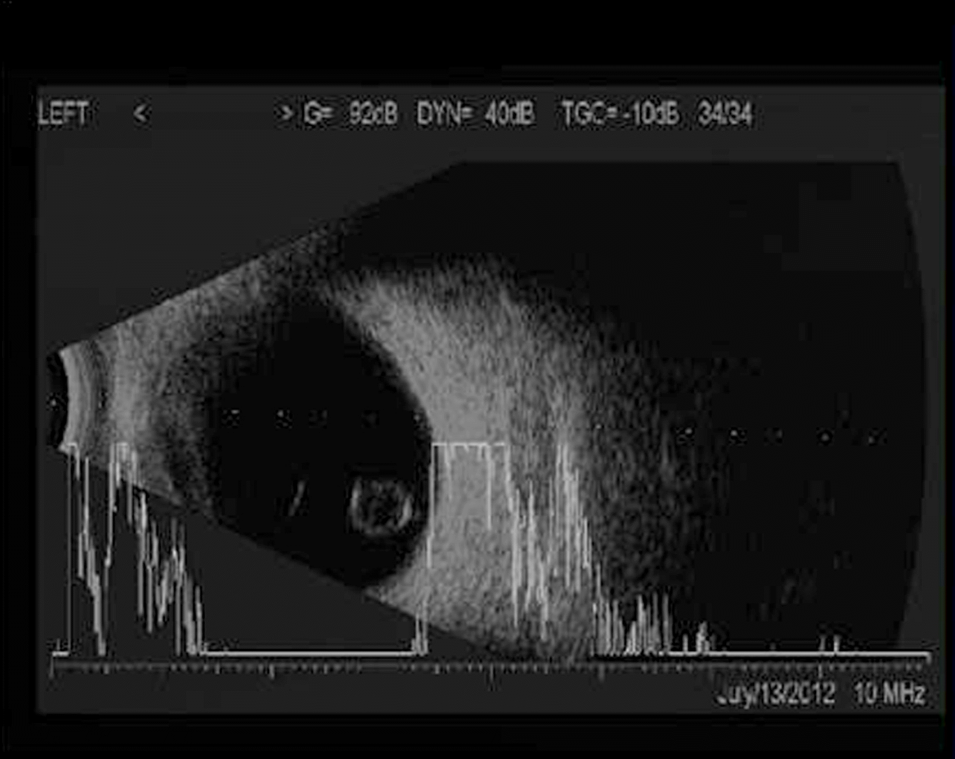
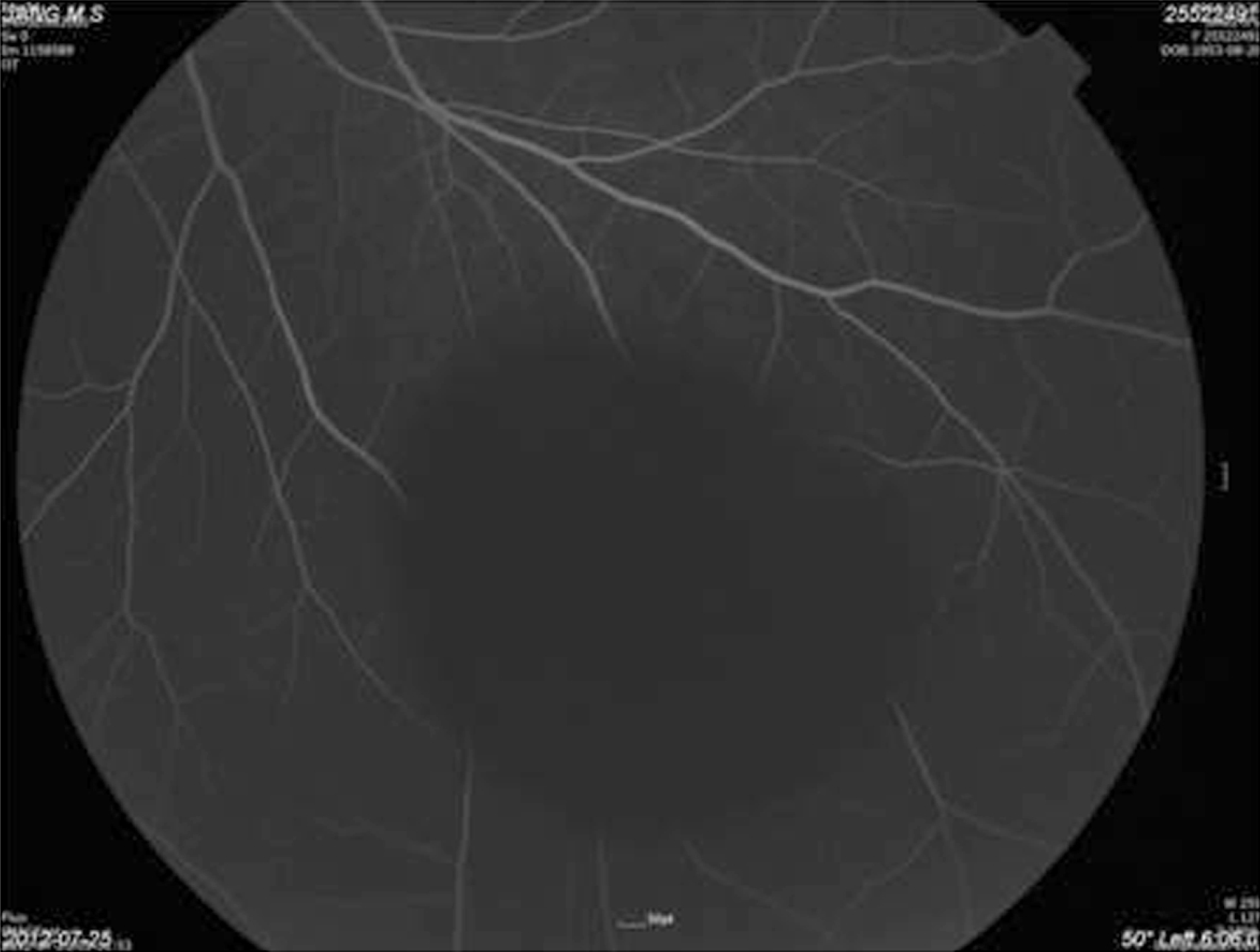
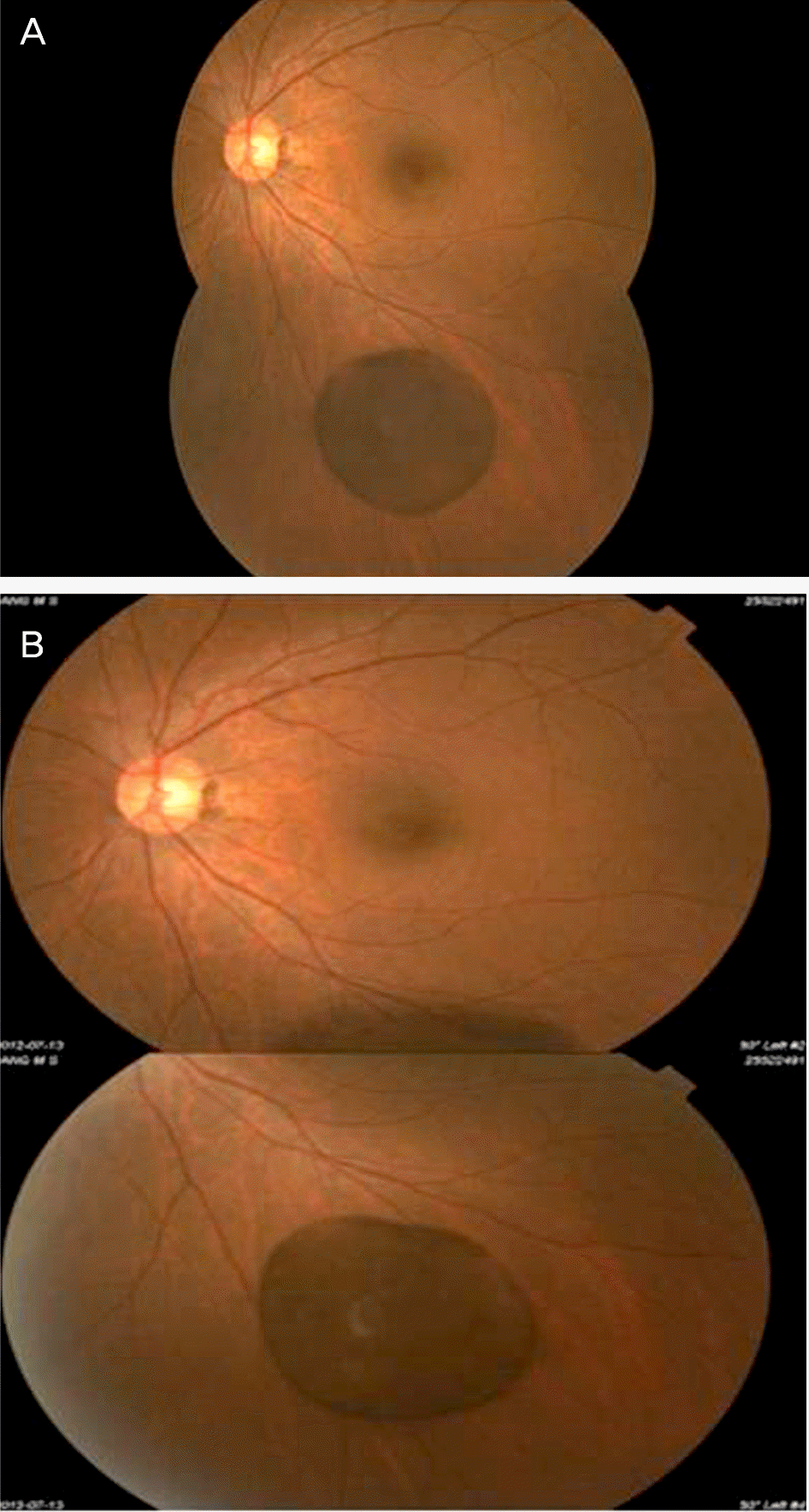
A 59-year-old female who complained of an intermittent floater in the left eye for 3 months visited our clinic. She had been suffering from visual disturbance for approximately 3 months. There was no previous history of trauma, infection, or inflammatory disorders. The best corrected visual acuity was 20/20 in both eyes. On fundoscopic exam, a 3-4 disc diameter (DD) sized, brown-colored pigmented vitreous cyst was detected at the inferior temporal side of the posterior vitreous in her left eye. B-scan ultrasound confirmed the presence of an echo-free cystic formation that was free from surrounding vitreous strands or other adhesions located at the posterior vitreous. No specific findings or leakage were observed on fluorescein angiography. We followed-up the patient periodically (1 month, 3 months, and 6 months after the initial visit) and monitored whether the size or location of the cyst had changed. At every follow-up exam, the size or location of the cyst was stationary and the patient's visual acuity was 20/20 in the affected eye, thus we suggested she should be followed-up periodically for her cyst without any intervention.
Conclusions
We report a case of a patient with no previous ocular history or impaired vision who had a free-floating vitreous cyst localized in the posterior vitreous in the left eye. The disease did not appear to progress or become aggravated over a short-term follow-up period and no specific treatment was required.
References
1. Tansley JO. Cyst of the vitreous. Trans Am Ophthalmol Soc. 1899; 8:507–9.
2. Aydin E, Demir HD, Tasliyurt T. Idiopathic pigmented free-floating posterior vitreous cyst. Int Ophthalmol. 2009; 29:299–301.

3. Yang JE, Baek TM, Kim JH, Lee JH. A case of free-floating vitreous cyst. J Korean Ophthalmol Soc. 1990; 31:1218–20.
4. Cruciani F, Santino G, Salandri AG. Monolateral idiopathic cyst of the vitreous. Acta Ophthalmol Scand. 1999; 77:601–3.
5. Orellana J, O'Malley RE, McPherson AR, Font RL. Pigmented free-floating vitreous cysts in two young adults. Electron microscopic observations. Ophthalmology. 1985; 92:297–302.
6. Bayraktar Z, Kapran Z, Ozdogan S. Pigmented congenital vitreous cyst. Eur J Ophthalmol. 2004; 14:156–8.
7. Wolter JR, Martony CL, Smith C. A free-floating vitreous cyst in the otherwise normal eye of a young man. J Pediatr Ophthalmol. 1975; 12:243–5.
8. Awan KJ. Biomicroscopy and argon laser photocystotomy of free-floating vitreous cysts. Ophthalmology. 1985; 92:1710–1.
9. Nork TM, Millecchia LL. Treatment and histopathology of a congenital vitreous cyst. Ophthalmology. 1998; 105:825–30.
10. Ruby AJ, Jampol LM. Nd:YAG treatment of a posterior vitreous cyst. Am J Ophthalmol. 1990; 110:428–9.
Figure 1.
There were fundus photographs of both eyes (A, B, C, D). Compared to the fundus photograph, which is normal, in right eye (A), a pigmented free-floating vitreous cyst (about 3-4 DD sized) at inferotemporal side in left eye was observed (B, C, D).
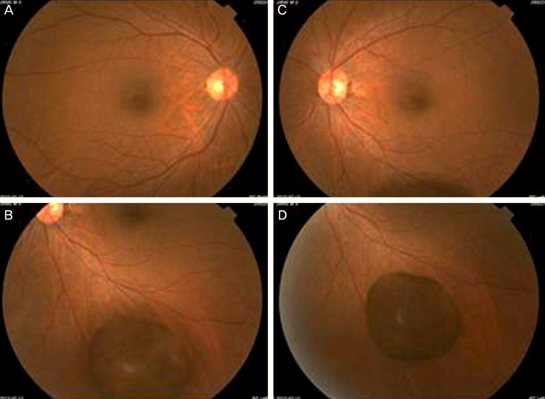
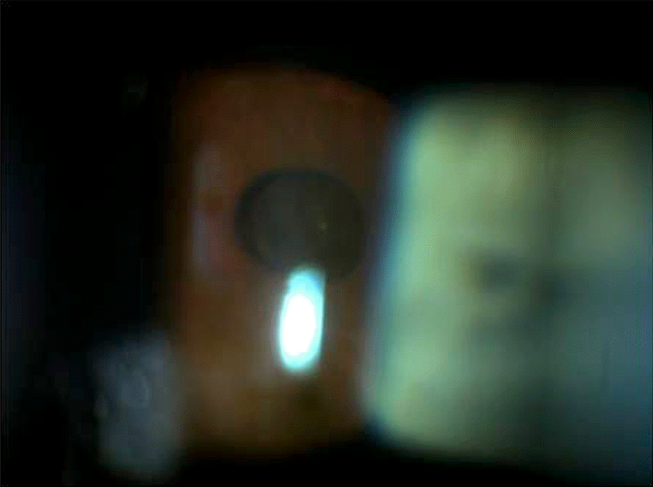
Figure 2.
Slit-lamp photograph of the free-floating vitreus cyst. It is observed to be round, and to have a smooth surface and brown-pigments in its wall.
 XML Download