

 PDF
PDF ePub
ePub Citation
 Print
Print


Abstract
Purpose
To analyze the clinical features and prognostic factors of ocular surface squamous neoplasia (OSSN).
Methods
A total of 31 eyes of 31 patients with suspected OSSN who were treated with excision and biopsy were retrospectively reviewed. Clinical features such as patient symptoms, invasion type, morphologic feature and size of lesion were analyzed. All patients were treated with simple excision or mitomycin C combination therapy. Factors affecting recurrence were evaluated.
Results
The most common symptom, invasion type, morphological feature, lesion size and histological diagnosis were foreign body sensation (38.7%), combined conjunctiva and cornea type (71.0%), gelatinous type (74.2%), 5 - 10 mm in largest diameter (45.2%) and intraepithelial neoplasia (51.6%), respectively. Age, sex, morphological features, lesion size and histological diagnosis had no effect on recurrence. Mitomycin C combination therapy was significantly associated with decreased recurrence compared to simple excision (p < 0.05).
References
1. Basti S, Macsai MS. Ocular surface squamous neoplasia: a review. Cornea. 2003; 22:687–704.
2. Zaki AA, Farid SF. Management of intraepithelial and invasive neoplasia of the cornea and conjunctiva: a long-term follow up. Cornea. 2009; 28:986–8.

3. Shields CL, Shields JA. Tumors of the conjunctiva and cornea. Surv Ophthalmol. 2004; 49:3–24.
4. Yang J, Foster CS. Squamous cell carcinoma of the conjunctiva. Int Ophthalmol Clin. 1997; 37:73–85.
5. Karp CL, Galor A, Chhabra S, et al. Subconjunctival/perilesional recombinant interferon α2b for ocular surface squamous neoplasia: a 10-year review. Ophthalmology. 2010; 117:2241–6.
6. Midena E, Angeli CD, Valenti M, et al. Treatment of conjunctival squamous cell carcinoma with topical 5-fluorouracil. Br J Ophthalmol. 2000; 84:268–72.
7. Frucht-Pery J, Rozenman Y, Pe'er J. Topical mitomycin-C for parti-ally excised conjunctival squamous cell carcinoma. Ophthalmology. 2002; 109:548–52.
8. Graue GF, Tena LB, Finger PT. Electron beam radiation therapy for conjunctival squamous carcinoma. Ophthal Plast Reconstr Surg. 2011; 27:277–81.
9. Finger PT. Radiation therapy for orbital tumors: concepts, current use, and ophthalmic radiation side effects. Surv Ophthalmol. 2009; 54:545–68.
10. Kim HT, Lee JS, Lee JY, Chung SK. A case of sessile papilloma in-vading the cornea. J Korean Ophthalmol Soc. 1999; 40:287–92.
11. Lee JS, Oh KG, Kim JD. A case of corneal squamous cell carcinoma in situ. J Korean Ophthalmol Soc. 1994; 35:206–9.
12. Song YW, Choi YH, Oh JH. Squamous cell carcinoma of the cornea. J Korean Ophthalmol Soc. 1983; 24:919–23.
13. Cheon HC, Lee DY, Park WC, Ahn HB. Ocular sSurface Ssquamous Nneoplasia. J Korean Ophthalmol Soc. 2006; 47:1920–8.
14. Yousef YA, Finger PT. Squamous carcinoma and dysplasia of the conjunctiva and cornea: an analysis of 101 cases. Ophthalmology. 2012; 119:233–40.
15. Sun EC, Fears TR, Goedert JJ. Epidemiology of squamous cell conjunctival cancer. Cancer Epidemiol Biomarkers Prev. 1997; 6:73–7.
16. Lee GA, Hirst LW. Ocular surface squamous neoplasia. Surv Ophthalmol. 1995; 39:429–50.
17. Pizzarello LD, Jakobiec FA. Bowen's disease of the conjunctiva: a misomer. Jakobiec FA, editor. Ocular Adnexal Tumors. Birmingham, AL: Aesculapius;1978. p. 553–71.
18. Finger PT, Tran HV, Turbin RE, et al. High-frequency ultrasonographic evaluation of conjunctival intraepithelial neoplasia and squamous cell carcinoma. Arch Ophthalmol. 2003; 121:168–72.
19. Illiff WJ, Marback R, Green R. Invasive squamous cell carcinoma of the conjunctiva. Arch Ophthalmol. 1975; 93:119–202.
20. Buuns DR, Tse DT, Folberg R, Buuns DR. Microscopically con-trolled excision of conjunctival squamous cell carcinoma. Am J Ophthalmol. 1994; 117:97–102.
21. Ash JE. Epibulbar tumors. Am J Ophthalmol. 1950; 33:1203–19.
22. Sjoö N, Heegaard S, Prause JU. Conjunctival papilloma. A: a his-topathologically based retrospective study. Acta Ophthalmol Scand. 2000; 78:663–6.
23. Artornsombudh P, Sanpavat A, Tinnungwattana U, et al. Prevalence and clinicopathologic findings of conjunctival epithelial neoplasia in pterygia. Ophthalmology. 2013; 120:1337–40.
24. Hirst LW, Axelsen RA, Schwab I. Pterygium and associated ocular surface squamous neoplasia. Arch Ophthalmol. 2009; 127:31–2.
25. Chui J, Coroneo MT, Tat LT, et al. Ophthalmic pterygium: a stem cell disorder with premalignant features. Am J Pathol. 2011; 178:817–27.
26. Oellers P, Karp CL, Sheth A, et al. Prevalence, treatment, and outcomes of coexistent ocular surface squamous neoplasia and pterygium. Ophthalmology. 2013; 120:445–50.
27. Soll DB, Harrison SE. Basic concepts and an overview of cryosurgery in ophthalmic plastic surgery. Ophthalmic Surg. 1979; 10:31–6.
28. Wilkes TD, Fraunfelder FT. Principles of cryosurgery. Ophthalmic Surg. 1979; 10:21–30.
29. Crooke ST, Bradner WT. Mitomycin C: a review. Cancer Treat Rev. 1976; 3:121–39.
30. Verweij J. Mitomycins. Pinedo HM, Longo DL, Chabner BA, editors. Cancer Chemotherapy and Biological Response Modifiers Annual 17. Amsterdam: Elsevier Science;1997. p. 76.
31. Shah SU, Kaliki S, Kim HJ, et al. Topical interferon alfa-2b for management of ocular surface squamous neoplasia in 23 cases: outcomes based on American Joint Committee on Cancer classification. Arch Ophthalmol. 2012; 130:159–64.
32. Krilis M, Tsang H, Coroneo M. Treatment of conjunctival and corneal epithelial neoplasia with retinoic acid and topical interferon alfa-2b: long-term follow-up. Ophthalmology. 2012; 119:1969–73.
33. Paul S, Stone DU. Intralesional bevacizumab use for invasive ocular surface squamous neoplasia. J Ocul Pharmacol Ther. 2012; 28:647–9.
34. Finger PT, Chin KJ. Refractory squamous cell carcinoma of the conjunctiva treated with subconjunctival ranibizumab (Lucentis): a two-year study. Ophthal Plast Reconstr Surg. 2012; 28:85–9.
35. Faramarzi A, Feizi S. Subconjunctival bevacizumab injection for ocular surface squamous neoplasia. Cornea. 2013; 32:998–1001.
36. Frucht-Pery J, Sugar J, Baum J, et al. Mitomycin C treatment for conjunctival-corneal intraepithelial neoplasia: a multicenter experience. Ophthalmology. 1997; 104:2085–93.
37. Heigle TJ, Stulting RD, Palay DA. Treatment of recurrent conjunctival epithelial neoplasia with topical mitomycin C. Am J Ophthalmol. 1997; 124:397–9.
38. Wilson MW, Hungerford JL, George SM, Madreperla SA. Topical mitomycin C for the treatment of conjunctival and corneal epithelial dysplasia and neoplasia. Am J Ophthalmol. 1997; 124:303–11.
39. Shields CL, Naseripour M, Shields JA. Topical mitomycin C for ex-tensive, recurrent conjunctival-corneal squamous cell carcinoma. Am J Ophthalmol. 2002; 133:601–6.
40. Haas K, Ben-Dor D, Levartovsky S. Treatment of conjunctival corneal intraepithelial neoplasia with topical mitomycin C. Arch Ophthalmol. 1999; 117:544–5.
41. Rozenman Y, Frucht-Pery J. Treatment of conjunctival intraepithelial neoplasia with topical drops of mitomycin C. Cornea. 2000; 19:1–6.
42. Erie JC, Campbell RJ, Leiesegang TJ. Conjunctival and corneal intraepithelial and invasive neoplasia. Ophthalmology. 1986; 93:176–83.
43. Tunc M, Char DH, Crawford B, Miller T. Intraepithelial and invasive squamous cell carcinoma of the conjunctiva: analysis of 60 cases. Br J Ophthalomol. 1999; 83:98–103.
44. McKelvie PA, Daniell M, McNab A, et al. Squamous cell carcinoma of the conjunctiva: a series of 26 cases. Br J Ophthalmol. 2002; 86:168–73.
45. Tabin G, Levin S, Snibson G, et al. Late recurrences and the neces-sity for long-term follow-up in corneal and conjunctival intraepithelial neoplasia. Ophthalmology. 1997; 104:485–92.
46. The AJCC Ophthalmic Oncology Task Force. Carcinoma of the conjunctiva. Edge SE, Byrd DR, Carducci MA, Compton CA, editors. AJCC Cancer Staging Manual. 7th ed.New York: Springer;2009. p. 531–3.
Figure 1.
Frequency of symptoms and signs of ocular surface squamous neoplasia. The patients’ most common symptom was foreign body sensation (38.7%).
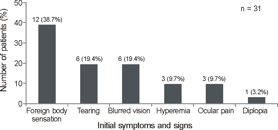
Figure 2.
Characteristics of ocular surface squamous neoplasia. (A) Frequency distribution of the tumor invasion type. The most common type was the combined conjunctiva and cornea type (71.0%). (B) Frequency distribution of macroscopic appearance. The most common type was the gelatinous type (74.2%). (C) Frequency distribution of the tumor size based on the largest diameter. The most common size was 5 to 10 mm in the largest diameter (45.2%). (D) Frequency distribution of the histologic diagnosis. The most common type was intraepithelial neoplasia (51.6%).
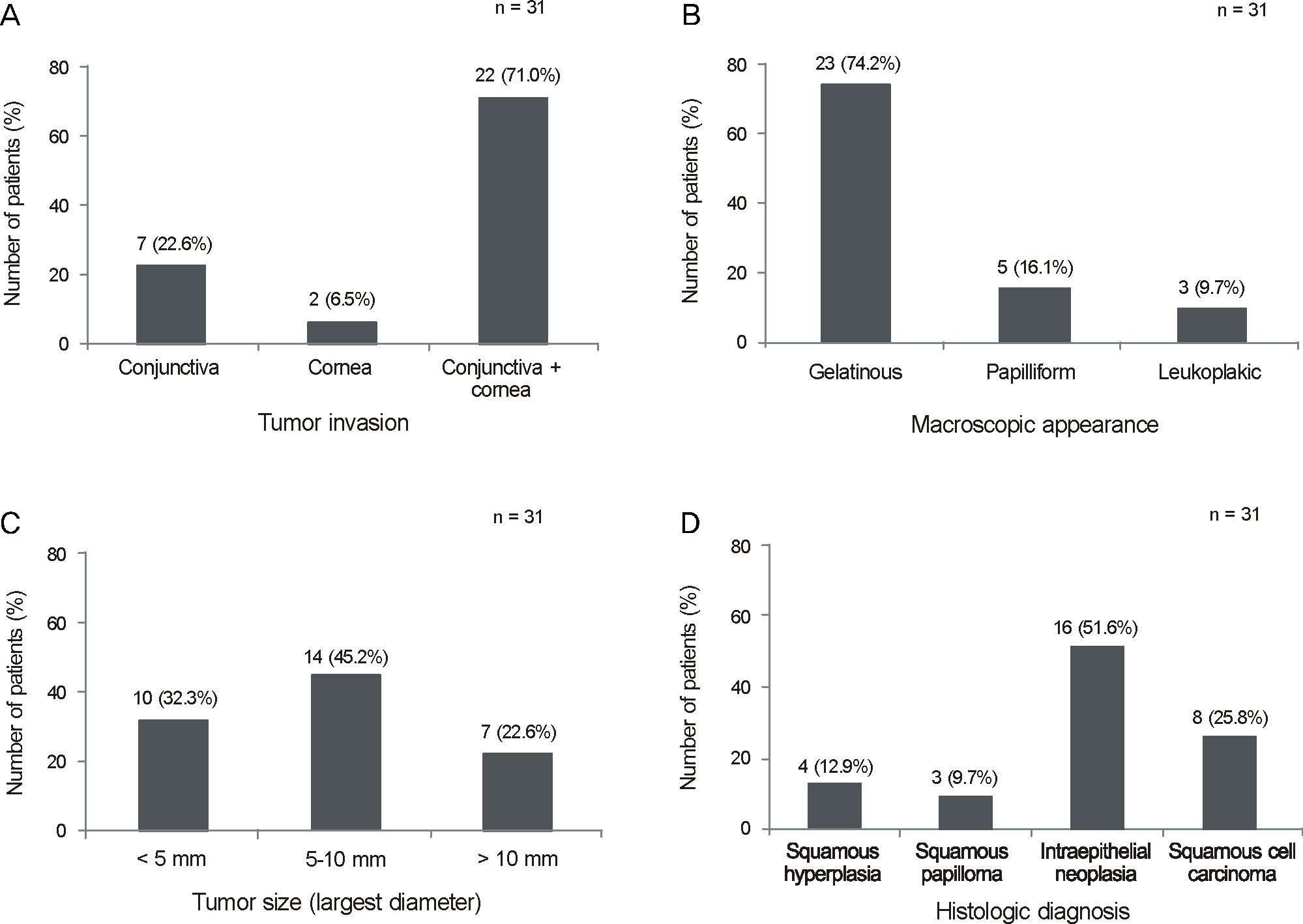
Table 1.
Demographics of 31 patients with ocular surface squamous neoplasia
Table 2.
Univariate analysis for factors affecting recurrence in patients with ocular surface squamous neoplasia
| Variables |
Number of patients |
p-value | |
|---|---|---|---|
| Recurrence group | Non-recurrence group | ||
| Age | 0.464* | ||
| Mean ± SD (years) | 69.9 ± 4.9 | 70.3 ± 11.7 | |
| Sex | 0.095† | ||
| Male/Female | 3/5 | 17/6 | |
| Macroscopic appearance | 0.915† | ||
| Gelatinous | 6 | 17 | |
| Papilliform | 1 | 4 | |
| Leukoplakic | 1 | 2 | |
| Tumor size (largest diameter) | 0.878† | ||
| <5 mm | 3 | 7 | |
| 5-10 mm | 3 | 11 | |
| >10 mm | 2 | 5 | |
| Operation method | 0.045† | ||
| Surgical excision | 7 | 10 | |
| Surgical excision + 0.02% mitomycin C chemotherapy | 1 | 13 | |
| Histologic diagnosis | 0.354† | ||
| Squamous hyperplasia | 0 | 4 | |
| Intraepithelial neoplasia | 5 | 11 | |
| Squamous cell carcinoma | 3 | 5 | |
| Squamous papilloma | 0 | 3 | |
 XML Download