

 PDF
PDF ePub
ePub Citation
 Print
Print


Abstract
Purpose
To investigate the relationship between photoreceptor inner/outer segment (IS/OS) integrity and macular sensi-tivity after epiretinal membrane (ERM) surgery using spectral-domain optical coherence tomography combined with microperimetry.
Methods
20 eyes of 20 patients with idiopathic ERM who underwent pars plana vitrectomy for ERM removal were pro-spectively reviewed. The IS/OS defect diameter, BCVA, macular sensitivity, and fixation stability were measured using spectral-domain optical coherence tomography combined with microperimetry. The correlation of these factors was analyzed.
Results
The macular sensitivity improved after successful ERM surgery (p < 0.001), but the IS/OS defect diameter has not improved. The preoperative and postoperative macular sensitivity were negatively correlated with preoperative IS/OS de-fect diameter (p = 0.035, p = 0.006). The fixation stability was not correlated with preoperative IS/OS defect diameter.
References
1. McDonald HR, Verre WP, Aaberg TM. Surgical management of idiopathic epiretinal membranes. Ophthalmology. 1986; 93:978–83.

2. Choi YK, Yoo JS, Kim MH. Result of surgery for epiretinal mem-brane and their recurrence. J Korean Ophthalmol Soc. 2000; 41:2357–62.
3. Margherio RR, Cox MS Jr, Trese MT. . Removal of epimacular membranes. Ophthalmology. 1985; 92:1075–83.
4. Kwon SI, Ko SJ, Park IW. The clinical course of the idiopathic epi-retinal membrane surgery. Korean J Ophthalmol. 2009; 23:249–52.
5. Rice TA, De Bustros S, Michels RG. . Prognostic factors in vi-trectomy for epiretinal membranes of the macula. Ophthalmology. 1986; 93:602–10.
6. Vujosevic S, Midena E, Pilotto E. . Diabetic macular edema: correlation between microperimetry and optical coherence tomog-raphy findings. Invest Ophthalmol Vis Sci. 2006; 47:3044–51.
7. Hwang DJ, Na KI, Kwon SI, Park IW. Long term changes in visual acuity and foveal thickness after vitrectomy for idiopathic epi-retinal membrane. J Korean Ophthalmol Soc. 2012; 53:434–9.
8. van Velthoven ME, Faber DJ, Verbraak FD. . Recent develop-ments in optical coherence tomography for imaging the retina. Prog Retin Eye Res. 2007; 26:57–77.
9. Ko TH, Fujimoto JG, Schuman JS. . Comparison of ultrahigh- and standard-resolution optical coherence tomography for imaging macular pathology. Ophthalmology. 2005; 112:1922.e1–15.
10. Schmidt-Erfurth U, Leitgeb RA, Michels S. . Three-dimensional ultrahigh-resolution optical coherence tomography of mac-ular diseases. Invest Ophthalmol Vis Sci. 2005; 46:3393–402.
11. Karacorlu M, Ozdemir H, Senturk F. . Correlation of retinal sensitivity with visual acuity and macular thickness in eyes with idiopathic epimacular membrane. Int Ophthalmol. 2010; 30:285–90.
12. Oster SF, Mojana F, Brar M. . Disruption of the photoreceptor inner segment/outer segment layer on spectral domain-optical co-herence tomography is a predictor of poor visual acuity in patients with epiretinal membranes. Retina. 2010; 30:713–8.
13. Lee JE, Kim EH, Oum BS. Relationship between visual acuity and photoreceptor layer or foveal thickness on optical coherence to-mography after macular hole surgery. J Korean Ophthalmol Soc. 2006; 47:1966–71.
14. Oh J, Smiddy WE, Flynn HW Jr. . Photoreceptor inner/outer segment defect imaging by spectral domain OCT and visual prog-nosis after macular hole surgery. Invest Ophthalmol Vis Sci. 2010; 51:1651–8.
15. Chang LK, Koizumi H, Spaide RF. Disruption of the photoreceptor inner segment-outer segment junction in eyes with macular holes. Retina. 2008; 28:969–75.
16. Chung H, Shin CJ, Kim JG. . Correlation of microperimetry with fundus autofluorescence and spectral-domain optical coher-ence tomography in repaired macular holes. Am J Ophthalmol. 2011; 151:128–36.e3.
17. Ooto S, Hangai M, Takayama K. . Photoreceptor damage and foveal sensitivity in surgically closed macular holes: an adaptive optics scanning laser ophthalmoscopy study. Am J Ophthalmol. 2012; 154:174–86.e2.
18. Kim YG, Baek SH, Moon SW. . Analysis of spectral domain optical coherence tomography findings in occult macular dystrophy. Acta Ophthalmol. 2011; 89:e52–6.
19. Lim JW, Cho JH, Kim HK. Assessment of macular function by multifocal electroretinography following epiretinal membrane sur-gery with internal limiting membrane peeling. Clin Ophthalmol. 2010; 4:689–94.
20. Kang JH, Choi ES, Yoon JM, Yoon HS. Multifocal electroretino-gram before and after epiretinal membrane surgery. J Korean Ophthalmol Soc. 2008; 49:104–10.
21. Okada K, Yamamoto S, Mizunoya S. . Correlation of retinal sensitivity measured with fundus-related microperimetry to visual acuity and retinal thickness in eyes with diabetic macular edema. Eye (Lond). 2006; 20:805–9.
22. Springer C, Völcker HE, Rohrschneider K. [Central serous cho-rioretinopathy–retinal function and morphology: microperimetry and optical coherence tomography]. Ophthalmologe. 2006; 103:791–7.
23. Amari F, Ohta K, Kojima H, Yoshimura N. Predicting visual out-come after macular hole surgery using scanning laser ophthalmo-scope microperimetry. Br J Ophthalmol. 2001; 85:96–8.
24. Klein R, Klein BE, Wang Q, Moss SE. The epidemiology of epi-retinal membranes. Trans Am Ophthalmol Soc. 1994; 92:403–25; discussion 425-30.
25. Gastaud P, Bétis F, Rouhette H, Hofman P. [Ultrastructural findings of epimacular membrane and detached posterior hyaloid in vitre-omacular traction syndrome]. J Fr Ophtalmol. 2000; 23:587–93.
26. Michels RG. Vitreous surgery for macular pucker. Am J Ophthalmol. 1981; 92:628–39.
27. Inoue M, Morita S, Watanabe Y. . Preoperative inner seg-ment/outer segment junction in spectral-domain optical coherence tomography as a prognostic factor in epiretinal membrane surgery. Retina. 2011; 31:1366–72.
28. Frisén L, Frisén M. Micropsia and visual acuity in macular edema. A study of the neuro-retinal basis of visual acuity. Albrecht Von Graefes Arch Klin Exp Ophthalmol. 1979; 210:69–77.
29. Ehrlich R, Mawer NP, Mody CH. . Visual function following photodynamic therapy for central serous chorioretinopathy: a com-parison of automated macular microperimetry versus best-cor-rected visual acuity. Clin Experiment Ophthalmol. 2012; 40:e32–9.
30. Suh MH, Seo JM, Park KH, Yu HG. Associations between macular findings by optical coherence tomography and visual outcomes af-ter epiretinal membrane removal. Am J Ophthalmol. 2009; 147:473–80.e3.
31. Tso MO. Animal modeling of cystoid macular edema. Surv Ophthalmol. 1984; 28:Suppl. 512–9.
32. Pesin SR, Olk RJ, Grand MG. . Vitrectomy for premacular fibroplasia. Prognostic factors, long-term follow-up, and time course of visual improvement. Ophthalmology. 1991; 98:1109–14.
33. Inoue M, Morita S, Watanabe Y. . Inner segment/outer segment junction assessed by spectral-domain optical coherence tomog-raphy in patients with idiopathic epiretinal membrane. Am J Ophthalmol. 2010; 150:834–9.
34. Mitamura Y, Hirano K, Baba T, Yamamoto S. Correlation of visual recovery with presence of photoreceptor inner/outer segment junction in optical coherence images after epiretinal membrane surgery. Br J Ophthalmol. 2009; 93:171–5.
35. Rohrschneider K, Bültmann S, Springer C. Use of fundus peri-metry (microperimetry) to quantify macular sensitivity. Prog Retin Eye Res. 2008; 27:536–48.
36. Sunness JS, Schuchard RA, Shen N. . Landmark-driven fun-dus perimetry using the scanning laser ophthalmoscope. Invest Ophthalmol Vis Sci. 1995; 36:1863–74.
37. Michalewski J, Michalewska Z, Cisiecki S, Nawrocki J. Mor- phologically functional correlations of macular pathology con-nected with epiretinal membrane formation in spectral optical coherence tomography (SOCT). Graefes Arch Clin Exp Ophthalmol. 2007; 245:1623–31.
38. Kinoshita T, Kovacs KD, Wagley S, Arroyo JG. Morphologic dif-ferences in epiretinal membranes on ocular coherence tomography as a predictive factor for surgical outcome. Retina. 2011; 31:1692–8.
Figure 1.
Representative figures of groups according to the integrity of junction between photoreceptor inner and outer segment (IS/OS) within 500 μ m from the center of fovea. (A) Group 1 (Preserved IS/OS). IS/OS is well preserved within the 500 μ m from the center of fovea. (B) Group 2 (Disrupted IS/OS). There is partially disrupted area of IS/OS (arrow). The disrupted IS/OS length was measured manually with caliper built in the spectral domain optical coherence tomography.

Figure 2.
A microperimetry combined with Spectral domain optical coherence tomographic image of a 76 year old male patient with idiopathic ERM. Preoperative fundus image of the right eye shows an epiretinal membrane (upper left). Optical coherence tomog-raphy (OCT) result shows epiretinal membrane delineated as highly reflective band and globally adherent to the retina. IS/OS dis-ruption (arrowheads) also presented. Microperimetry results show mean sensitivity in colors (upper right), and fixation stability (bottom right).

Figure 3.
(A) Preoperative macular sensitivity (dB) of the group 1 and 2. Preoperative macular sensitivity in the group 1 is significantly better than that in the group 2. (B) Preoperative best-corrected visual acuity (BCVA) (log MAR) of the group 1 and 2. However, there is no significant difference between the group 1 and the group 2. (C) Postoperative BCVA (log MAR) of the group 1 and 2. Postoperative BCVA in the group 1 is significantly better than that in the group 2. (p val-ue related to Kruskall-Wallis test). (* p < 0.05, ** p > 0.05) The box and whisker plots: horizontal lines within the box represent medians, the ends of the boxes represent the first and third quartiles, and the whiskers represent the smallest and largest non-outlier values.
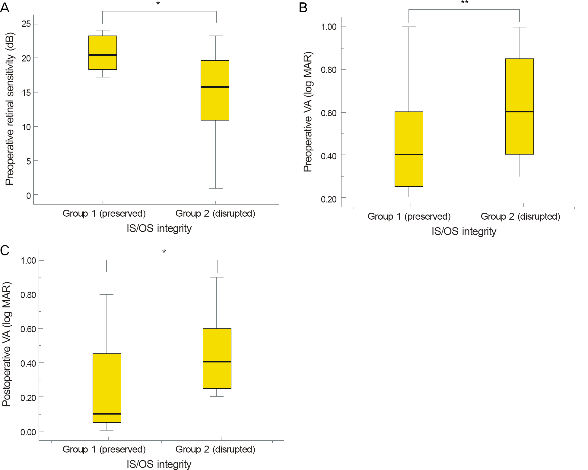
Figure 4.
Correlation between macular sensitivity (dB) and diame-ter of disrupted photoreceptor inner and outer segment junction (IS/OS defect diameter) (μ m) in the group 1 and 2 (marked with black triangle and white circle respectively) at the preoperative (A) and postoperative (B) visits. (A) shows that there is significant negative correlation between the preoperative IS/OS defect diame-ter (μ m) and macular sensitivity (dB) (r = -0.474, p = 0.035). (B) shows that there is significant negative correlation between the preoperative IS/OS defect diameter (μ m) and postoperative mac-ular sensitivity (dB) (r = -0.589, p = 0.006). In accordance with an increase in the IS/OS defect diameter at the preoperative visit, the postoperative macular sensitivity worsened. (C) shows there is significant negative correlation between the postoperative IS/OS defect diameter (μ m) and macular sensitivity (dB) (r = -0.647, p = 0.002). p-value related to linear logistic regression analysis.
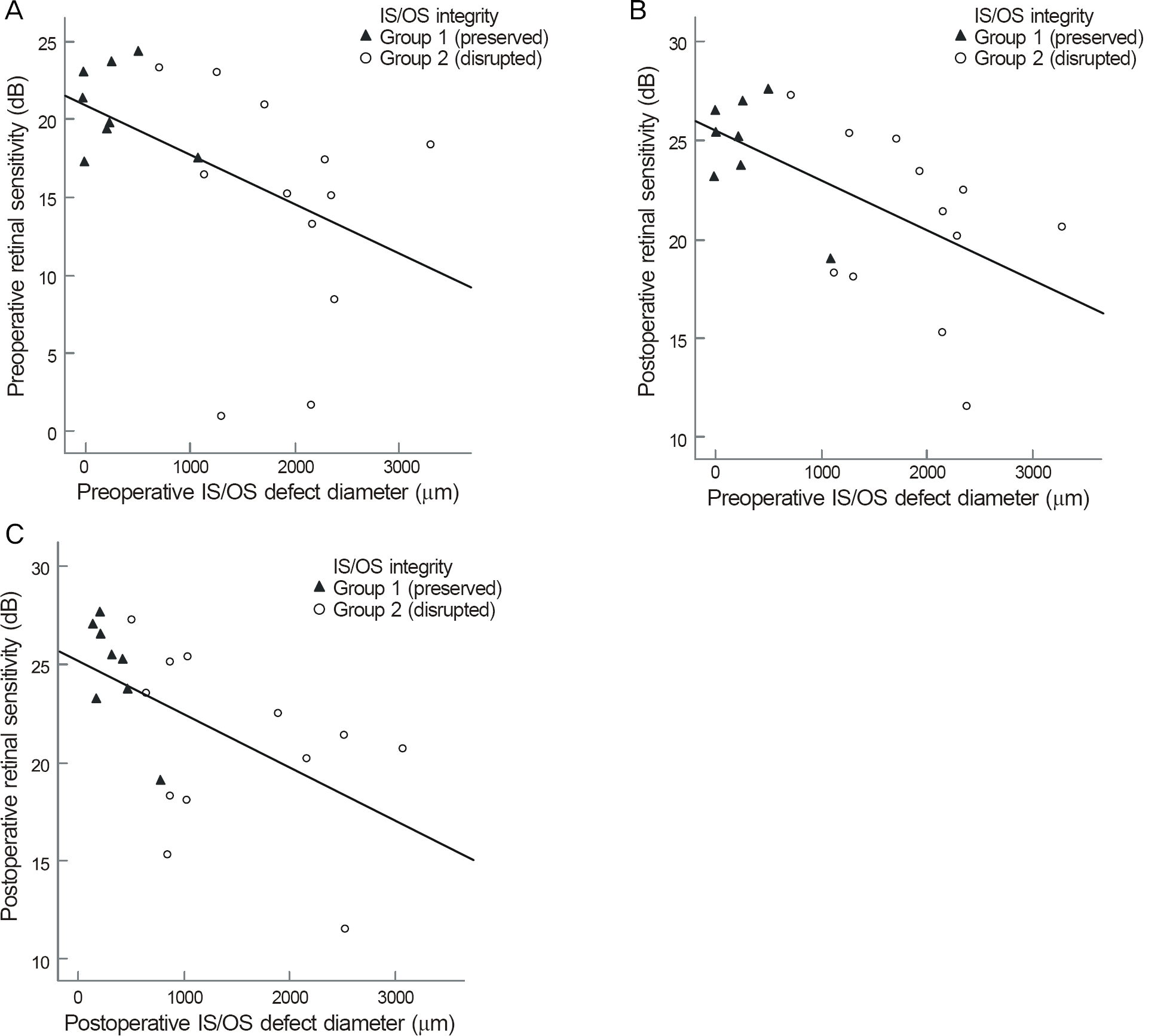
Table 1.
Summary of demographic data of all eyes with Idiopathic epiretinal membrane
| Characteristics | Value |
|---|---|
| Number of eyes (subjects) | 20 (20) |
| Sex (M:F) [n(%)] | 7 (35%) : 13 (65%) |
| Age (years) | 64.55 ± 2.41 |
| Refraction (S.E) | 0.63 ± 1.22 |
| Lens status* | |
| Mild cataract | 4 |
| Moderate cataract | 16 |
| Severe cataract | 0 |
Table 2.
Preoperative data and outcomes during follow up
| Characteristics | Base line | After surgery | p-value* |
|---|---|---|---|
| BCVA (log MAR) | 0.58 ± 0.05 | 0.39 ± 0.057 | 0.003 |
| IS/OS defect diameter (μ m) | 1247.8 ± 988.9 | 1023.4 ± 896.2 | 0.062 |
| Macular sensitivity (dB) | 16.95 ± 6.60 | 22.35 ± 4.29 | <0.001 |
| Fixation stability (%) | 61.0 ± 18.2 | 67.4 ± 23.5 | 0.059 |
Table 3.
Comparison of preoperative characteristics of patients with IS/OS preserved and disrupted epiretinal membrane
 XML Download