

 PDF
PDF ePub
ePub Citation
 Print
Print


Abstract
Purpose
We report a case of a patient with ptosis, lid swelling, limitation of ocular movement, and visual disturbance after a trauma. The patient was diagnosed with unilateral orbital cellulitis, ophthalmic vein thrombosis and bilateral septic cavernous thrombosis and treated.
Case summary
After head and facial area trauma that occurred 1 month earlier, a 56-year-old man suffered from ongoing visual loss, limitation of ocular movement in all directions, proptosis in the left eye and abduction limitation of the right eye. A week before admission, mild fever and chills were also present. At admission, visual acuity of the left eye was no light perception and pupil reflex was lost. Brain MRA and MRI indicated dilation and thrombosis of the left superior ophthalmic vein, left orbital cellulitis and inflammation in bilateral cavernous sinuses. The patient was immediately treated with systemic antibiotics and steroid injection. Coagulase negative staphylococci were detected in blood culture.
Conclusions
Infection caused by facial trauma spread through the facial area's venous plexus and caused orbital cellulitis. As a result, septic cavernous sinus thrombosis and ophthalmic vein thrombophlebitis occurred. Serious complications can occur after facial trauma, thus rapid differential diagnosis and appropriate treatment are important in determining prognosis.
J Korean Ophthalmol Soc 2013;54(4):662-666
References
1. Chandler JR, Langenbrunner DJ, Stevens ER. The pathogenesis of orbital complications in acute sinusitis. Laryngoscope. 1970; 80:1414–28.

2. Ebright JR, Pace MT, Niazi AF. Septic thrombosis of the cavernous sinuses. Arch Intern Med. 2001; 161:2671–6.
3. SHAW RE. Cavernous sinus thrombophlebitis: a review. Br J Surg. 1952; 40:40–8.
4. Neil R, Miller NJ, Newman WF, et al. Walsh and Hoyt's clinical neuro-ophthalmology. 4th ed. Baltimore: Williams & Wilkins;1991. p. 2734–7.
5. Tveterås K, Kristensen S, Dommerby H. Septic cavernous and lateral sinus thrombosis: modern diagnostic and therapeutic principles. J Laryngol Otol. 1988; 102:877–82.
6. Clifford-Jones RE, Ellis CJ, Stevens JM, Turner A. Cavernous sinus thrombosis. J Neurol Neurosurg Psychiatry. 1982; 45:1092–7.
7. Kraus CL, Culican SM. Challenging presentations of cavernous sinus thrombophlebitis. J Ophthalmic Inflamm Infect. 2012; 2:133–6.
8. Kang MH, Lee YJ, Shin SY. A case of bilateral cavernous sinus thrombosis secondary to right sphenoid sinusitis. J Korean Ophthalmol Soc. 2004; 45:1194–9.
9. Koo NK, Kim JH, Lee SY. A case of bilateral cavernous sinus thrombosis with intraorbital abscess. J Korean Ophthalmol Soc. 2005; 46:731–5.
10. Martin-Hirsch DP, Habashi S, Hinton AH, Kotecha B. Orbital cellulitis. Arch Emerg Med. 1992; 9:143–8.
11. Berge J, Louail C, Caillé JM. Cavernous sinus thrombosis diagnostic approach. J Neuroradiol. 1994; 21:101–17.
12. Solomon OD, Moses L, Volk M. Steroid therapy in cavernous sinus thrombosis. Am J Ophthalmol. 1962; 54:1122–4.
13. Chen YC, Cheng TT, Lai HM, Wu CH. Overwhelming septic cavernous sinus thrombosis in a woman after combination of high-dose steroid and intravenous cyclophosphamide therapy for lupus nephritis. Lupus. 2000; 9:78–9.
14. Desa V, Green R. Cavernous sinus thrombosis: current therapy. J Oral Maxillofac Surg. 2012; 70:2085–91.
15. Levine SR, Twyman RE, Gilman S. The role of anticoagulation in cavernous sinus thrombosis. Neurology. 1988; 38:517–22.
16. Coutinho JM, Ferro JM, Canhão P, et al. Unfractionated or low-molecular weight heparin for the treatment of cerebral venous thrombosis. Stroke. 2010; 41:2575–80.
Figure 1.
Patient's photographs of initial presentation show (A) exophthalmos and (B) chemosis of the left eye. Orbit M RI before treatment shows (C) heterogeneous enhancement in both cavernous sinus which means inflammatory change (arrow) and (D, E) engorged left superior ophthalmic vein with contrast filling defect which means thrombophlebitis (arrow).
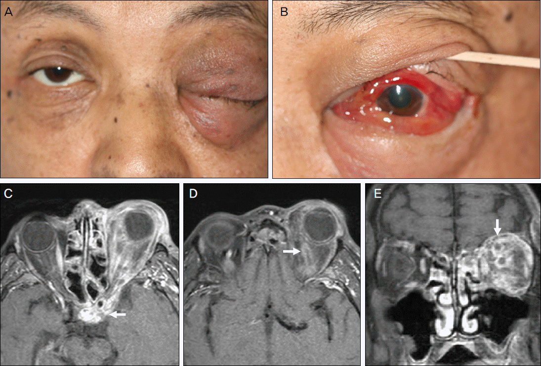
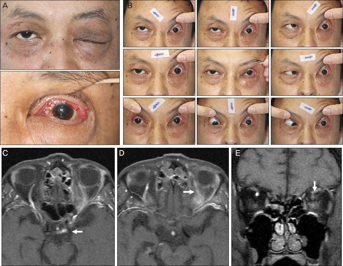
Figure 2.
Patient had been treated with steroid and antibiotics for 5 days. Patient's photographs show that (A) exophthalmos and chemosis of the left eye improved, but, (B) ocular movement limitation remains in both eyes. At 2 weeks after treatment, orbit M RI shows improvement of (C) cavernous sinus thrombosis, (D) left superior ophthalmic vein thrombosis and (E) left orbital cellulitis (arrow).
 XML Download