

 PDF
PDF ePub
ePub Citation
 Print
Print


Abstract
Purpose
To report clinical manifestations including neurocutaneous and ocular findings and to evaluate outcomes of tra- beculectomy in patients with Sturge-Weber syndrome.
Methods
The medical records of 10 eyes of 8 glaucoma patients with Sturge-Weber syndrome who were followed up for at least 1 year after trabeculectomy were reviewed retrospectively. We analyzed neurocutaneous and ocular findings, cumulative surgical success rates, and complications in patients with Sturge-Weber syndrome.
Results
The mean patient age at the time of surgery was 12.6 ± 13.0 years and mean follow-up period was 71.6 ± 81.8 months. All patients showed various clinical findings including facial hemangioma (8 patients), seizure (6 patients), intra- cranial lesion (6 patients), developmental delay (4 patients), conjunctival/episcleral hemangioma (4 eyes), and choroidal hemangioma (4 eyes). Postoperative success was achieved in 8 out of 10 eyes (80.0%). Postoperatively, serous retinal detachment occurred in 2 out of 4 eyes with preoperative diffuse choroidal hemangioma.
Conclusions
Management of glaucoma associated with Sturge-Weber syndrome requires multidisciplinary treatment because of systemic involvement. Trabeculectomy appears to be an effective and relatively safe surgical option for glaucoma associated with Sturge-Weber syndrome. However, serious complications such as serous retinal detachment should be considered when planning trabeculectomy for patients with diffuse choroidal hemangioma.
References
1. Hunt HB, Moore RC. Encephalotrigeminal angiomatosis; (Sturge- Weber syndrome). Med Radiogr. 1951; 27:53–60.
2. Cibis GW, Tripathi RC, Tripathi BJ. Glaucoma in Sturge-Weber syndrome. Ophthalmology. 1984; 91:1061–71.

3. Sujansky E, Conradi S. Outcome of Sturge-Weber syndrome in 52 adults. Am J Med genet. 1995; 57:35–45.
4. Sullivan TJ, Clarke MP, Morin JD. The ocular manifestations of the Sturge-Weber syndrome. J Pediatr Ophthalmol Strabismus. 1992; 29:349–56.
5. Kim JW, Park CH, Lee CH. Clinical experience of treatment in a case of Sturge-Weber syndrome with bilateral glaucoma. J Korean Ophthalmol Soc. 1996; 908–12.
6. Bae JH, Cho HD, Woo SC. Serous retinal detachment following trabeculectomy in a case of Sturge-Weber syndrome with glaucoma. J Korean Ophthalmol Soc. 1996; 37:2150–3.
7. Lee SH, Jung DY. Choroidal effesuion after trabeculetomy in glau- coma with Sturge-Weber syndrome. J Korean Ophthalmol Soc. 1999; 40:1164–8.
8. Kim HB, Hong YJ, Kim YJ, Kim SW. A case of Sturge - Weber syndrome. J Korean Ophthalmol Soc. 1981; 22:273–6.
9. Yoo HM, Chung I, Chang SK. A case of Sturge - Weber syndrome. J Korean Ophthalmol Soc. 1986; 27:723–8.
10. Choi JS, Yi KP, Hong KY. A case of Sturge - Weber syndrome. J Korean Ophthalmol Soc. 1989; 30:459–64.
11. Chung IH, Kim MH. Sturge - Weber syndrome with congenital oc- ular anomaly. J Korean Ophthalmol Soc. 1995; 36:2266–70.
12. Lee CW, Choi DY, Oh YG, et al. An infantile case of Sturge-Weber syndrome in association with Klippel-Trenaunay-Weber syndrome and phakomatosis pigmentovascularis. J Korean Med Sci. 2005; 20:1082–4.
13. Cairns JE. Trabeculectomy. Preliminary report of a new method. Am J Ophthalmol. 1968; 66:673–9.
14. Sharan S, Swamy B, Taranath DA, et al. Port-wine vascular mal- formations and glaucoma risk in Sturge-Weber syndrome. J AAPOS. 2009; 13:374–8.
15. Parsa CF. Sturge-weber syndrome: a unified pathophysiologic mechanism. Curr Treat Options Neurol. 2008; 10:47–54.
16. Patrianakos TD, Nagao K, Walton DS. Surgical management of glaucoma with the sturge weber syndrome. Int Ophthalmol Clin. 2008; 48:63–78.
17. Ali MA, Fahmy IA, Spaeth GL. Trabeculectomy for glaucoma as- sociated with Sturge-Weber syndrome. Ophthalmic surgery. 1990; 21:352–5.
18. Fontana H, Nouri-Mahdavi K, Lumba J, et al. Trabeculectomy with mitomycin C: outcomes and risk factors for failure in phakic open-angle glaucoma. Ophthalmology. 2006; 113:930–6.
19. Keverline PO, Hiles DA. Trabeculectomy for adolescent onset glaucoma in the Sturge-Weber syndrome. J Pediatr Ophthalmol. 1976; 13:144–8.
20. Yang Q, Guo WY, Sun XH, et al. Clinical analysis on sixteen cases of Sturge-Weber syndrome-associated glaucoma. Zhonghua Yan Ke Za Zhi. 2009; 45:883–7.
21. Bellows AR, Chylack LT Jr, Epstein DL, Hutchinson BT. Choroidal effusion during glaucoma surgery in patients with prom- inent episcleral vessels. Arch Ophthalmol. 1979; 97:493–7.
22. Susac JO, Smith JL, Scelfo RJ. The “tomato-catsup” fundus in sturge-weber syndrome. Arch Ophthalmol. 1974; 92:69–70.
23. Tripathi BJ, Tripathi RC. Neural crest origin of human trabecular meshwork and its implications for the pathogenesis of glaucoma. Am J Ophthalmol. 1989; 107:583–90.
24. Dorairaj S, Ritch R. Encephalotrigeminal Angiomatosis (Sturge- Weber Syndrome, Klippel-Trenaunay-Weber Syndrome): A Review. Asia-Pac J Ophthalmol. 2012; 1:226–34.
25. Beck RW, Hanno R. The phakomatoses. Int Ophthalmol Clin. 1985; 25:97–116.
26. Pascual-Castroviejo I, Díaz-Gonzalez C, García-Melian RM, et al. Sturge-Weber syndrome: study of 40 patients. Pediatr Neurol. 1993; 9:283–8.
27. Bebin EM, Gomez MR. Prognosis in Sturge-Weber disease: com- parison of unihemispheric and bihemispheric involvement. J Child Neurol. 1988; 3:181–4.
28. Sujansky E, Conradi S. Sturge-Weber syndrome: age of onset of seizures and glaucoma and the prognosis for affected children. J Child Neurol. 1995; 10:49–58.
29. Klapper J. Headache in Sturge-Weber syndrome. Headache. 1994; 34:521–2.
Figure 1.
Conjunctival and/or episcleral hemangioma of patient. (A) Mildly engorged conjunctival and/or episcleral vessel with hypovascular bleb. (B) Moderately engorged conjunctival and/ or episcleral vessel with avascular bleb. (C, D) Severely engorged conjunctival and/or episcleral vessel.
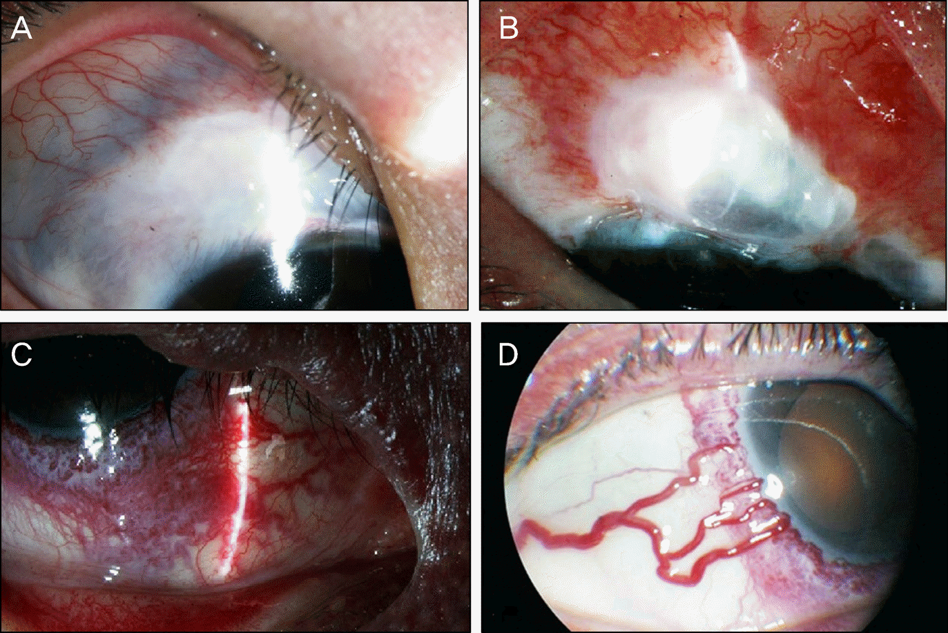
Figure 2.
Diffuse choroidal hemangioma in patients with Sturge-Weber syndrome and Klippel-Trenaunay- Weber syndrome. (A) Right eye of patient 6. (B) Left eye of patient 2. (C) Left eye of patient 8.
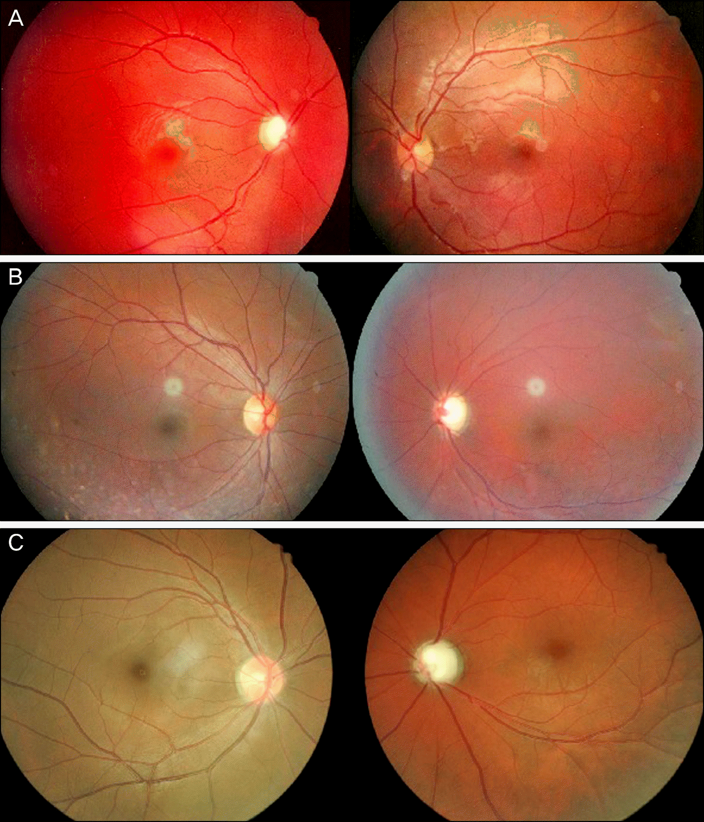
Figure 3.
Kaplan-Meier survival curve showing the cumulative success rates of trabeculectomy for patients with Sturge-Weber syndrome.
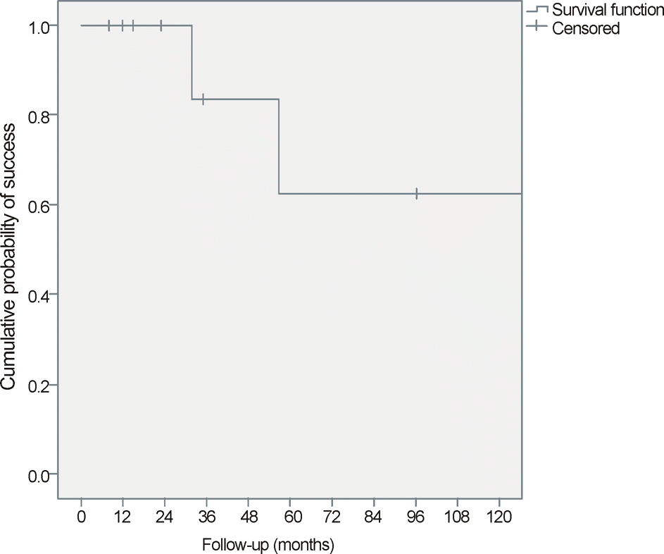
Figure 4.
Patient 6 presented choroidal detachment with serous retinal detachment and optic disc swelling after trabeculectomy. (A, B) Severe choroidal detachment with serous retinal detachmnet and optic disc swelling on th 19th postoperative day. (C, D) Decreased choroidal detachment with serous retinal detachemnt and remained optic disc swelling on the 60th postoperative day. (E) No choroidal effusion or retinal detachment was noted 5 years after trabeculectomy.
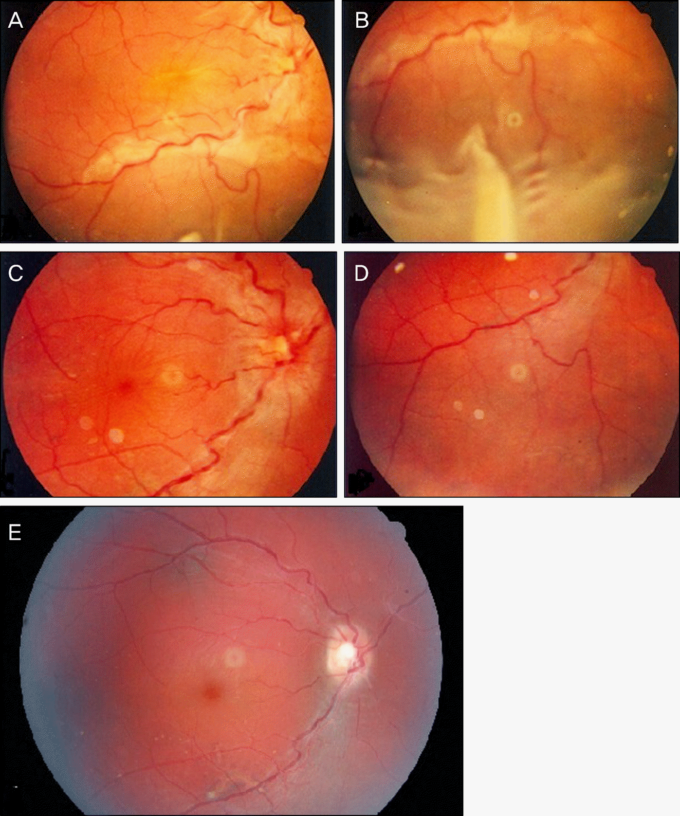
Figure 5.
Immediately after removal of releasable suture at postoperative third days (Patient 1), (A, B) almost total hyphema was developed, and (C) massive subconjuntival hemorrahge was seen. (D) Hemorrhagic bleb was noted. (E, F) Two-year postoperative appearance of the bleb on the right eye of patient 1. The figures show diffuse moderately elevated, hypovascular bleb.
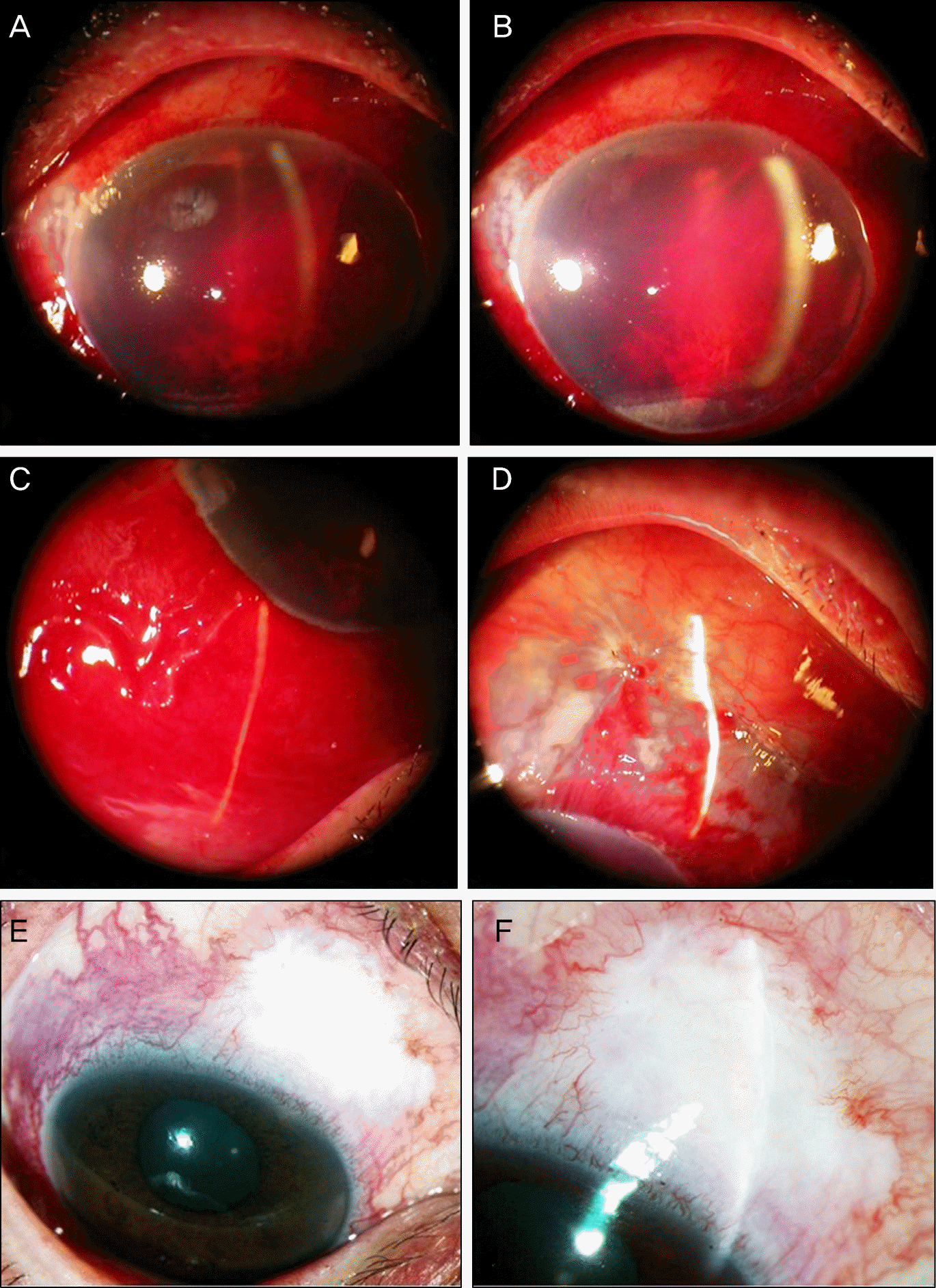
Figure 6.
Brain MRI in a patient with Sturge- Weber syndrome. (A) Leptomeningeal angioma (arrow) and choroid plexus hemangioma (arrow head) of patient 1. (B) Dominant choroidal plexus hemangioma (arrow) of patient 3. (C) Diffuse cortical atrophy (arrow) of patinet 8.
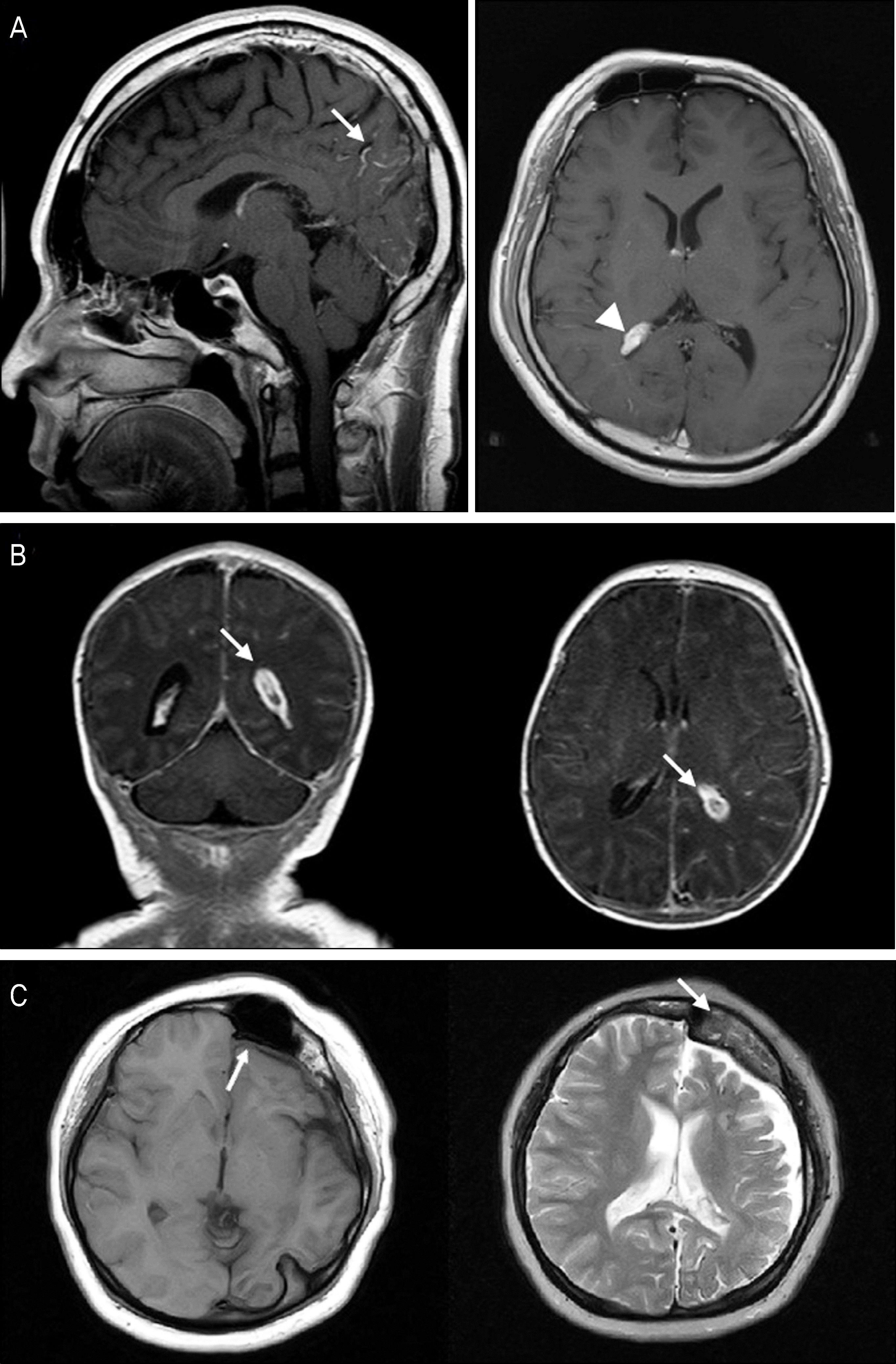
Figure 7.
Cutaneous manifestations of Sturge-Weber syndrome and Klippel- Trenaunay-Weber syndrome. (A) Portwine stain in patient 2 with Sturge- Weber syndrome. Note the hypertrophy and nodularity of facial angioma. (B) Bilateral involvement of facial hemangioma (Patient 5) (C) Limbal and truncal hemangioma in patient 7 with Klippel- Trenaunay-Weber syndrome. (D) Limbal soft tissue hypertrophy in patient 7 with Klippel-Trenaunay-Weber syndrome.

Table 1.
Patient demographics and clinical characteristics
Table 2.
Distribution of facial hemangioma in patients with Sturge-Weber syndrome
Table 3.
Ocular manifestations in Sturge-Weber syndrome (N = 8)
| Ocular manifestation | Number (%) |
|---|---|
| Conjunctival and/or Episcleral hemangioma | 4 (50) |
| Choroidal hemangioma | 4 (50) |
| Ocular melanosis | 3 (37.5) |
| Iris heterochromia | 0 (0) |
Table 4.
Details of patients with Sturge-Weber syndrome treated with trabeculectomy
| Diagnosis | Age* | Sex | Side | Onset | Preop. IOP | Final IOP | Preop C/D | Final C/D | Postoperative complications | |
|---|---|---|---|---|---|---|---|---|---|---|
| Patient 1 | SWS | 39 years | M | OD | J | 24 | 8 | 0.95 | 0.95 | Hyphema |
| Patient 2 | SWS, KTW | 27 years | M | OS | I | 23 | 19 | 0.9 | 0.9 | Wound leak |
| Patient 3 | SWS | 1 month | M | OS | I | 25 | 27 | 0.85 | 0.8 | SRD, Shallow A.C. |
| Patient 4 | SWS, KTW | 9 years | M | OS | J | 23 | 16 | 0.95 | Total cup | (-) |
| Patient 5 | SWS, KTW | 32 months | M | OD | I | 23 | 33 | 0.95 | 0.7 | Shallow A.C. |
| 7 years | M | OS | I | 32 | 14 | 0.8 | 0.55 | Shallow A.C. | ||
| Patient 6 | SWS, KTW | 5 years | F | OD | I | 28 | 8 | 0.7 | 0.6 | SRD, Shallow A.C., Hypotony |
| Patient 7 | SWS, KTW | 6 years | M | OD | I | 35 | 12 | 0.8 | 0.6 | Shallow A.C. |
| 6 years | M | OS | I | 36 | 13 | 0.8 | 0.6 | Shallow A.C. | ||
| Patient 8 | SWS | 25 years | F | OS | J | 26 | 10 | 0.95 | 0.95 | (-) |
Table 5.
Neurologic manifestations in Sturge-Weber syndrome (N = 8)
 XML Download