

 PDF
PDF ePub
ePub Citation
 Print
Print


Abstract
Purpose
To report a case of bilateral acute myopia and angle-closure with ciliochoroidal detachment in Vogt-Koyanagi-Harada (VKH) syndrome.
Case summary
A 43-year-old Korean woman diagnosed with VKH syndrome underwent intravenous methylprednisolone steroid pulse treatment. After oral medication was given for 2 days, the anterior chambers became shallow in both eyes. Intraocular pressure (IOP) increased to 25 mm Hg in the right eye and 23 mm Hg in the left eye. Subretinal fluid increased and visual acuity decreased with myopic shift in both eyes. IOP did not decrease despite maximum tolerated medical therapy. Ultrasound biomicroscopy (UBM) revealed that ciliochoroidal effusion caused forward displacement of the lens-iris diaphragm, which resulted in anterior chamber shallowing and angle closure in both eyes. The patient was treated with cycloplegic eyedrops and intravenous steroid pulse therapy. After intravenous steroid pulse treatment was given for 2 days, the anterior chambers became deep in both eyes. IOP reduced to 13 mm Hg and 14 mm Hg in the right and left eye respectively. Visual acuity increased with normalization of myopic shift. UBM revealed that the ciliochoroidal effusion had resolved in both eyes.
Figures and Tables
Figure 1
Fundus photographs showing bilateral multiple serous retinal detachment (A). Late phase of fluorescein angiography showing multiple cobble stone like hyperfluorescent spots and dye pooling (B). Optical coherence tomography demonstrates serous retinal detachment in the both eyes (C).
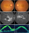
Figure 2
Ultrasound biomicroscopic images of the anterior segment before second steroid pulse therapy in the right eye (A) and the left eye (B). The anterior chamber angle is narrow, and a layer of fluid has accumulated external to the ciliary body and choroid (ciliochoroidal detatchment-asterisk) with forward displacement of the ciliary body in the both eyes. Ultrasound biomicroscopic images of the anterior segment on day 2 of the second steroid pulse therapy. The anterior chamber has deepened to normal depth and ciliochoroidal detachment has disappeared in the right eye (C) and the left eye (D).
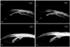
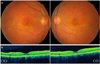
Figure 3
Fundus photographs show bilateral multiple depigmentation and sunset glow appearance around the fovea in both eyes at 2 months after the second steroid pulse therapy (A). Optical coherence tomography shows that serous retinal detachment has resolved 2 months after the second steroid pulse therapy in the both eyes (B).
References
1. Moorthy RS, Inomata H, Rao NA. Vogt-Koyanagi-Harada syndrome. Surv Ophthalmol. 1995. 39:265–292.
2. Sugiura S. Vogt-Koyanagi-Harada disease. Jpn J Ophthalmol. 1978. 22:9–35.
3. Forster DJ, Rao NA, Hill RA, et al. Incidence and management of glaucoma in Vogt-Koyanagi-Harada syndrome. Ophthalmology. 1993. 100:613–618.
4. Kimura R, Sakai M, Okabe H. Transient shallow anterior chamber as initial symptom in Harada's syndrome. Arch Ophthalmol. 1981. 99:1604–1606.
5. Kimura R, Kasai M, Shoji K, Kanno C. Swollen ciliary processes as an initial symptom in Vogt-Koyanagi-Harada syndrome. Am J Ophthalmol. 1983. 95:402–403.
6. Tokuoka S, Iwasaki Y, Sugasawa J. Harada's disease with shallow anterior chamber, myopia and intraocular pressure rise in a pair of siblings. Jpn Clin Ophthalmol. 1988. 42:365–368.
7. Rathinam SR, Namperumalsamy P, Nozik RA, Cunningham ET Jr. Angle closure glaucoma as a presenting sign of Vogt-Koyanagi-Harada syndrome. Br J Ophthalmol. 1997. 81:608–609.
8. Eibschitz-Tsimhoni M, Gelfand YA, Mezer E, Miller B. Bilateral angle closure glaucoma: an unusual presentation of Vogt-Koyanagi-Harada syndrome. Br J Ophthalmol. 1997. 81:705–706.
9. Ashizuka S, Nishioka Y, Kawano Y, et al. Ciliochoroidal detachment in Vogt-Koyanagi-Harada syndrome. Jpn J Clin Ophthalmol. 1997. 51:1067–1070.
10. Kawano Y, Tawara A, Nishioka Y, et al. Ultrasound biomicroscopic analysis of transient shallow anterior chamber in Vogt-Koyanagi-Harada syndrome. Am J Ophthalmol. 1996. 121:720–723.
11. Maruyama Y, Kimura Y, Kishi S, Shimizu K. Serous detachment of the ciliary body in Harada disease. Am J Ophthalmol. 1998. 125:666–672.
12. Wada S, Kohno T, Yanagihara N, et al. Ultrasound biomicroscopic study of ciliary body changes in the post-treatment phase of Vogt-Koyanagi-Harada disease. Br J Ophthalmol. 2002. 86:1374–1379.
13. Gohdo T, Tsukahara S. Ultrasound biomicroscopy of shallow anterior chamber in Vogt-Koyanagi-Harada syndrome. Am J Ophthalmol. 1996. 122:112–114.
14. Ahn JK. Morphologic changes in the anterior segment in patients with initial-onset or recurrent Vogt-Koyanagi-Harada disease. Ocul Immunol Inflamm. 2010. 18:314–318.
15. Nakamura H, Tsukamoto H, Shibahara R, et al. Ultrasound biomicroscopy in the management of Vogt-Koyanagi-Harada disease. Acta Ophthalmol Scand. 2000. 78:718–719.
16. Kishi A, Nao-i N, Sawada A. Ultrasound biomicroscopic findings of acute angle-closure glaucoma in Vogt-Koyanagi-Harada syndrome. Am J Ophthalmol. 1996. 122:735–737.
17. Ikeda N, Ikeda T, Nagata M, Mimura O. Pathogenesis of transient high myopia after blunt eye trauma. Ophthalmology. 2002. 109:501–507.
 XML Download