

 PDF
PDF ePub
ePub Citation
 Print
Print


Abstract
Purpose
To evaluate spectral-domain optical coherence tomography (SD-OCT) images of acute central retinal artery occlusion (CRAO).
Methods
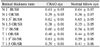
Six eyes of 6 patients who were diagnosed with acute CRAO were enrolled in the present study. The main SD-OCT measurements included macular thickness in the macular cube map and retinal thickness in the 5-line raster map.
Results
In acute CRAO, macular thickness increased more than that in the normal fellow eye in all cases. SD-OCT images demonstrated increased reflectivity and thickness in the inner retinal layer, especially in the nerve fiber layer and ganglion cell layer. However, in the outer retinal layer, SD-OCT images disclosed decreased reflectivity and increased thickness. The central subfield thickness area image showed normal reflectivity and thickness. The horizontal cross-sectional image showed relatively the same edema ratio between the inner retinal layer and outer retinal layer thickness. There was no cystoid or serous foveal detachment-type edema.
Figures and Tables
Figure 1
ETDRS subfields within standard three concentric circles of diameters 1,3 and 6 mm in the right eye used for reporting retinal thickness. F = fovea; SI = superior inner; TI = temporal inner; II = inferior inner; NI = nasal inner; SO = superior outer; TO = temporal outer; IO = inferior outer; NO = nasal outer.
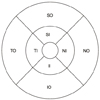
Figure 2
Double-headed arrows indicate various thickness. SRT = sensory retinal thickness; IRT = inner retinal thickness; MFT = minimal foveal thickness.
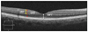
Figure 3
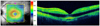
Optical coherence tomography images of case 1. The macular cube map demonstrates increased thickness on the macular area. The horizontal cross sectional image demonstrates increased reflectivity and thickness of the inner retinal layers, but decreased reflectivity in the outer retinal layer. Beneath the fovea, there is an area of normal reflectivity of the IS/OS line and RPE layer.


References
1. Otani T, Kishi S, Maruyama Y. Patterns of diabetic macular edema with optical coherence tomography. Am J Ophthalmol. 1999. 127:688–693.
2. Kang SW, Park CY, Ham DI. The correlation between fluorescein angiographic and optical coherence tomographic features in clinically significant diabetic macular edema. Am J Ophthalmol. 2004. 137:313–322.
3. Song YB, Park SP. Short-term effects of intravitreal Bevacizumab injection and macular edema patterns in branch retinal vein occlusion. J Korean Ophthalmol Soc. 2010. 51:379–385.
4. Hayreh SS, Kolder HE, Weingeist TA. Central retinal artery occlusion and retinal tolerance time. Ophthalmology. 1980. 87:75–78.
5. Hayreh SS, Zimmerman MB, Kimura A, Sanon A. Central retinal artery occlusion. Retinal survival time. Exp Eye Res. 2004. 78:723–736.
6. Kroll AJ. Experimental central retinal artery occlusion. Arch Ophthalmol. 1968. 79:453–469.
7. Early Treatment Diabetic Retinopathy Study Research Group. Photocoagulation for diabetic macular edema. Early Treatment Diabetic Retinopathy Study report number 1. Arch Ophthalmol. 1985. 103:1796–1806.
8. Dahrling BE 2nd. The histopathology of early central retinal artery occlusion. Arch Ophthalmol. 1965. 73:506–510.
9. Ritter M, Sacu S, Deák GG, et al. In vivo identification of alteration of inner neurosensory layers in branch retinal artery occlusion. Br J Ophthalmol. 2012. 96:201–207.
10. Kaur C, Foulds WS, Ling EA. Blood-retinal barrier in hypoxic ischaemic conditions: basic concepts, clinical features and management. Prog Retin Eye Res. 2008. 27:622–647.
11. Antcliff RJ, Marshall J. The pathogenesis of edema in diabetic maculopathy. Semin Ophthalmol. 1999. 14:223–232.
12. Yanoff M, Fine BS, Brucker AJ, Eagle RC Jr. Pathology of human cystoid macular edema. Surv Ophthalmol. 1984. 28:Suppl. 505–511.
13. Jalkh AE, Trempe CL. Macular edema in branch retinal vein occlusion: types and treatment. Ophthalmic Surg. 1989. 20:26–32.
14. Yanoff M, Fine BS. Ocular Pathology: A Text and Atlas. 1989. 3rd ed. Philadelphia: Lippincott;386–389.
 XML Download