

 PDF
PDF ePub
ePub Citation
 Print
Print


Abstract
Purpose
To report specific spectral domain OCT findings of Oguchi disease diagnosed with fundoscopic examination and electrophysiological study.
Case summary
A 14-year-old patient visited our clinic with a complaint of night blindness for ten years. Fundoscopic examination showed a golden-yellow fundus reflex. After three hours of dark adaptation, the fundus color returned to normal (Mizuo-Nakamura phenomenon). In full-field ERG, rod b-wave was not detectable. The a-wave amplitude in maximal combined response increased after three hours of dark adaptation, although the b-wave amplitude was similar to the amplitude before dark adaptation, demonstrating a negative waveform. In the spectral domain OCT images of the perifoveal area, no gap between the retinal pigment epithelium and the inner segment/outer segment (IS/OS) junction was detected before prolonged dark adaptation, and a highly reflective band was shown. However, the gap appeared after three hours of dark adaptation, and two highly reflective bands were detected in the OCT images.
Figures and Tables
Figure 1
Fundus photographs with montage view. (A) Before dark adaptation, golden metallic sheen is widely observed through the central and peripheral retina. (B) After 3 hours of dark adaptation, the fundus color returns to normal.
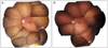
Figure 2
(A) Full-field ERG findings recorded with ISCEV standard protocol. In maximal combined response, b-wave (Amplitude : 75.7 µV) smaller than a-wave (76.3 µV ) was observed. The cone response and the 30Hz flicker response were normal. (B) Full-field ERG responses with (1) 30 minutes and with (2) 3 hour dark adaptation. After 3 hours of dark adaptation, rod response was still absent. The amplitude of a-wave (105.6 µV) in maximal combined response increased, but the amplitude of b-wave (78.1 µV) was similar to that before the dark adaptation. Any changes in oscillatory potential was not detected, either. Arrowheads in each response indicate stimulus onset.
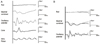
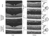
Figure 3
Horizontal cross sectional OCT images (fovea and perifovea) of normal subject (A,B) and the patient with Oguchi disease (C,D,E,F). The graph to the right of the images shows the reflective intensity at each vertical point of the retina (arrowhead). (A, B) The retinal outer layer is made up of two reflective band, IS/OS (Inner segment/Outer segment) junction and the retinal pigment epithelium (RPE) at both fovea and perifoveal region. (C, D) With dim light adaptation, the retinal outer layer is made up of two reflective band at the fovea (IS/OS junction and RPE) (C) as shown in the normal subject (A). But at the perifoveal region, a single highly reflective thick band is seen on the center of the graph (circle) (D). (E, F) After 3 hours of dark adaptation, the fovea and perifovea had two reflective band, as shown in the IS/OS junction and RPE band of the normal subject.
References
1. Usui T, Tanimoto N, Ueki S, et al. ERG rod a-wave in Oguchi disease. Vision Res. 2004. 44:535–540.
2. Lee JH, Kim SJ, Jo HI. A case of Oguchi's disease. J Korean Ophthalmol Soc. 1986. 27:265–270.
3. Kim HT, Lim SJ, Kim JW. A case of Oguchi's disease. J Korean Ophthalmol Soc. 1990. 31:105–109.
4. Miyake Y, Horiguchi M, Suzuki S, et al. Electrophysiological findings in patients with Oguchi's disease. Jpn J Ophthalmol. 1996. 40:511–519.
5. Hashimoto H, Kishi S. Shortening of the rod outer segment in Oguchi disease. Graefes Arch Clin Exp Ophthalmol. 2009. 247:1561–1563.
6. Yamada K, Motomura Y, Matsumoto CS, et al. Optical coherence tomographic evaluation of the outer retinal architecture in Oguchi disease. Jpn J Ophthalmol. 2009. 53:449–451.
7. Takada M, Otani A, Ogino K, Yoshimura N. Spectral-domain optical coherence tomography findings in the Mizuo-Nakamura phenomenon of Oguchi disease. Retina. 2011. 31:626–628.
8. Carr RE, Gouras P. Oguchi's disease. Arch Ophthalmol. 1965. 73:646–656.
9. ten Doesschate J, Alpern M, Lee GB, Heyner F. Some visual characteristics of Oguchi's disease. Doc Ophthalmol. 1966. 20:406–419.
10. Yamamoto S, Sippel KC, Berson EL, Dryja TP. Defects in the rhodopsin kinase gene in the Oguchi form of stationary night blindness. Nat Genet. 1997. 15:175–178.
11. Fuchs S, Nakazawa M, Maw M, et al. A homozygous 1-base pair deletion in the arrestin gene is a frequent cause of Oguchi disease in Japanese. Nat Genet. 1995. 10:360–362.
 XML Download