

 PDF
PDF ePub
ePub Citation
 Print
Print


Abstract
Case summary
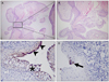
A 37-year-old woman presented with a mass around the punctum in the right lower eyelid margin, which had developed several years earlier and was increasing in size. The mass was a light pinkish nodule consisting of a few smooth-surface papillary lobules. The pathologic findings of the specimen obtained from a shaving biopsy showed a papillary projection which was covered with epithelium and communication with duct-like structures showing glandular configuration in the deep portion of the lesion. EMA-positive cells were found at the invaginated epithelium, implying a glandular epithelial origin. GCDFP-15-positive cells were found in the deep portion of the lesion, implying apocrine differentiation. The findings were compatible with syringocystadenoma papilliferum.
Figures and Tables
Figure 1
(A, B) Pinkish nodule consisting of a few smooth-surface papillary lobules around the punctum in the right lower eyelid.
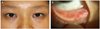
Figure 2
(A, B) Papillary projection which is covered with epithelium, is communicating with duct-like structures. (H&E, ×40). (C) EMA-positive cells are found at the superficially-located epithelium (arrowheads). Glandular configuration is also seen in the deep portions of the lesion (asterisk) (×400). (D) GCDFP-15-positive cells (black arrow) are found in the deep portions of the lesion, implying apocrine differentiation (×400).
References
1. Barbarino S, McCormick SA, Lauer SA, Milman T. Syringocystadenoma papilliferum of the eyelid. Ophthal Plast Reconstr Surg. 2009. 25:185–188.
2. Jakobiec FA, Streeten BW, Iwamoto T, et al. Syringocystadenoma papilliferum of the eyelid. Ophthalmology. 1981. 88:1175–1181.
3. Mammino JJ, Vidmar DA. Syringocystadenoma papilliferum. Int J Dermatol. 1991. 30:763–766.
4. Stokes JH. A clinico-pathologic study of an unusual cutaneous neoplasm combining naevus syringadenomatous papilliferus and a granuloma. J Cutan Dis. 1917. 35:411–425.
5. Helmi A, Alaraj AM, Alkatan H. Report of 3 histopathologically documented cases of syringocystadenoma papilliferum involving the eyelid. Can J Ophthalmol. 2011. 46:287–289.
6. Feuerstein RC, Mims LC. Linear nevus sebaceus with convulsions and mental retardation. Am J Dis Child. 1962. 104:675–679.
7. Jancar J. Naevus syringocystadenomatosus papilliferus with skull and brain lesions, hemiparesis, epilepsy and mental retardation. Br J Dermatol. 1970. 82:402–405.
8. Helwig EB, Hackney VC. Syringadenoma papilliferum; lesions with and without naevus sebaceous and basal cell carcinoma. AMA Arch Derm. 1955. 71:361–372.
9. Shams PN, Hardy TG, El-Bahrawy M, Joshi N. Syringocystadenoma papilliferum of the eyelid in a young girl. Ophthal Plast Reconstr Surg. 2006. 22:67–69.
10. Yoon DH, Jang IG, Kim TY, et al. Syringocystadenoma papilliferum, basal cell carcinoma and trichilemmoma arising from nevus sebaceus of Jadassohn. Acta Derm Venereol. 1997. 77:242–243.
11. Hsu PJ, Liu CH, Huang CJ. Mixed tubulopapillary hidradenoma and syringocystadenoma papilliferum occurring as a verrucous tumor. J Cutan Pathol. 2003. 30:206–210.
12. Ishida-Yamamoto A, Sato K, Wada T, et al. Syringocystadenocarcinoma papilliferum: case report and immunohistochemical comparison with its benign counterpart. J Am Acad Dermatol. 2001. 45:755–759.
13. Yamamoto O, Doi Y, Hamada T, et al. An immunohistochemical and ultrastructural study of syringocystadenoma papilliferum. Br J Dermatol. 2002. 147:936–945.
14. Nowak M, Pathan A, Fatteh S, et al. Syringocystadenoma papilliferum of the male breast. Am J Dermatopathol. 1998. 20:422–424.
15. Coyne JD, Fitzgibbon JF. Mixed syringocystadenoma papilliferum and papillary eccrine adenoma occurring in a scrotal condyloma. J Cutan Pathol. 2000. 27:199–201.
 XML Download