

 PDF
PDF ePub
ePub Citation
 Print
Print


Abstract
Purpose
To evaluate the change in multifocal visual evoked potential (mfVEP) in unilateral anisometropic amblyopia before and after occlusion treatment.
Methods
The mfVEP was performed using RETIscan® (Roland,Brandenburg, Germany) for patients with unilateral anisometropic amblyopia before and after occlusion treatment. Amplitude and latency values of mfVEP were analyzed according to the field divided by 6 rings or 4 sectors.
Results
The each amplitude of values of all 6 rings were not significantly different before treatment (p = 0.077) in amblyopic eyes. However, the value of ring 1 (p = 0.00) was significantly higher than the value of other rings after treatment. In fellow eyes, the values of ring 1 was consistently significantly higher than the value of other rings before (p = 0.014) and after (p = 0.049) occlusion treatment. Additionally, the amplitudes of ring 1 (p = 0.005) and ring 3 (p = 0.007) were significantly increased in amblyopic eyes after occlusion treatment. In fellow eyes, the values of all rings did not change significantly. The analysis of amplitudes with 4 sectors revealed no significant result. The analysis of latencies with 6 rings and 4 sectors revealed no significant result.
Figures and Tables

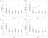
Figure 2
Amplitudes of 6rings at before (A) and after (B) treatment in amblyopic eyes, at before (C) and after (D) treatment in fellow eyes. In amblyopic eyes, the values of each rings were not different significantly before treatment. However, the values of ring1 (central field) were significantly higher than those of other rings after treatment (p = 0.00). In fellow eyes, the values of ring1 (central field) were significantly higher than those of other rings before treatment (p = 0.014) and after treatment (p = 0.049).
References
1. Noorden GK. Mechanisms of amblyopia. Adv Ophthalmol. 1977. 34:93–115.
2. Sen DK. Anisometropic amblyopia. J Pediatr Ophthalmol Strabismus. 1980. 17:180–184.
3. Kiorpes L. Visual processing in amblyopia: animal studies. Strabismus. 2006. 14:3–10.
4. Sokol S. Abnormal evoked potentiallatencies in amblyopia. Br J Ophthalmol. 1983. 67:310–314.
5. Davis ET, Bass SJ, Sherman J. Flash visual evoked potential (VEP) in amblyopia and optic nerve disease. Optom Vis Sci. 1995. 72:612–618.
6. Kirschen DG, Flom MC. Visual acuity at different retinal loci of eccentrically fixating functional amblyopes. Am J Optom Physiol Opt. 1978. 55:144–150.
7. Levi DM, Klein SA, Aitsebaomo P. Detection and discrimination of the direction of motion in central and peripheral vision of normal and amblyopic observers. Vision Res. 1984. 24:789–800.
8. Harding GF, Odom JV, Spileers W, Spekreijse H. International Society for Clinical Electrophysiology of Vision. Standard for visual evoked potentials 1995. Vision Res. 1996. 36:3567–3572.
9. Fortune B, Hood DC. Conventional pattern-reversal VEPs are not equivalent to summed multifocal VEPs. Invest Ophthalmol Vis Sci. 2003. 44:1364–1375.
10. Winn BJ, Shin E, Odel JG, et al. Interpreting the multifocal visual evoked potential: the effects of refractive errors, cataracts, and fixation errors. Br J Ophthalmol. 2005. 89:340–344.
11. Sutter EE. Imaging visual function with the multifocal m-sequence technique. Vision Res. 2001. 41:1241–1255.
12. Baseler HA, Sutter EE, Klein SA, Carney T. The topography of visual evoked response properties across the visual field. Electroencephalogr Clin Neurophysiol. 1994. 90:65–81.
13. Hood DC, Zhang X, Greenstein VC, et al. An interocular comparison of the multifocal VEP: a possible technique for detecting local damage to the optic nerve. Invest Ophthalmol Vis Sci. 2000. 41:1580–1587.
14. Fortune B, Zhang X, Hood DC, et al. Normative ranges and specificity of the multifocal VEP. Doc Ophthalmol. 2004. 109:87–100.
15. Graham SL, Klistorner AI, Grigg JR, Billson FA. Objective VEP perimetry in glaucoma: asymmetry analysis to identify early deficits. J Glaucoma. 2000. 9:10–19.
16. Hood DC, Odel JG, Zhang X. Tracking the recovery of local optic nerve function after optic neuritis: a multifocal VEP study. Invest Ophthalmol Vis Sci. 2000. 41:4032–4038.
17. Yu M, Brown B, Edwards MH. Investigation of multifocal visual evoked potential in anisometropic and esotropic amblyopes. Invest Ophthalmol Vis Sci. 1998. 39:2033–2040.
18. Shan Y, Moster ML, Roemer RA, Siegfried JB. Abnormal function of the parvocellular visual system in anisometropic amblyopia. J Pediatr Ophthalmol Strabismus. 2000. 37:73–78.
19. Donahue SP. The relationship between anisometropia, patient age, and the development of amblyopia. Trans Am Ophthalmol Soc. 2005. 103:313–336.
20. Hood DC. Assessing retinal function with the multifocal technique. Prog Retin Eye Res. 2000. 19:607–646.
 XML Download