

 PDF
PDF ePub
ePub Citation
 Print
Print


Abstract
Purpose
To report a case of superior oblique muscle tenotomy in a patient with suspected bilateral inferior oblique muscle overaction. The patient showed secondary superior oblique muscle overaction and inferior oblique muscle underaction after inferior oblique muscle myectomy.
Case summary
The patient showed V-pattern exotropia with suspected bilateral inferior oblique muscle overaction. After bilateral lateral rectus muscle recession with bilateral inferior oblique muscle myectomy, the patient showed secondary esotropia and inferior oblique underaction. After the surgery, progressive secondary superior oblique muscle overaction continued and finally, a superior oblique muscle tenotomy was performed. After the superior oblique muscle tenotomy, the superior oblique muscle overaction was corrected but the inferior oblique muscle underaction continued.
References
1. Isenberg SJ, Leonard APT. Inferior oblique weakening procedures: technique and indications. Rosenbaum AL, Santiago AP, editors. Clinical Strabismus Management Principles and Surgical Techniques. Philadelphia: W.B. Saunders Company;1999. chap.p. 34.
2. Biglan AW. Pattern strabismus. Rosenbaum AL, Santiago AP, editors. Clinical Strabismus Management Principles and Surgical Techniques. Philadelphia: W.B. Saunders Company;1999. chap.p. 14.
3. McNeer KW, Scott AB, Jampolsky A. A technique for surgically weakening the inferior oblique muscle. Arch Ophthalmol. 1965; 73:87–8.

4. Stuart JA. Myectomy of the inferior oblique muscle. Am J Ophthalmol. 1964; 57:118–21.
5. White JW. Surgery of the inferior oblique at or near the insertion. Tr Am Ophthalmol Soc. 1942; 40:118.
6. Chang BL, Chang MH, Yang SW. Inferior oblique overaction. J Korean Ophthalmol Soc. 1988; 29:1065–9.
7. Rosenbaum AL, Santiago AP. Clinical Strabismus Management: Principles and Surgical Techniques. Philadelphia: W.B. Saunders Company;1999. p. 453.
8. Toosi SH, Von Noorden GK. Effect of isolated Inferior oblique muscle myectomy in the management of superior oblique muscle palsy. Am J Ophthalmol. 1979; 88:602–8.
9. Jones TW, Lee DA, Dyer JA. Experience at the Mayo Clinic from 1960 to 1981. Arch Ophthalmol. 1984; 102:714–6.
10. Parks MM. Inferior oblique weakening procedures. Int Ophthalmol Clin. 1985; 25:107–17.
11. Dyer JA, Duke DG. Inferior oblique weakening procedures. Int Ophthalmol Clin. 1976; 16:103–12.
12. Davis G, McNeer KW, Spencer RF. Myectomy of the inferior oblique muscle. Arch Ophthalmol. 1986; 104:855–8.
13. Min BM, Park JH. Comparison between myectomy and anterior transposition in inferior oblique overaction. J Korean Ophthalmol Soc. 1992; 33:977–82.
14. Shin YJ. Lee TS. Effect of myectomy on overaction inferior oblique muscle. J Korean Ophthalmol Soc. 1984; 25:347–51.
15. Shipman T, Burke J. Unilateral inferior oblique muscle myectomy and recession in the treatment of inferior oblique muscle overaction: a longitudinal study. Eye. 2003; 17:1013–8.
16. Scott AB. Round table discussion. Helvestone EM, editor. Symposium on Strabismus. Transaction of the New Orleans Academy of Ophthalmology. St. Louis: CV Mosby;1978. p. 533.
Figure 1.
Preoperative fundus photos. Preoperatively, authors misdiagnosed bilateral excyclotorsions.

Figure 2.
(A) Postoperative 7 month 9-cardinal photos; Bilateral lateral rectus muscle recession (7.0 mm) with bilateral inferior oblique muscle myectomy. Consecutive esotropia and A-pattern esotropia developed. Right eye showed inferior oblique muscle underaction (−1.5) and left eye showed (−1.0) inferior oblique muscle underaction. (B) Postoperative fundus photo showed bilateral incyclotorsions.
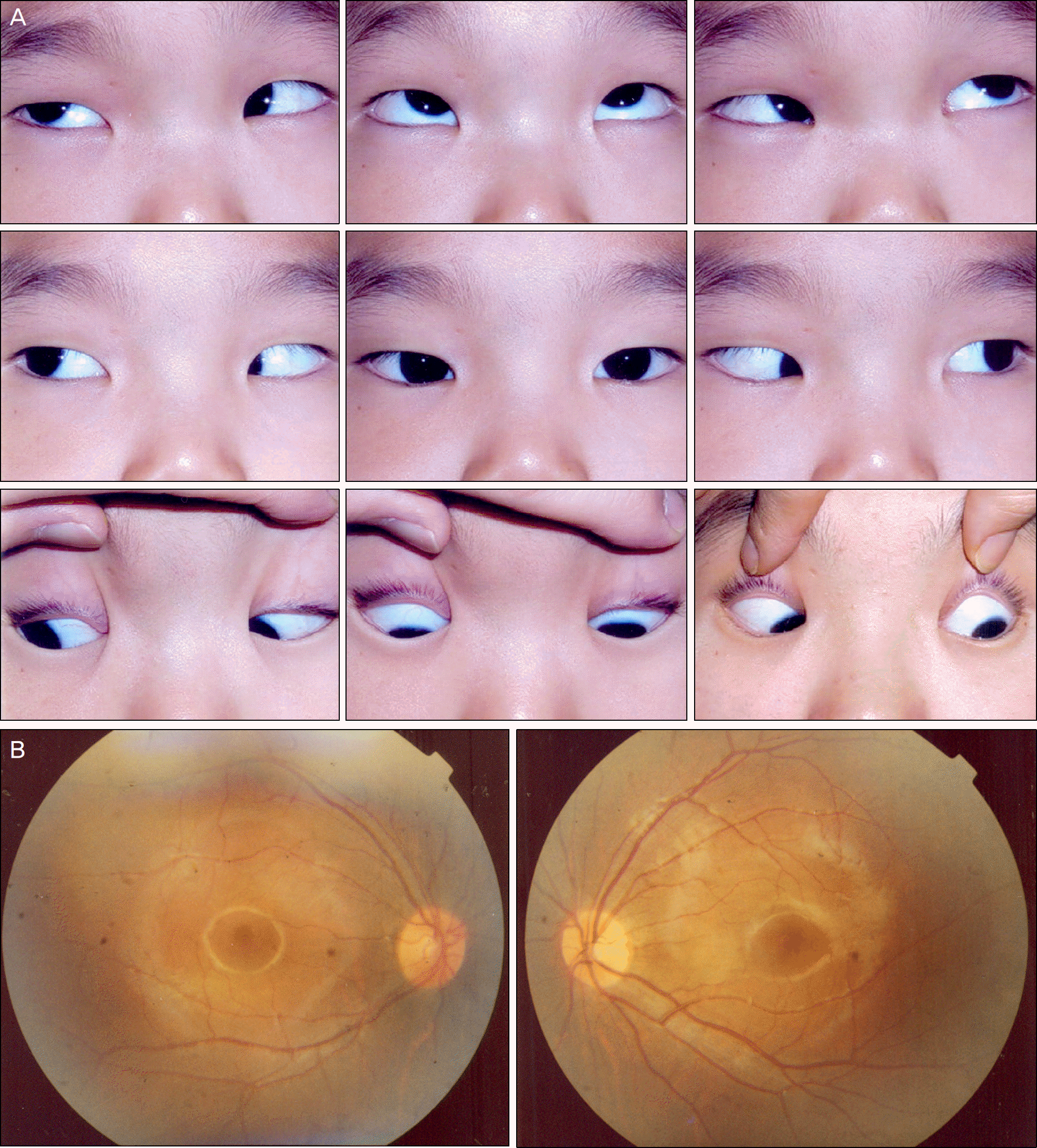
Figure 3.
(A) Postoperative 3 year 5 months 9-cardinal photos after secondary operation; Right lateral rectus muscle advancement (5.0 mm) and right inferior oblique muscle exploration. Consecutive esotropia and A-pattern esotropia still remained. Both eyes showed inferior oblique muscle underaction (−2.0) and superior oblique muscle overaction (+3.0). (B) Postoperative fundus photo showed marked bilateral incyclotorsions.
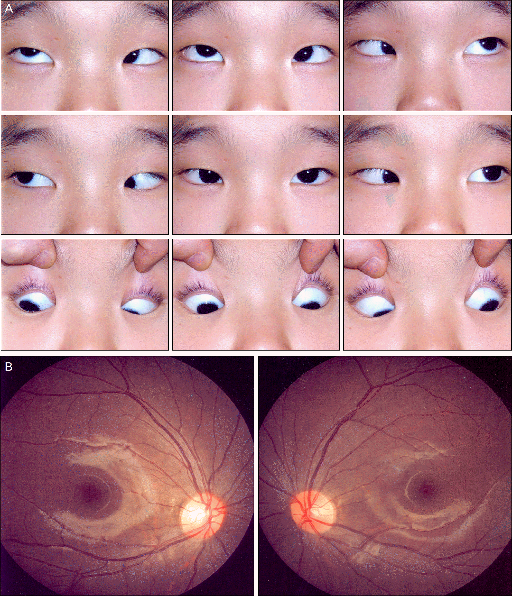
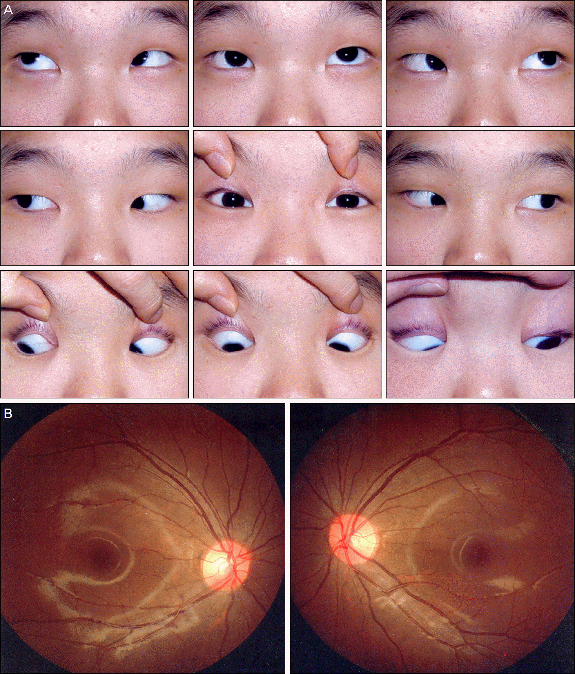
Figure 4.
(A) Postoperative 6 months 9-cardinal photos after tertiary operation; Bilateral superior oblique muscle tenotomy. In primary position, mild esophoria and left hypotropia was noticed. Both eyes still showed inferior oblique muscle underaction (−1.0). Right eye showed mild and left eye had no superior oblique muscle overaction. (B) Postoperative fundus photo showed mild in-cyclotorsion in the right eye and no torsion in the left eye.
 XML Download