

 PDF
PDF ePub
ePub Citation
 Print
Print


Abstract
Purpose
To report a new modified method using a mixture of sodium hyaluronate and indocyanine green solution to facilitate the complete removal of a large conjunctival cyst.
Case summary
Two patients with a large conjunctival cyst on the bulbar conjunctiva were treated. In order to achieve complete removal, a mixture of 1% sodium hyaluonate and indocyanine green solution was injected through a 27-G needle into the cyst. The procedure provided excellent visualization of the cyst boundaries while maintaining cyst integrity allowing for an easy and complete resection. Apocrine hidrocystoma and a simple retention cyst were confirmed on histopathologic examination, respectively.
References
1. Shields CL, Shields JA. Tumors of the conjunctiva and cornea. Surv Ophthalmol. 2004; 49:3–24.

2. Cherrick GR, Stein SW, Leevy CM, Davidson CS. Indocyanine green: observations on its physical properties, plasma decay, and hepatic extraction. J Clin Invest. 1960; 39:592–600.
3. Kogure K, David NJ, Yamanouchi U, Choromokos E. Infrared absorption angiography of the fundus circulation. Arch Ophthalmol. 1970; 83:209–14.
4. Horiguchi M, Miyake K, Ohta I, Ito Y. Staining of the lens capsule for circular continuous capsulorrhexis in eyes with white cataract. Arch Ophthalmol. 1998; 116:535–7.
5. Pandey SK, Werner L, Escobar-Gomez M, et al. Dye-enhanced cataract surgery. Part 1: anterior capsule staining for capsulorhexis in advanced/white cataract. J Cataract Refract Surg. 2000; 26:1052–9.
6. Da Mata AP, Burk SE, Riemann CD, et al. Indocyanine green-as-sisted peeling of the retinal internal limiting membrane during vitrectomy surgery for macular hole repair. Ophthalmology. 2001; 108:1187–92.
7. Li K, Wong D, Hiscott P, et al. Trypan blue staining of internal limiting membrane and epiretinal membrane during vitrectomy: visual results and histopathological findings. Br J Ophthalmol. 2003; 87:216–9.
8. Hahm IR, Tae KS, Cho SW, et al. The outcomes after indocyanine green-assisted peeling of the internal limiting membrane in macular hole surgery. J Korean Ophthalmol Soc. 2005; 46:1361–7.
9. Auffarth GU, Holzer MP, Vissesook N, et al. Removal times and techniques of a viscoadaptive ophthalmic viscosurgical device. J Cataract Refract Surg. 2004; 30:879–83.
10. Kobayashi A, Saeki A, Nishimura A, et al. Visualization of conjunctival cyst by indocyanine green. Am J Ophthalmol. 2002; 133:827–8.
11. Hoffman RS, Fine IH, Packer M. Stabilization of flat anterior chamber after trabeculectomy with Healon5. J Cataract Refract Surg. 2002; 28:712–4.
12. Kobayashi A, Sugiyama K. Visualization of conjunctival cyst using Healon V and trypan blue. Cornea. 2005; 24:759–60.
13. Kobayashi A, Sugiyama K. Successful removal of a large conjunctival cyst using colored 2.3% sodium hyaluronate. Ophthalmic Surg Lasers Imaging. 2007; 38:81–3.
14. Kodjikian L, Richter T, Halberstadt M, et al. Toxic effects of indocyanine green, infracyanine green, and trypan blue on the human retinal pigmented epithelium. Graefes Arch Clin Exp Ophthalmol. 2005; 243:917–25.
Figure 1.
(A) 6×5 mm-sized large conjunctival cyst is seen in the bulbar conjunctiva of the right eye.(B) The cyst margin are clearly visible through the conjunctiva after stained with a mixture of sodium hyaluronate and indocyanine green solution. (C, D) Successful removal of the conjunctival cyst with delineated capsule and preserved integrity.
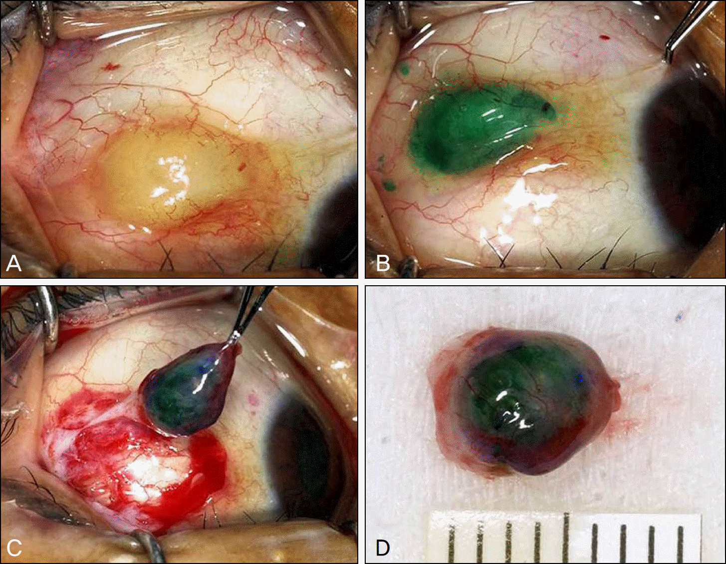
Figure 2.
Double layered lining of apocrine hidrocytoma (H&E stain, ×40) (A), consisting of large columnar cells with eosinophilic cytoplasm with luminal decapitation secretion in the inner layer and flat myoepithelial cells in the outer layer (H&E stain, ×100).(B) Arrows indicate PAS-positive, diastase-resistant granule on the apical surfaces of the inner layer (PAS stain, ×100).
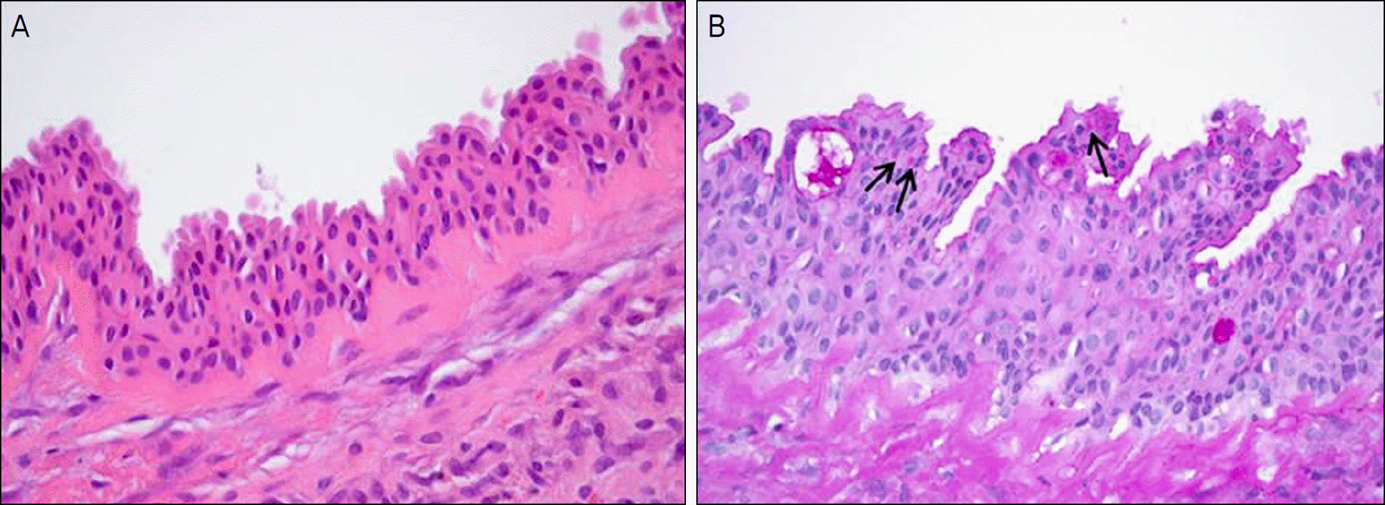
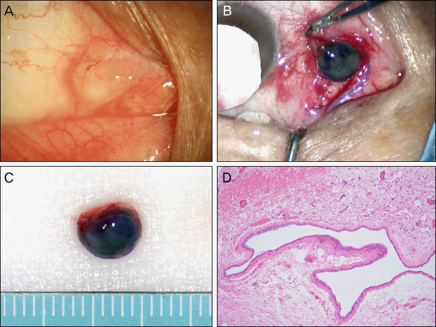
Figure 3.
(A) 6×4 mm-sized large conjunctival cyst is seen in the medial conjunctiva adjacent to the caruncle of the right eye. (B) The cyst is stained using a mixture of sodium hyaluronate and indocyanine green solution. (C) Succesful removal of the conjunctival cyst with delineated capsule and preserved integrity. (D) A solitary uniocular cyst is lined by non-keratinizing cuboidal epithelium. Goblet cells are often included (H&E stain, ×20).
 XML Download