Abstract
Purpose
The authors report a case of Rubinstein-Taybi syndrome with optic disc coloboma and chorioretinal coloboma.
Case summary
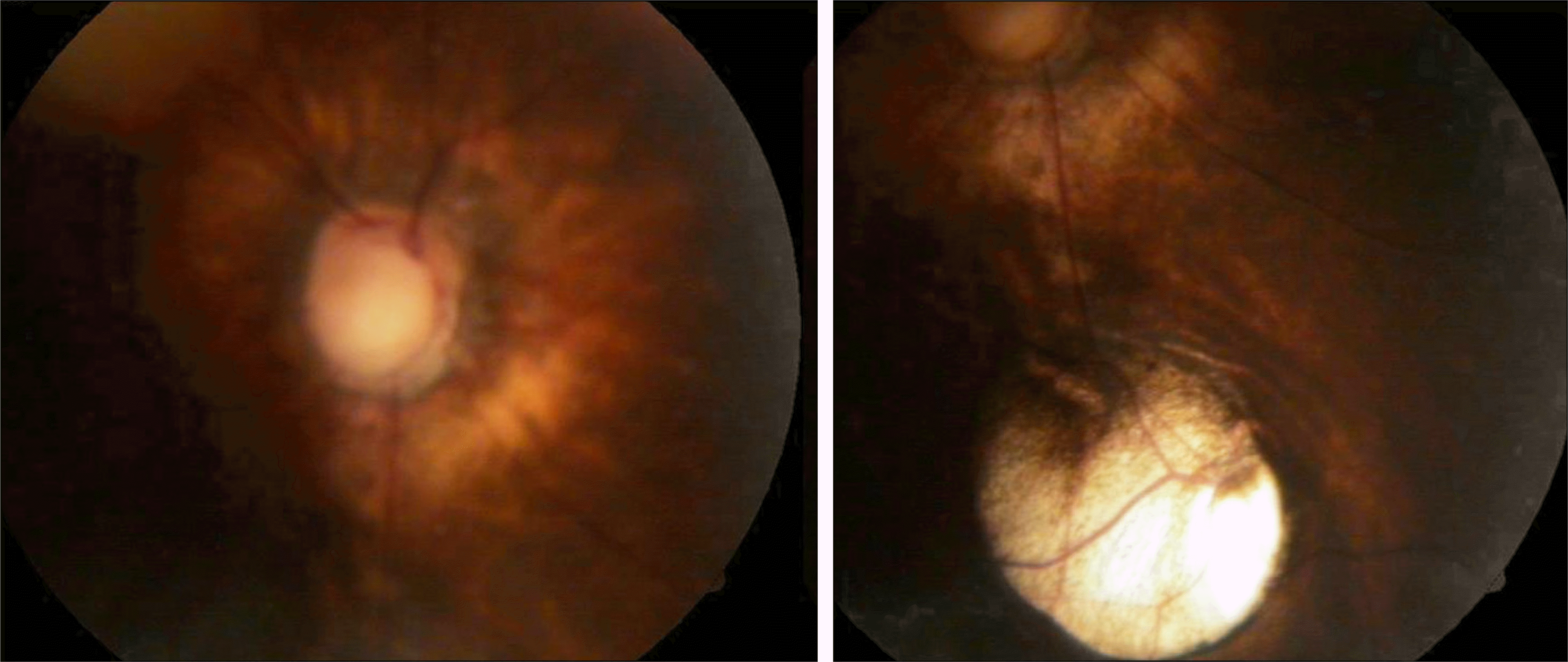
A 17-month-old female infant was brought to our clinic presenting exodeviation in the right eye. On cycloplegic refraction, her refractive power was −5.50 D sph −2.50 D cyl axis 180° in the right eye and +0.50 D sph in the left eye. On ophthalmologic examination, exotropia of 60 prism diopters with no limitation of ocular movement was observed. Fundus examination showed optic disc coloboma and chorioretinal coloboma in the right eye. The patient's physical characteristics were downward slanted palpebral fissures, long eyelashes, low set ears, and the thumb and the big toe were disproportionately broad. The patient also demonstrated delayed gait abilities. The clinical diagnosis of Rubinstein-Taybi syndrome was given.
References
1. Rubinstein JH, Taybi H. Broad thumbs and toes and facial abnormalities. A possible mental retardation syndrome. Am J Dis Child. 1963; 105:588–608.
2. Hennekam RC, Stevens CA, Van de Kamp JJ. Etiology and recurrence risk in Rubinstein-Taybi syndrome. Am J Med Genet Suppl. 1990; 6:56–64.

5. Goodfellow A, Emmerson RW, Calvert HT. Rubinstein-Taybi syndrome and spontaneous keloids. Clin Exp Dermatol. 1980; 5:369–70.

6. van Genderen MM, Kinds GF, Riemslag FC, Hennekam RC. Ocular features in Rubinstein-Taybi syndrome: investigation of 24 patients and review of the literature. Br J Ophthalmol. 2000; 84:1177–84.


 PDF
PDF ePub
ePub Citation
 Print
Print


 XML Download