

 PDF
PDF ePub
ePub Citation
 Print
Print


Abstract
Purpose
To compare the changes in angle parameters after laser peripheral iridotomy (LPI) alone versus LPI and ALPI (argon laser peripheral iridoplasty) in primary angle closure patients by using anterior segment optical coherence tomography (AS-OCT).
Methods
A total of 25 eyes from 17 patients with narrow angles were enrolled in this present study. Eleven eyes in the LPI treatment group and 14 eyes in the LPI and ALPI combined treatment group were evaluated using AS-OCT. The anterior chamber depth (ACD), angle opening distance at 500 μm (AOD 500) and 750 μm (AOD 750), angle recess area at 500 μm (ARA 500) and 750 μm (ARA 750), trabecular-iris space area at 500 μm (TISA 500) and 750 μm (TISA 750), trabecular-iris angle (TIA) were measured. The pre- and post-treatment parameters were compared in each group. The parameter changes after laser treatment were also compared.
Results
AOD 500, AOD 750, ARA 500, ARA 750, TISA 500 and TISA 750 except ACD significantly increased following LPI treatment (p = 0.013, p = 0.010, p = 0.008, p = 0.003, p = 0.006, p = 0.003, p = 0.013, respectively, Wilcoxon signed rank test) and LPI and ALPI combined therapy (p = 0.001, p = 0.001, p = 0.001, p = 0.001, p = 0.001, p = 0.001, p = 0.001, respectively, Wilcoxon signed rank test). The AOD 500 difference, TISA 500 difference, and TISA 750 difference were significantly greater after LPI and ALPI combined therapy than after LPI treatment alone (p = 0.112, p = 0.147, p = 0.049, p =0.037, respectively, Mann-Whitney U-test).
References
1. Erie JC, Hodge DO, Gray DT. The incidence of primary angle-clo-sure glaucoma in Olmstead County, Minnesota. Arch Ophthalmol. 1997; 115:177–81.
2. Foster PJ. Glaucoma in China: how big is the problem? Br J Ophthalmol. 2001; 85:1277–82.

3. Dandona L, Dandona R, Mandal P, et al. Angle-closure glaucoma in an urban population in southern India. The Andhra Pradesh eye disease study. Ophthalmology. 2000; 107:1710–6.
4. Foster PJ, Baasanhu J, Alsbirk PH, et al. Glaucoma in Mongolia: A population-based survey in Hövsgöl Province, Northern Mongolia. Arch Ophthalmol. 1996; 114:1235–41.
5. Kimbrough RL, Trempe CS, Brockburst RJ, Simmon RJ. Angle-closure glaucoma in nanophthalmos. Am J Ophthalmol. 1979; 88:572–9.
6. Iwata K, Abe , Sugiyama J. Argon laser iridotomy in primary chronic angle-closure glaucoma. Glaucoma. 1985; 7:103–6.
7. Del Price LV, Robin AL, Pollack IP. Neodymium:YAG and argon laser iridotomy. Long-term follow-up in a prospective, randomized clinical trial. Ophthalmology. 1988; 95:1207–11.
8. Ritch R. Argon laser treatment for medically unresponsive attacks of angle closure glaucoma. Am J Ophthalmol. 1982; 94:194–204.
9. Saunders DC. Acute closured-angle glaucoma and Nd-YAG laser iridotomy. Br J Ophthalmol. 1990; 74:523–5.
10. Lam DS, Lai JS, Tham CC. Immediate argon laser peripheral iridoplasty as treatment for acute attack of primary angle-closure glaucoma. Ophthalmology. 1998; 105:2231–6.
11. Ritch R, Podos SM. Argon laser treatment of angle-closure glaucoma. Perspectives in Ophthalmology. 1980; 4:129–38.
12. Ritch R. Argon laser peripheral iridoplasty: an overview. J Glaucoma. 1992; 1:206–13.
13. Ritch R, Tham CC, Lam DS. Argon laser peripheral iridoplasty (ALPI): an update. Surv Ophthalmol. 2007; 52:279–88.
14. Hung T, Chou LH. Provocation and mechanism of angle-closure glaucoma after iridectomy. Arch Ophthalmol. 1979; 97:1862–4.
15. Hong C, Park KH, Hyung SM, et al. Evaluation of pupillary block component in angle-closure glaucoma. Jpn J Ophthalmol. 1996; 40:239–43.
16. Aung T, et al. FRCS, FRCOphth. Acute primary angle-closure: long-term intraocular pressure outcome in Asian eyes. Am J Ophthalmol. 2001; 131:7–12.
17. Ningli W, Heping WU, Zhigang F. Primary angle closure glaucoma in Chinese and Western populations. Chin Med J. 2002; 115:1706–15.
18. Robert R, Jeffrey M. Liebmann. Argon laser peripheral iridoplasty. Ophthalmic Surg Lasers. 1996; 27:289–300.
19. Izatt JA, Hee MR, Swanson EA, et al. Micrometer-scale resolution imaging of the anterior eye in vivo with optical coherence tomography. Arch Ophthalmol. 1994; 112:1584–9.
20. Radhakrishnan S, Rollins AM, Roth JE, et al. Real-time optical coherence tomography of the anterior segment at 1310 nm. Arch Ophthalmol. 2001; 119:1179–85.
21. See JL, Chew PT, Smith SD, et al. Changes in anterior segment morphology in response to illumination and after laser iridotomy in Asian eyes: an anterior segment OCT study. Br J Ophthalmol. 2007; 91:1485–9.
22. Yip JL, Foster PJ, Gilbert CE, et al. Incidence of occludable angles in a high-risk Mongolian population. Br J Ophthalmol. 2008; 92:30–3.
23. Pavlin CJ, Harasiewiez K, Foster FS. Ultrasound biomicroscopy of anterior segment structures in normal and glaucomatous eyes. Am J Ophthalmol. 1992; 113:381–9.
24. Radhakrishnan S, Goldsmith J, Huang D, et al. Comparison of optical coherence tomography and ultrasound biomicroscopy for detection of narrow anterior chamber angles. Arch Ophthalmol. 2005; 123:1053–9.
25. Müler M, Dahmen G, Pörksen E, et al. Anterior chamber angle measurement with optical coherence tomography: Intraobserver and interobserver variability. J Cataract Refract Surg. 2006; 32:1803–8.
26. Alsagoff Z, Aung T, Ang LP, Chew PT. Long-term clinical course of primary angle-closure glaucoma in an Asian population. Ophthalmology. 2000; 107:2300–4.
27. Aung T, Ang LP, Chan SP, et al. Acute primary angle-closure: long-term intraocular pressure outcome in Asian eyes. Am J Ophthalmol. 2001; 131:7–12.
28. Krasnov MM. Q-switched laser iridectomy and Q-switched laser goniopuncture. Adv Ophthalmol. 1977; 34:192–6.
29. Sassani JW, Ritch R, McCormick S, et al. Histopathology of argon laser peripheral iridoplasty. Ophthalmic Surg. 1993; 24:740–5.
30. Wirbelauer C, Gochmann R, Pham DT. Imaging of the anterior eye chamber with optical coherence tomography. Klin Monatsbl Augenheilkd. 2005; 222:856–62.
31. Tomlinson A, Leighton DA. Ocular dimensions in the heredity of angle-closure glaucoma. Br J Ophthalmol. 1973; 57:475–86.
32. Palvin CJ, Ritch R, Foster FS. Ultrasound biomicroscopy in pla-teau iris syndrome. Am J Ophthalmol. 1992; 113:390–5.
33. Chalita MR, Li Y, Patil C, et al. High-speed optical coherence tomography of laser iridotomy. Am J Ophthalmol. 2005; 140:1133–6.
34. Ishikawa H, Liebmann JM, Ritch R. Quantitative assessment of the anterior segment using ultrasound biomicroscopy. Curr Opin Ophthalmol. 2000; 11:133–9.
35. Kim HS, Kim YY, Jung HR. Effect of combined argon laser peripheral iridoplasty and laser iridotomy in primary angle-closure glaucoma. J Korean Ophthalmol Soc. 2003; 44:2565–70.
Figure 1.
(A) Optical coherence tomography image (Visante OCT) with graphic tools for measurement of different anterior chamber dimensions. Anterior chamber depth is measured between the corneal endothelium and a line joining the two opposite iris recesses. (B) Optical coherence tomography (Visante OCT) cross-sectional view through the anterior chamber angle region. Anterior chamber parameters such as AOD 500, AOD 750, ARA 500, ARA 750, TISA 500, TISA 750 and TIA are automatically measured.
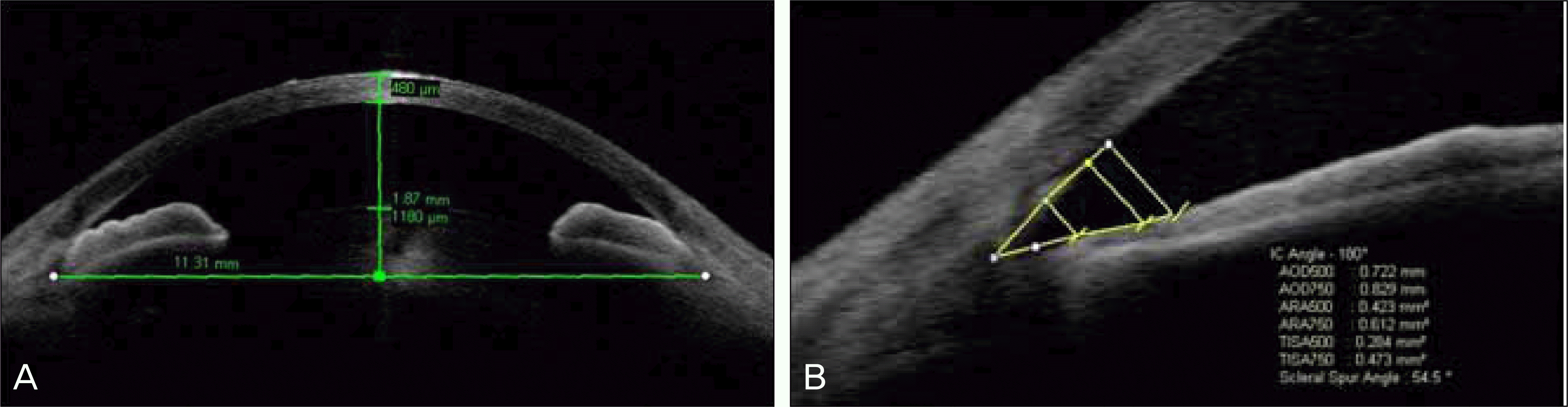
Figure 2.
Angle changes of AS-OCT before and after laser peripheral iridotomy. (A) AS-OCT image showing angle closure before laser peripheral iridotomy. (B) AS-OCT image showing the same angle open after laser peripheral iridotomy.
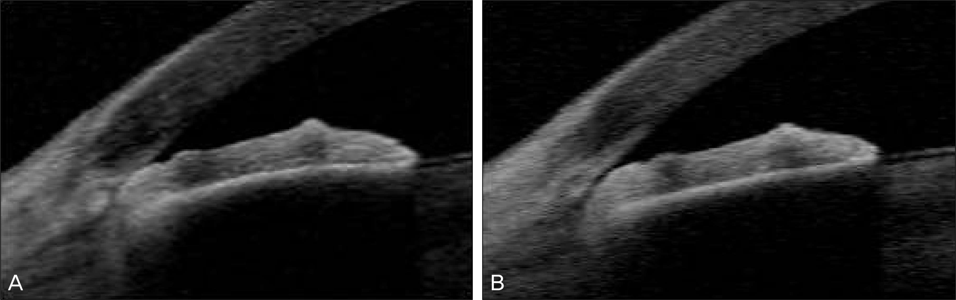
Figure 3.
Angle changes of AS-OCT before and after laser peripheral iridotomy and argon laser peripheral iridoplasty. (A) AS-OCT image showing angle closure before laser peripheral iridotomy and argon laser peripheral iridoplasty. (B) AS-OCT image showing the same angle open after laser peripheral iridotomy and argon laser peripheral iridoplasty.
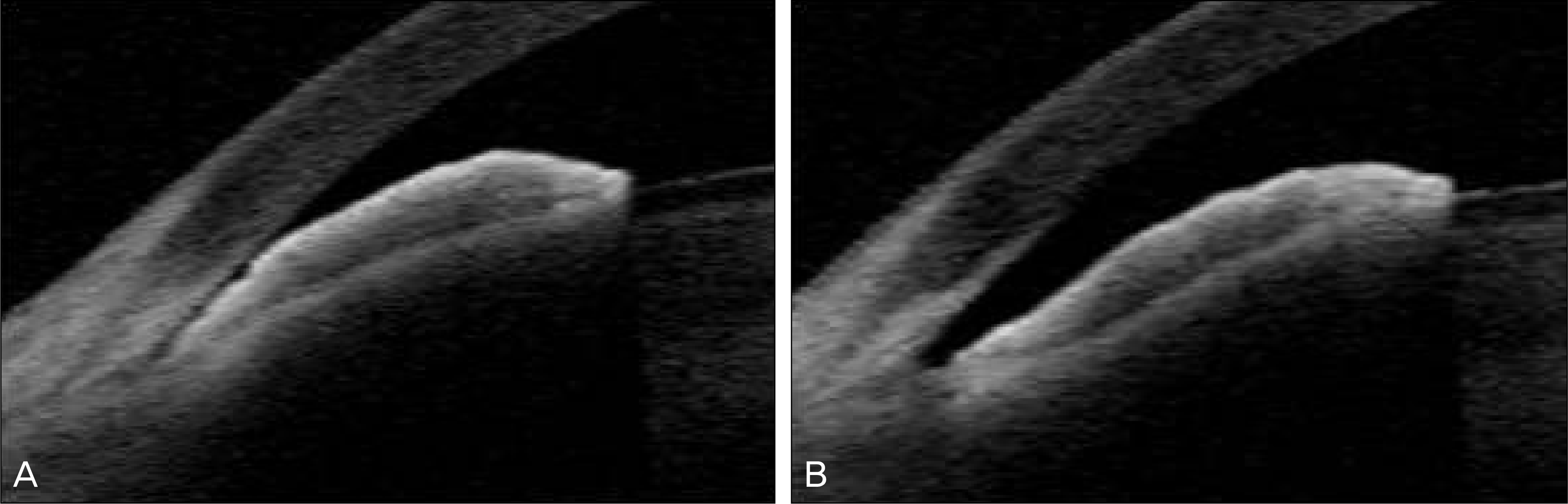
Table 1.
Comparison of anterior segment-optical coherence tomography (AS-OCT) parameters before only laser peripheral iridot-omy (LPI) versus LPI and argon laser peripheral iridoplasty (ALPI)
| Parameter | Pre-LPI (mean ± SD) | Pre-LPI &ALPI (mean ± SD) | p-value* |
|---|---|---|---|
| ACD† (mm) | 1.89 ± 0.07 | 1.90 ± 0.33 | 0.344 |
| AOD‡ 500 (μm) | 58.1 ± 17.3 | 137.0 ± 57.0 | 0.000 |
| AOD 750 (μm) | 89.5 ± 32.4 | 155.1 ± 89.7 | 0.095 |
| ARA§ 500 (×10-2 mm2) | 7.3 ± 2.4 | 11.2 ± 5.1 | 0.058 |
| ARA 750 (×10-2 mm2) | 9.1 ± 2.4 | 14.8 ± 6.4 | 0.033 |
| TISAΠ 500 (×10-2 mm2) | 4.0 ± 1.1 | 6.7 ± 2.8 | 0.015 |
| TISA 750 (×10-2 mm2) | 5.7 ± 1.2 | 10.3 ± 4.4 | 0.005 |
| TIA# (degrees) | 6.6 ± 1.9 | 15.0 ± 5.9 | 0.000 |
Table 2.
Changes in anterior segment-optical coherence tomography (AS-OCT) parameters before and after laser peripheral iridotomy (LPI)
| Parameter | Pre-LPI (mean ± SD) | Post-LPI (mean ± SD) | p-value* |
|---|---|---|---|
| ACD† (mm) | 1.89 ± 0.07 | 1.89 ± 0.06 | 1.000 |
| AOD‡ 500 (μm) | 58.1 ± 17.3 | 143.6 ± 124.8 | 0.013 |
| AOD 750 (μm) ARA§ 500 (×10-2 mm2) | 89.5 ± 32.4 7.3 ± 2.4 | 202.6 ± 136.4 11.8 ± 5.1 | 0.010 0.008 |
| ARA 750 (×10-2 mm2) | 9.1 ± 2.4 | 16.1 ± 7.6 | 0.003 |
| TISAΠ 500 (×10-2 mm2) | 4.0 ± 1.1 | 7.1 ± 3.7 | 0.006 |
| TISA 750 (×10-2 mm2) | 5.7 ± 1.2 | 11.4 ± 6.6 | 0.003 |
| TIA# (degrees) | 6.6 ± 1.9 | 15.0 ± 11.5 | 0.013 |
Table 3.
Changes in anterior segment-optical coherence tomography (AS-OCT) parameters before and after laser peripheral iridot-omy (LPI) and argon laser peripheral iridoplasty (ALPI)
| Parameter | Pre-LPI & ALPI (mean ± SD) | Post-LPI & ALPI (mean ± SD) | p-value* |
|---|---|---|---|
| ACD† (mm) | 1.90 ± 0.33 | 1.92 ± 0.25 | 0.656 |
| AOD‡ 500 (μm) | 137.0 ± 57.0 | 303.4 ± 128.9 | 0.001 |
| AOD 750 (μm) | 155.1 ± 89.7 | 373.6 ± 169.8 | 0.001 |
| ARA§ 500 (×10-2 mm2) | 11.2 ± 5.1 | 18.5 ± 7.6 | 0.001 |
| ARA 750 (×10-2 mm2) | 14.8 ± 6.4 | 26.8 ± 11.1 | 0.001 |
| TISAΠ 500 (×10-2 mm2) | 6.7 ± 2.8 | 12.3 ± 5.0 | 0.001 |
| TISA 750 (×10-2 mm2) | 10.3 ± 4.4 | 20.6 ± 8.6 | 0.001 |
| TIA# (degrees) | 15.0 ± 5.9 | 29.9 ± 9.7 | 0.001 |
Table 4.
Comparison in difference of anterior segment-optical coherence tomography (AS-OCT) parameters before and after only laser peripheral iridotomy (LPI) versus laser peripheral iridotomy (LPI) and argon laser peripheral iridoplasty (ALPI)
| Parameter | LPI (mean ± SD) | LPI &ALPI (mean ± SD) | p-value* |
|---|---|---|---|
| Δ ACD† (mm) | 0.00 ± 0.29 | 0.02 ± 0.13 | 0.609 |
| Δ AOD‡ 500 (μm) | 85.6 ± 120.4 | 166.4 ± 108.2 | 0.033 |
| Δ AOD 750 (μm) | 113.1 ± 124.0 | 218.5 ± 164.3 | 0.112 |
| Δ ARA§ 500 (×10-2 mm2) | 4.6 ± 5.4 | 7.3 ± 5.9 | 0.147 |
| Δ ARA 750 (×10-2 mm2) | 7.0 ± 7.8 | 12.1 ± 8.9 | 0.055 |
| Δ TISAΠ 500 (×10-2 mm2) | 3.1 ± 3.8 | 5.5 ± 4.0 | 0.049 |
| Δ TISA 750 (×10-2 mm2) | 5.7 ± 6.5 | 10.3 ± 7.2 | 0.037 |
| Δ TIA# (degrees) | 8.4 ± 11.1 | 14.9 ± 8.3 | 0.052 |
Table 5.
Changes in intraocular pressure (IOP) before and after only laser peripheral iridotomy (LPI) versus laser peripheral iridot-omy (LPI) and argon laser peripheral iridoplasty (ALPI)
| IOP before treatment (mmHg) |
IOP after treatment (mmHg) |
|||||
|---|---|---|---|---|---|---|
| Immediately | 1 wk | 1 mon | 3 mon | 6 mon | ||
| LPI (mean ± SD) | 15.0 ± 3.5 | 14.1 ± 4.4 | 13.5 ± 2.2 | 11.8 ± 0.8 | 17.0 ± 3.5 | 13.5 ± 2.6 |
| LPI & ALPI (mean ± SD) | 18.8 ± 5.1 | 16.3 ± 4.3 | 14.4 ± 3.8 | 15.0 ± 3.9 | 14.4 ± 2.6 | 17.8 ± 3.8 |
| p-value* | 0.087 | 0.222 | 0.767 | 0.143 | 0.181 | 0.059 |
 XML Download