

 PDF
PDF ePub
ePub Citation
 Print
Print


Abstract
Purpose
To report a case of chronic central serous chorioretinopathy treated with an intravitreal injection of 2.5 mg of bevacizumab.
Case summary
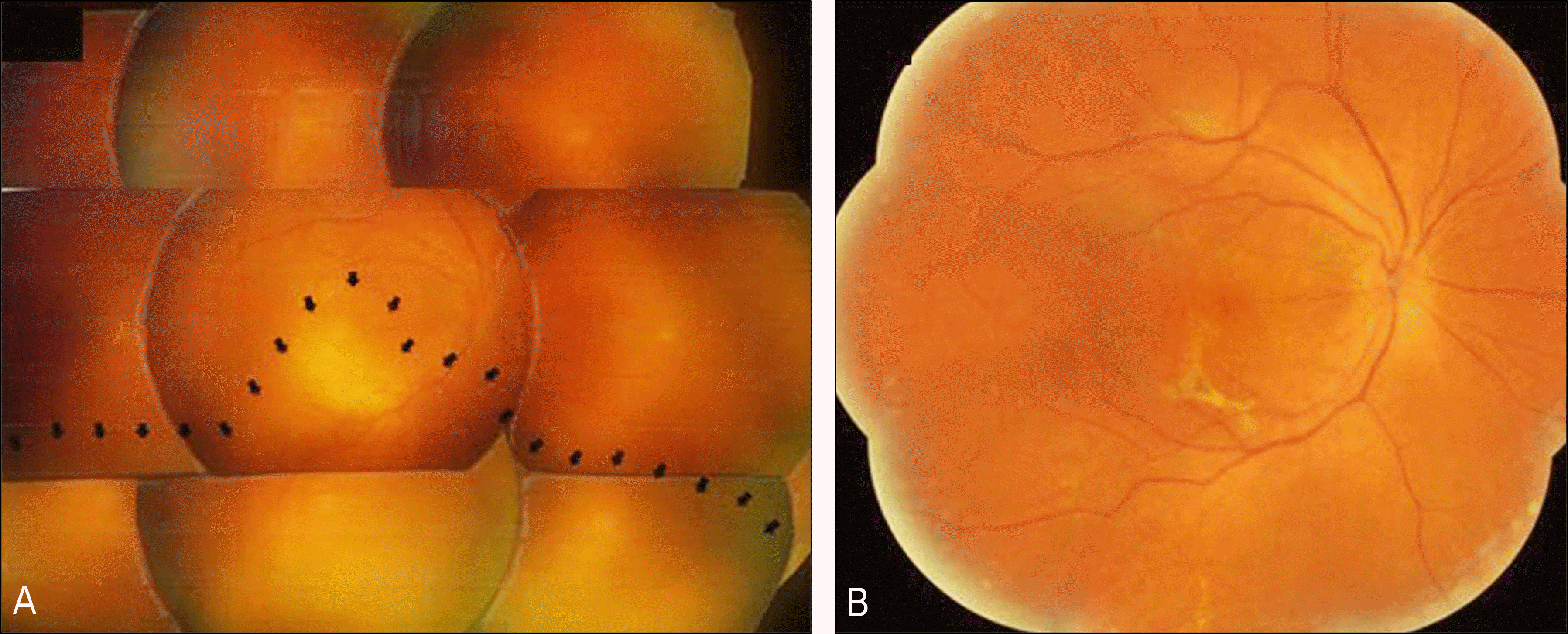
A 38-year-old male complained of visual disturbance in the right eye. He had a history of central serous chorioretinopathy which improved without treatment. Since the patient did not feel any ocular discomfort, he did not visit our clinic for approximately 11 months. At the time of presentation, fundus examination revealed exudative retinal detachment involving the macular area. Additionally, fluorescein angiography revealed multiple early phase hyperfluorescent lesions. Neurosensory detachment around the macula and increased central macular thickness were also observed using optical coherence tomography. Under the diagnosis of chronic central serous chorioretinoapthy, the patient was treated with 2.5 mg of intravitreal bevacizumab. Visual acuity improved two months after treatment, with improvement in both neurosensory retinal detachment and fluorescein leakage. No changes were observed at the six-month follow-up.
References
1. Spitznas M. Pathogenesis of central serous retinopathy: a new working hypothesis. Graefes Arch Clin Exp Ophthalmol. 1986; 224:321–4.

2. Gass JD. Pathogenesis of disciform detachment of the aberrations. Am J Ophthalmol. 1967; 63(Suppl):1–139.
3. Lafaut BA, Salati C, Priem H, De Laey JJ. Indocyanine green angiographic is of value for the diagnosis of chronic serous chorioretinopathy in elderly patients. Graefes Arch Clin Exp Ophthalmol. 1998; 236:513–21.
4. Piccolino FC, Borgia L, Zinicola E, Zingirian M. Indocyanine green angiographic findings in central serous chorioretinopathy. Eye. 1995; 9:324–32.
5. Schaal KB, Hoeh AE, Scheuerle A, et al. Intravitreal bevacizumab for treatment of chronic central serous chorioretinopathy. Eur J Ophthalmol. 2009; 19:613–7.
6. Artunay O, Yuzbasioglu E, Rasier R, et al. Intravitreal bevacizumab in treatment of idiopathic persistent central serous chorioretinopathy: A prospective, controlled clinical study. Curr Eye Res. 2010; 35:91–8.
7. Seong HK, Bae JH, Kim ES, et al. Intravitreal Bevacizumab to treat acute central serous chorioretinopathy: Short-term effect Ophthalmologica. 2009; 223:343–7.
8. Torres-Soriano ME, García-Aguirre G, Kon-Jara V, et al. A pilot study of intravitreal bevacizumab for the treatment of central serous chorioretinopathy (Case report). Graefes Arch Clin Exp Ophthalmol. 2008; 246:1235–9.
9. Lim SJ, Roh MI, Kwon OW. Intravitreal bevacizumab injection for central serous chorioretinopathy. Retina. 2010; 30:100–6.
10. Kim M, Chung M. The result of photodynamic therapy in chronic central serous chorioretinopathy. J Korean Ophthalmol Soc. 2009; 50:1035–45.
11. Guyer DR, Yannuzzi LA, Slakter JS, et al. Digital indocyanine green videoangiography of central serous chorioretinopathy. Arch Ophthalmol. 1994; 112:1057–62.
12. Azad RV, Rani A, Pal N, et al. Current and future role of photo-dynamuc therapy in chronic central serous chorioretinopathy. Am J Ophthalmol. 2005; 139:393–4.
13. Colucciello M. Choroidal neovascularization complicating photodynamic therapy for central serous retinopathy. Retina. 2006; 26:239–42.
14. Heiduschka P, Fietz H, Hofmeister S, et al. Penetration of bevacizumab through the retina after intravitreal injection in the monkey. Invest Ophthalmol Vis Sci. 2007; 48:2814–23.
15. Amselem L, Cervera E, Diaz-Llopis M, et al. Intravitreal bevacizumab (Avastin) for choroidal metastasis secondary to breast carcinoma: short-term follow-up. Eye. 2007; 21:566–7.
Figure 1.
Fundus photographs, fluorescein angiographs (FAG) and optical coherence tomographs (OCT) of the patient's right eye with CSC. Two months after observation without treatment (A, B, C), fundus photograph reveals a yellowish membranous lesion around the macular area (A). FAG reveals hyperfluorescent lesion around the macular area (B). Neurosensory retinal detachment is observed by OCT (C). On the day of severely decreased visual acuity (D, E, F). Fundus photograph shows exudative retinal detachment involving the macular area (D), and FAG reveals increased number and size of the hyperfluorescent lesion (E), Intraretinal edema was observed by OCT (F). Two months after intravitreal 2.5-mg bevacizumab injection (G, H, I). Fundus photograph shows resolution of the retinal detachment. (G) and hyperfluorescent lesion improved on FAG (H). OCT reveals complete resolution of SRF (I).

 XML Download