

 PDF
PDF ePub
ePub Citation
 Print
Print


Abstract
Purpose
To describe the characteristics of the disabled patients visiting the eye clinic in our institute.
Methods
We carried out a retrospective analysis of 35 cases in our clinic from April 2004 to June 2008 using the McBride disability evaluation. We investigated the clinical features and the causes of disorders through visual acuity, visual field and ocular motility.
Results
Thirty-three (94.3%) of the 35 patients had disabilities due to trauma; twelve (34.3%) of them were caused by traffic accidents, and 21 (60%) of them were due to blows or lacerations. Other causes of disability were glaucoma and retinal break (5.8%). Nine patients (25.7%) had abnormal findings in the visual field examination, and nine other patients (25.7%) had limitations in ocular motility. Twenty-eight patients (80%) had decreased visual acuity, and nine (25.7%) had multiple symptoms.
Conclusions
Considering the contribution of disability estimation of visual field and ocular motility in McBride disability evaluations, we considered the importance of repetitive examinations and evaluations. When patients complained of unexplained decreased visual acuity with no anatomical abnormalities, multifocal ERG and multifocal VEP should be considered in order to distinguish it from malingering or functional visual loss.
References
1. Kim SY, Lee DH, Park SH. An analysis of visual fields in patients with posttraumatic functional visual loss. J Korean Ophthalmol Soc. 2004; 45:469–79.
2. Kim CH. A Study on the Reform of the Physical Disability aberrations System of Korea. Seoul: Kunkuk University Graduate School;2005. p. 9–11.
3. Massof RW. The measurement of vision disability. Optom Vis Sci. 2002; 79:516–52.

4. McBride DE. Disability Evaluation and Principles of Treatment of compensable Injuries: Injury of The eye. 6th ed.Philadelphia: Lippincott;1968. p. 465–79.
5. Ohn YH, Ahn YS. Clinical applications of multifocal aberrations (mfERG). J Korean Ophthalmol. 2002; 43:1901–17.
6. Park YT, Park SH, Shin HH. Problems of application to McBride disability evaluation in loss of visual efficiency patients. J Korean Ophthalmol. 1997; 38:1273–9.
7. Park MJ, Lim SH, Lee SJ, Park SH. The effectiveness of visual evoked potentials in disability evaluation. J Korean Ophthalmol. 2006; 47:283–91.
8. Xu S, Meyer D, Yoser S, et al. Pattern visual evoked potential in the diagnosis of functional visual loss. Ophthalmology. 2001; 108:76–81.
9. False M, Mohn G. Assessment of visual function in suspected aberrations malingering. Br J Ophthalmol. 1989; 73:651–4.
10. Gruber H. Decrease of visual acuity in patients with clear media and normal fundi. Objective screening methods for differentiation and documentation. Doc Ophthalmol. 1984; 56:327–35.
11. Hood DC. Assessing Retinal Function with the Multifocal aberrations. Prog Retin Eye Res. 2000; 19:607–46.
12. Baek SC, Kim DK, Kang SM, Ohn YH. Multifocal electroretinograms in amblyopic patients. J Korean Ophthalmol Soc. 2005; 46:1313–20.
13. Sutter EE, Tran D. The field topography of ERG components in man-I. The Photopic luminance response. Vision Res. 1992; 32:433–6.
14. Kim DK, Park TK, Ohn YH. Changes of multifocal electroretinograms after macular hole surgery. J Korean Ophthalmol Soc. 2005; 46:1351–60.
15. Hood DC, Seiple W, Holopigian K, Greenstein V. A Comparison of the components of the multifocal and full-field ERGs. Vis Neurosci. 1997; 14:533–44.
Figure 1.
Fundus findings of case 1. The color fundus photos (A, B) and fluorescein angiographs (C, D) show nonspecific findings.

Figure 2.
The visual evoked potential findings of case 1. The pattern visual evoked potential of the patient's left eye shows slightly delayed latency compared to that of his right eye.
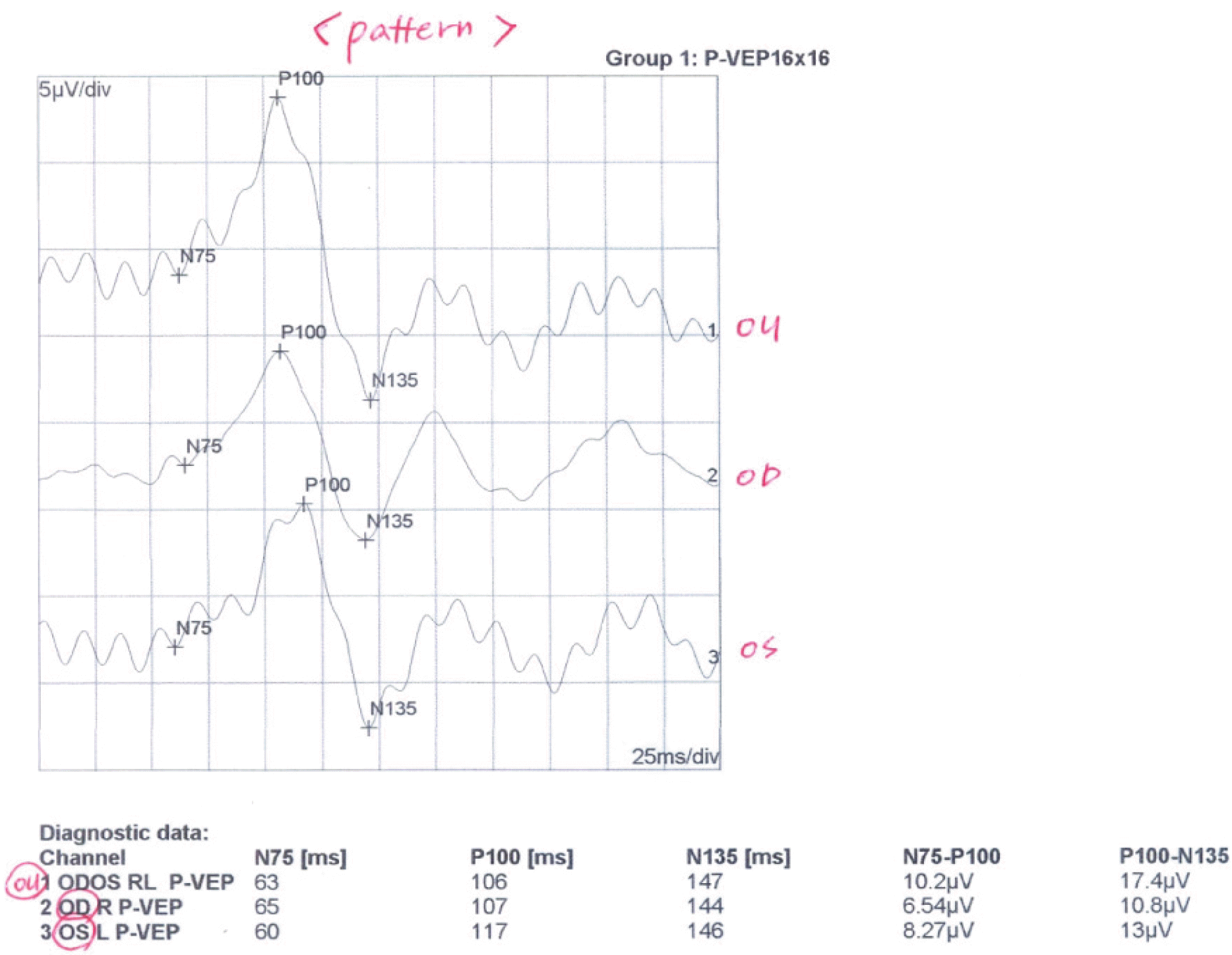
Figure 3.
The optical coherent tomography findings of case 1. The patient's retinal nerve fiber layer analysis shows normal findings.
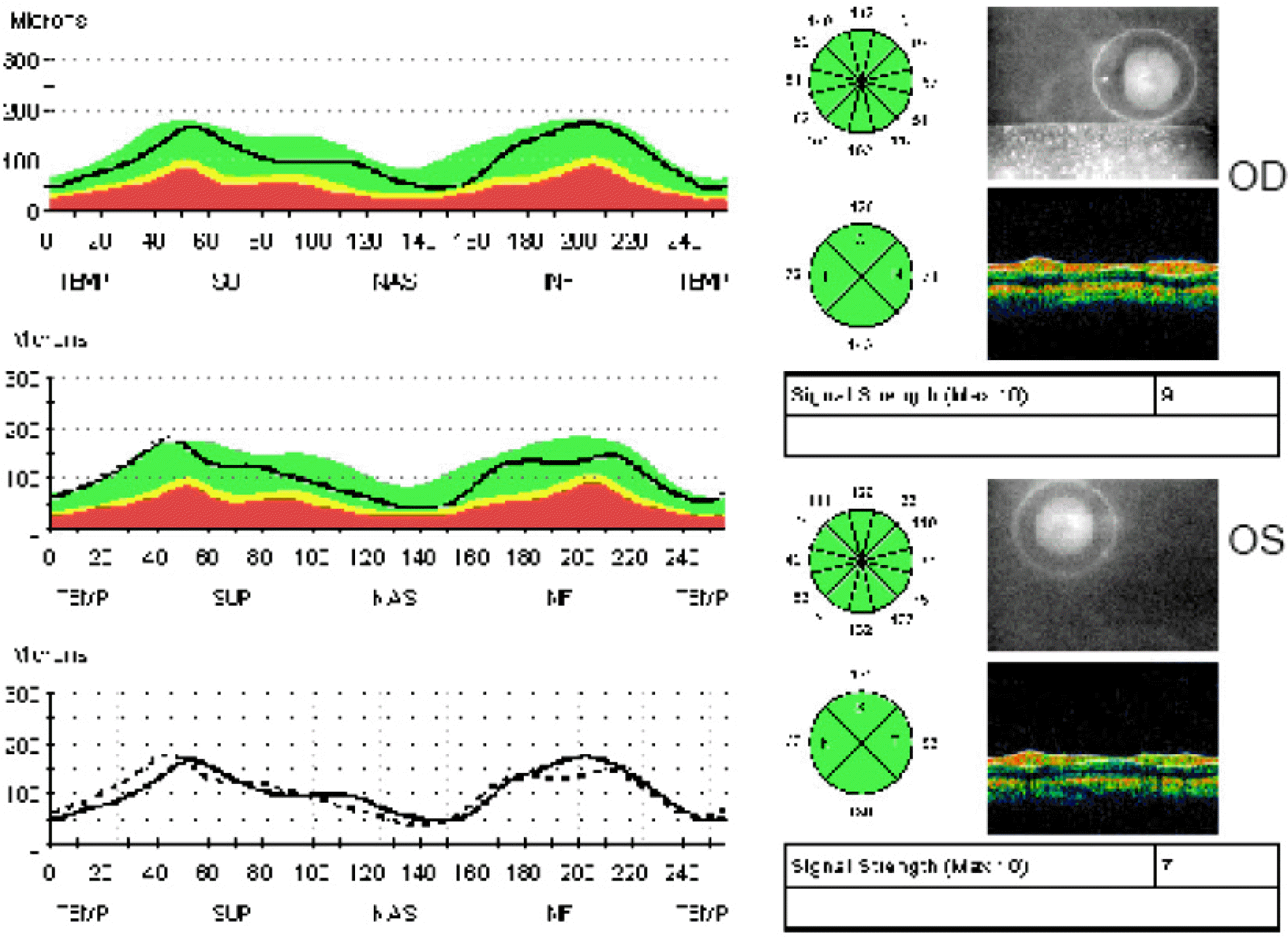
Figure 4.
The visual field findings of case 2. The visual field shows peripheral constriction and suggestive malingering.
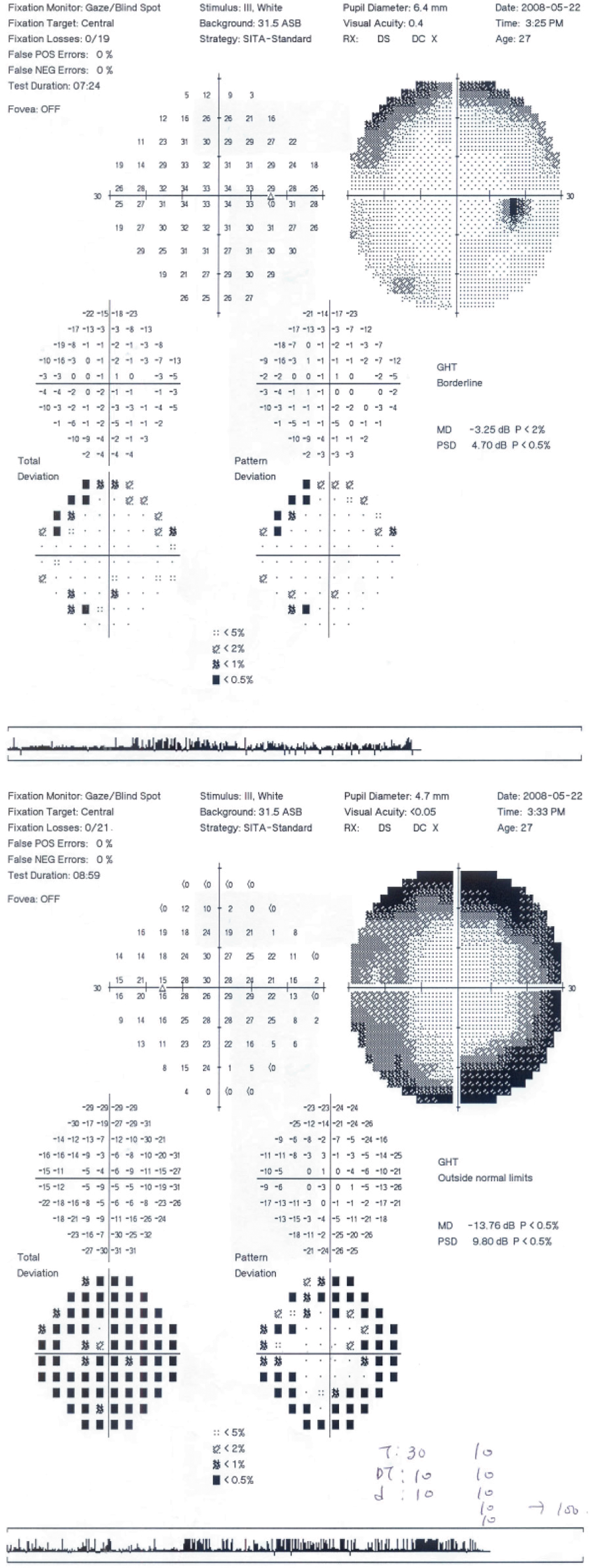
Figure 5.
The visual evoked potential findings of case 2. The pattern visual evoked potential of the left eye shows slightly decreased amplitude compared to that of his right eye.
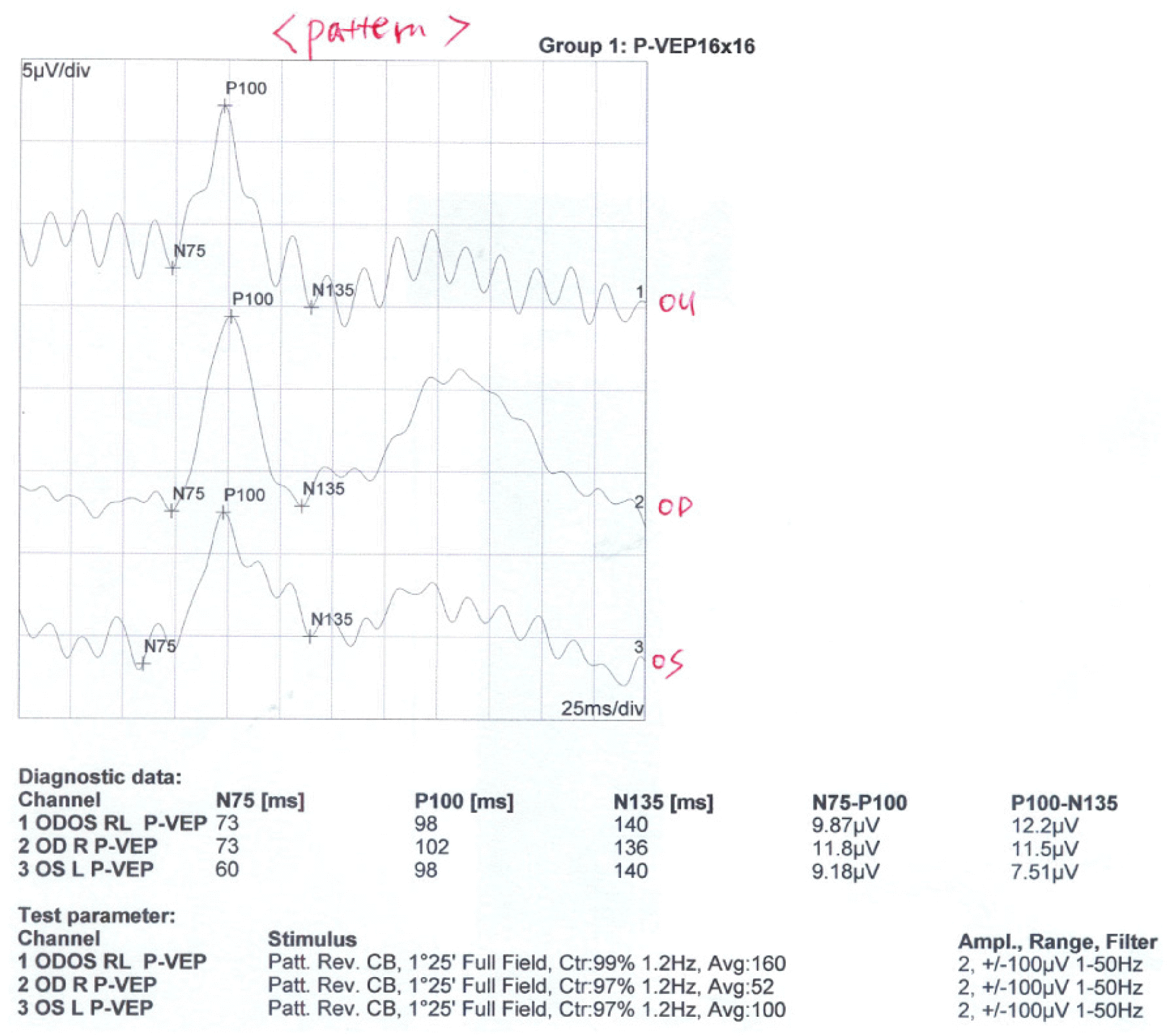
Figure 6.
The optical coherent tomography findings of case 2. The patient's retinal nerve fiber layers analysis shows normal findings.
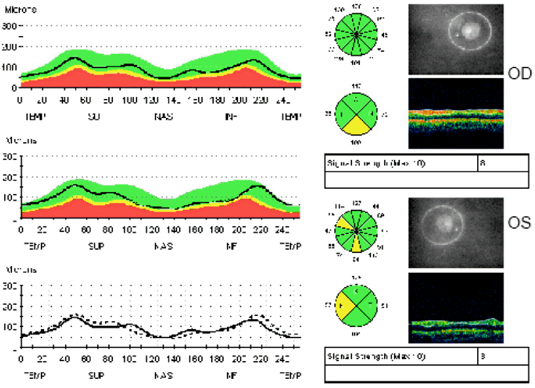
Figure 7.
The fundus findings of case 2. The color fundus photos (A, B) and fluorescein angiography (C, D) show nonspecific findings.

Table 1.
Clinical data of 35 patients diagnosed by McBride disability evaluation
| | BCVA* (far & near) | V/F† | diplopia | Anatomical finding |
|---|---|---|---|---|
| 1 | 0.2/0.15 & 0.2/0.15 | | under at central 20° | L)‡ esotropia |
| 2 | 0.8/0.02 & 0.5/0.02 | B)§ peripheral constriction | | L)‡ corneal opacity |
| | | | | & pseudophakia |
| 3 | 1.2/1.2 | | over at superior 20° | R)∏ superior oblique |
| | | | | muscle palsy |
| 4 | 0.8/1.0 & 0.7/1.0 | R)∏ peripheral constriction | at right gaze | |
| 5 | LP#(−)/0.3 & | L)‡ sparing under central 5° | | R)∏ optic atrophy & brain |
| | LP#(−)/0.4 | | | parietotemporal area lesion |
| 6 | 0.1/1.0 & 0.1/1.0 | | | R)∏ macular hole |
| 7 | 1.0/1.2 & 1.0/1.0 | L)‡ hemianopsia | under at central 10° | R)∏ exotropia |
| | | R)∏ total scotoma | | |
| 8 | 1.5/0.8 & 1.5/1.0 | L)‡ quadrantanopia | | L)‡ pale optic disc |
| 9 | LP#(−)/0.7 | | | R)∏ pale optic disc |
| 10 | 0.8/FC**5 cm | | | L)‡ choroidal rupture |
| | | | | L)‡ subfoveal fibrosis |
| 11 | 1.5/0.6 & 1.0/0.5 | L)‡ superior altitudinal field | | L)‡ pale optic disc |
| | | defect | | |
| 12 | 0.5/LP#(−) & 0.5/LP#(−) | | | L)‡ corneoscleral laceration |
| | | | | & pale optic disc |
| 13 | FC**5 cm / 0.5 | | | R)∏ macular hole |
| 14 | 1.0/HM††(+) | | | L)‡ pale optic disc |
| 15 | 0.9/0.5 & 0.5/0.5 | | | |
| 16 | LP#(−)/1.0 | | | R)∏ band keratopathy |
| | | | | R)∏ macular hole, PVR‡‡ |
| 17 | 1.2/0.01 | | | L)‡ macular scar |
| 18 | 1.2/0.01 | | | L)‡ corneoscleral laceration |
| 19 | 0.06/1.0 & 0.1/1.0 | | | R)∏ corneal opacity |
| 20 | 1.0/0.9 & 1.0/1.0 | | at 1° gaze | B)§6th. nerve palsy |
| 21 | 0.9/LP#(+)&0.8/LP#(+) | | | L)‡ macular hole & scar, |
| | | | | corneal opacity |
| 22 | 0.2/1.5 & 0.2/1.5 | R)∏ peripheral constriction | | R)∏ pale optic disc |
| 23 | 0.6/1.0 & 0.8/1.0 | R)∏ peripheral constriction | at 1° gaze | R)∏ exotropia |
| 24 | 0.4/1.2 | | at 1° gaze | R)∏ superor oblique muscle palsy |
| 25 | 1.2/0.9 | | at 1° gaze | R)∏ inferior oblique muscle palsy |
| 26 | 1.0/1.2 & 1.0/1.0 | | at 1° gaze | R)∏ superior oblique muscle palsy |
| 27 | 0.2/1.2 & 0.16/1.2 | | | R)∏ corneal opacity |
| 28 | 1.5/0.6 & 1.5/0.1 | | | L)‡ corneal opacity & optic atrophy |
| 29 | 1.5/0.2 & 1.5/0.1 | | | L)‡ corneal opacity |
| 30 | 0.02/0.9 & 0.02/1.0 | | | R)∏ corneal opacity |
| 31 | 1.5/LP#(−) | | | L)‡ corneal opacity & optic atrophy |
| 32 | LP#(−)/1.0 | | | R)∏ pale optic disc |
| 33 | 1.5/LP#(−) | | | L)‡ optic atrophy |
| 34 | 0.5/LP#(+) | | | L)‡ pale optic disc |
| 35 | 1.0/1.0 | L)‡ sparing under central 5° | | L)‡ primary open-angle glaucoma |
Table 2.
Data of multifocal electroretinogram of case 1 (right eye)
| Ring | Amp.* P1[nV/deg2] | Amp.* P1[nV] | Amp.* N1[nV] | Impl.† P1[ms] | Impl.† N1[ms] |
|---|---|---|---|---|---|
| 1 | 134.2 | 1.66 | 0.62 | 41.0 | 18.5 |
| 2 | 82.4 | 1.45 | 0.54 | 40.0 | 20.5 |
| 3 | 51.4 | 1.31 | 0.56 | 37.1 | 19.5 |
| 4 | 35.3 | 1.25 | 0.51 | 38.1 | 18.5 |
| 5 | 25.7 | 1.21 | 0.46 | 37.1 | 19.5 |
Table 3.
Data of multifocal electroretinogram of case 1 (left eye)
| Ring | Amp.* P1[nV/deg2] | Amp.* P1[nV] | Amp.* N1[nV] | Impl.† P1[ms] | Impl.† N1[ms] |
|---|---|---|---|---|---|
| 1 | 100.7 | 1.24 | 0.43 | 41.0 | 14.6 |
| 2 | 55.7 | 0.98 | 0.39 | 40.0 | 22.5 |
| 3 | 33.4 | 0.85 | 0.43 | 39.0 | 19.5 |
| 4 | 18.3 | 0.65 | 0.27 | 39.8 | 21.5 |
| 5 | 17.6 | 0.83 | 0.38 | 40.0 | 20.5 |
Table 4.
The difference of average amplitude P1 of both eyes (case 1)
| Ring | Diff.* of Both Ave.† Amp.‡ P1[nV/deg2] | p-value |
|---|---|---|
| 1 | 33.5 | |
| 2 | 21.1±28.2 | 0.127 |
| 3 | 11.3±13.5 | 0.014 |
| 4 | 10.8±9.9 | 0.000 |
| 5 | 3.6±9.7 | 0.086 |
Table 5.
The difference of average implicit P1 of both eyes (case 1)
| Ring | Diff.* of Both Ave.† Impl.‡ P1[ms] | p-value |
|---|---|---|
| 1 | 0.0 | |
| 2 | 2.0±3.9 | 0.270 |
| 3 | 2.4±4.3 | 0.074 |
| 4 | 4.7±4.6 | 0.000 |
| 5 | 5.6±4.2 | 0.000 |
Table 6.
Data of multifocal electroretinogram of case 2 (right eye)
| Ring | Amp.* P1[nV/deg2] | Amp.* P1[nV] | Amp.* N1[nV] | Impl.† P1[ms] | Impl.† N1[ms] |
|---|---|---|---|---|---|
| 1 | 107.7 | 1.33 | 0.42 | 39.0 | 19.5 |
| 2 | 67.7 | 1.19 | 0.53 | 36.1 | 20.5 |
| 3 | 41.1 | 1.05 | 0.48 | 34.2 | 18.5 |
| 4 | 30.1 | 1.07 | 0.50 | 35.1 | 18.5 |
| 5 | 25.1 | 1.18 | 0.56 | 36.1 | 19.5 |
Table 7.
Data of multifocal electroretinogram of case 2 (left eye)
| Ring | Amp.* P1[nV/deg2] | Amp.* P1[nV] | Amp.* N1[nV] | Impl.† P1[ms] | Impl.† N1[ms] |
|---|---|---|---|---|---|
| 1 | 136.2 | 1.68 | 0.21 | 40.0 | 11.7 |
| 2 | 86.5 | 1.52 | 0.69 | 37.1 | 21.5 |
| 3 | 51.8 | 1.32 | 0.52 | 35.1 | 17.6 |
| 4 | 39.2 | 1.39 | 0.56 | 36.1 | 19.5 |
| 5 | 28.6 | 1.35 | 0.65 | 36.1 | 20.5 |
Table 8.
The difference of average amplitude P1 of both eyes (case 2)
| Ring | Diff.* of Both Ave.† Amp.‡ P1[nV/deg2] | p-value |
|---|---|---|
| 1 | 28.5 | |
| 2 | 26.5±32.3 | 0.101 |
| 3 | 18.0±15.4 | 0.002 |
| 4 | 11.3±11.1 | 0.000 |
| 5 | 8.4±8.2 | 0.000 |
Table 9.
The difference of average implicit P1 of both eyes (case 2)
| Ring | Diff.* of Both Ave.† Impl.‡ P1[ms] | p-value |
|---|---|---|
| 1 | 1.0 | |
| 2 | 0.5±3.0 | 0.711 |
| 3 | 0.1±2.9 | 0.874 |
| 4 | 0.9±2.7 | 0.157 |
| 5 | 2.0±3.7 | 0.014 |
Table 10.
The amplitude of multifocal electroretinogram of control group
| Ring | Average of Amp.* P1[nV/deg2] | Stdev† of Amp.* P1 | 95% Confidence Range |
|---|---|---|---|
| 1 | 172.63 | 49.44 | 134.62∼210.63 |
| 2 | 97.17 | 34.51 | 70.64∼123.71 |
| 3 | 59.01 | 16.88 | 46.03∼71.99 |
| 4 | 41.93 | 10.27 | 34.03∼49.83 |
| 5 | 32.70 | 10.96 | 24.27∼41.12 |
 XML Download