Abstract
Purpose
The schwannoma is a tumor originating from Schwann cell proliferation. Schwannoma is a rare disease, making up only 1% of all the tumors that develop in the orbit. Schwannomas usually arise from the choroids or the ciliary body, and occurrence in the conjunctiva is especially rare. Few cases have been reported worldwide, and no cases have been reported in Korea thus far. We report our case along with a literature review.
Case summary
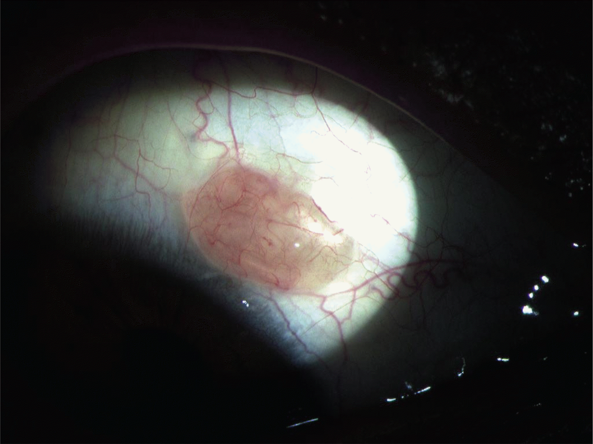
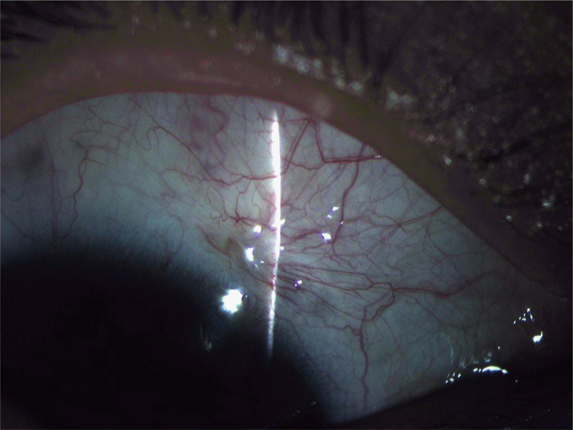
A 31-year-old male patient visited our department with a history of discomfort of in his right eye for the past 5 years caused by a bulbar conjunctival mass. On ophthalmologic examination, a 5×3×3 mm, elevated, yellowish mass with a smooth surface was observed on the bulbar conjunctiva adjacent to the superonasal limbus near the 2 o' clock area of the right eye. We performed excision and biopsy for diagnosis and treatment of the bulbar conjunctival mass and confirmed the pathologic report for the diagnosis of schwannoma.
References
1. Jacobiec FA, Albert DM. Peripheral nerve sheath tumors of the orbit In: Jacobiec FA, Levin LA. Principle and practice of ophthalmology. 2nd ed.Philadelphia: WB Saunders;2000. 4:chap.p. 242.
2. Kim JP, Lee MK, Park BI. A case of orbital neurilemoma. J Korean Ophthalmol Soc. 1986; 27:937–941.
3. Yoon SW, Ahn BC. A case of orbital neurilemoma associated with neurofibromatosis. J Korean Ophthalmol Soc. 1999; 40:1993–7.
4. Byon DS, Kim SK, Byun KD. A clinical experience of postoperative double elevator palsy after surgical removal of schwannoma in orbit and carvernous sinus. J Korean Ophthalmol Soc. 1999; 40:1732–7.
5. Kim SH, Hoang HR, Park BI. A case of intraocular neurilemoma. J Korean Ophthalmol Soc. 1982; 23:1135–7.
6. Beom JS, Yoon HM, Kim YH. A case of intraocular neurilemoma. J Korean Ophthalmol Soc. 1986; 27:127–31.
7. Lee JK, Song JS, Lee TW. A case of the ciliary body neurilemoma. J Korean Ophthalmol Soc. 1998; 39:1294–9.
8. Andreoli CM, Hatton M, Semple JP, et al. Perilimbal conjunctival schwannoma. Arch Ophthalmol. 2004; 122:388–9.

9. Chales NC, Fox DM, Avendano JA, et al. Conjunctival neurilemoma: report of 3 cases. Arch Ophthalmol. 1997; 115:547–9.
10. Le Marc'hadour F, Romanet JP, Fdili A, et al. Schwannoma of the bulbar conjunctiva. Arch Opththalmol. 1996; 114:1258–60.
11. Spencer WH. Ophthalmic pathology: An atlas and textbook. 4th ed.Philadelphia: WB Saunders;1995. 1653–89. p. 2645–50.
12. Riccardi VM. Neurofibromatosis: An overview and new directions in clinical investigations. Adv Neurol. 1982; 22:1–21.
13. Jacobiec FA, Albert DM. Mesenchymal, fibroosseous, and cartilaginous orbital tumors. Jacobiec FA, Bilyk JR, Hidayat AA, editors. Principle and practice of ophthalmology. 2nd ed.Philadelphia: WB Saunders;2000. 4:chap.p. 248.


 PDF
PDF ePub
ePub Citation
 Print
Print


 XML Download