

 PDF
PDF ePub
ePub Citation
 Print
Print


Introduction
Skin aging is a complicated biological alteration induced by miscellaneous factors. The pathway of skin aging is based on the combination of endogenous or intrinsic factors (genetics, metabolism, and hormone) and exogenous or extrinsic factors. One of the major contributing factors in intrinsic skin aging is hormonal changes typically in postmenopausal women due to decreasing of estrogen level.1 The hormonal modifications in postmenopausal women intensify the facial skin aging as a result of collagen reduction, lack of hydration, and loss of elasticity that are classified as estrogen deficient skin.2
Menopause is the end of women's fertile period and reproductive ability which is a natural process undergone by every woman. It is indicated by permanent cessation of menstrual cycle preceded by the absence of menstruation for 12 consecutive months as a result of decreased ovarian follicle activity, which decrease the body estrogen level.3 Decreased estrogen level causes not only climacteric complaints, but also changes in the skin.4
Estrogen influences the balance of collagen synthesis and degradation in the skin. It stimulates the synthesis, maturation, and turnover of collagen, increases hyaluronic acid synthesis, and maintains water content.5 Low estrogen level could cause changes in the skin such as skin thinning due to decreased collagen, elasticity, vascularization, and moisture, as well as increased wrinkles.56 Immunohisto-chemistry examination of postmenopausal women's skin biopsy showed decreased collagen type I alpha 1 (COL1A1), collagen type III alpha 1 (COL3A1), and COL3A1/COL1A1 ratio compared to premenopausal women.7 Because of structural and physiological changes in every layer of the skin due to a decrease of estrogen, a lot of women consult physicians to slow the aging process.
Estrogen is sometimes used to treat skin aging in postmenopausal women. Studies showed that oral, topical and transdermal estrogen affects collagen content and induces the production of procollagen I and III which is a precursor of COL1A1 and COL3A1 in collagen synthesis.8 Decreased estrogen is also linked in menopausal-related collagen production in both sun protected skin and photodamaged skin.9 Oral estrogen was one of the first regimen used, however, due to its numerous side effects (headache, nausea, vomiting, breast pain, vaginal bleeding, endometrium thickening, ovarian cancer, and breast cancer), it is not wise to give oral estrogen as hormonal therapy for postmenopausal women with just skin aging problems.5 Therefore, it is thought to switch to topical estrogen. However, topical estrogen still could cause systemic side effects like oral estrogen, although less severe. Currently, phytoestrogen is starting to gain attention because it is safer and has fewer side effects compared to oral and topical estrogen.10
Phytoestrogen is a nonsteroidal organic chemical compound from plants, fruits, and seeds.11 It is synthesized from phenylpropanoid and phenol, with partial agonist effect towards endogenic estrogen.10 The sources of phytoestrogens can be used in several forms, such as: fresh, dried simplicia, extract, or fraction. Those forms enable synergistic relationship between several natural chemical compounds from those plants, fruits, and seeds to have phytoestrogenic effect.12 Phytoestrogen is classified as natural selective estrogen receptor modulators (SERMs).1013 Its mechanism of action is similar to 17β-estradiol sex hormone, by binding to estrogen receptor (ER) α and β, causing a conformational change in the receptors, which will result in recruiting essential cofactors which may be co-activators or co-repressors act on the target tissue.1014 Studies on phytoestrogen have been performed in the past decade, especially from soybean, which gave satisfactory results. Other sources of phytoestrogen include Korean ginseng, paddy seeds, red grape skin, mulberry, blackberry, Trigonella foenum-graceum L. (fenugreek) seeds, and others.12 However, a study on the efficacy of other sources of phytoestrogen, such as fenugreek plant, is needed to broaden the knowledge regarding herbal plants and to develop safe, effective, and affordable herbal cosmetics.
Fenugreek (family: Fabaceae, genus: Trigonella, species: Trigonella foenum-graecum L.) is easy to obtain and often used in herbal medicine. This plant exists all around the world, has various uses, and is easy to grow. In Indonesia, fenugreek seeds are used as cooking spices to make curry and also used as one of ingredients to make jamu. In India, fenugreek leaves are consumed as vegetable.15 Fenugreek seed consists of mucilage fiber polysaccharide (45 - 60%) mostly composed of galactomannans (25 - 45%), protein (30%) including lysine and tryptophan, lipid or essential oils (5 - 10%), saponin (4.8%), ash (3.9%) and other secondary metabolites.16 Active compounds found in fenugreek seed are protoalkaloid including trigonelline (0.2 - 0.38%), choline (0.05%), gentianine and carpaine; sterols including β-sitosterol and hydrolyzed steroidal sapogenins such as diosgenin, yamogenin, tiogenin, and neotiogenin; several flavonoids consist of apigenin, luteolin, quercetin, orientin, isoorientin, and isovitexin; free amino acids including 4-hydroxyisoleucine (0.09%), arginine, histidine, and lysine; minerals such as calcium and iron; cholesterol and sitosterol; vitamins A, B1, C and nicotinic acid; volatile oils including n-alkanes and sesquiterpenes (0.015%).17 It is challenging to identify exact component of a herbal preparation, therefore, a single marker based of trigonelin concentration has been established to obtain detailed formulation of fenugreek seed extract.18
Fenugreek seeds extract is used as oral supplement to lower LDL, triglycerides, blood sugar, and has anti-inflammatory effect in asthma/allergy cases.19 In addition, other secondary metabolites found in fenugreek have been reported to perform several benefits including antioxidant and antiradical by protecting cellular compound oxidative damage. There are also studies that indicate the use of fenugreek as antidiabetic, antifertility, antifungal, analgesic, anti-inflammatory, antipyretic, and enhance immunomodulatory activities.20 Different approach is also known in topical form in order to aid severe skin inflammation, chapped lips and skin aging.21 There are three studies in India and Pakistan to prove the effect of topical fenugreek seeds extract towards skin elasticity, moisture, and its side effects involving both young female and male participants as the subjects.222324 An in vitro study in India found estrogenic effects of fenugreek seeds mimics the estradiol effects and boost cell proliferation.25
There is no previous study evaluating the role of fenugreek seeds extract as anti-aging therapy for postmenopausal women. Based on the explanation above, this study aims to be the first to molecularly assess the role of fenugreek seeds extract effect towards COL1A1 and COL3A1.
Experimental
In vitro experimental study
This in vitro experimental study is using fibroblast culture from leftover tissue of blepharoplasty on a 60 year-old woman, who has been in menopausal state for 5 years (estradiol level <5 pg/mL). The patient was selected based on inclusion criteria: women who have been in menopause for at least 2 years and maximum 5 years, estradiol level <30 pg/mL, wrinkles on face and consented to donate their leftover skin tissue from surgery. Exclusion criteria includes women with facial dermatitis, hormonal supplement consumption in the last 12 months, usage of retinoic acid/hydroquinone or its derivatives products, radiation/facelift/face implant therapy in the last 5 years, and history of ovariectomy. Fibroblasts obtained were then designated as old Human Dermal Fibroblast (HDF). As a control comparison, young fibroblasts from preputium of YARSI University's biorepository were used and designated as young HDF. Cell isolation methods used were explant and enzymatic techniques, following research procedure reported by Hadi26 and Kusuma.27 All chemicals and medium were purchased from Gibco (Grand Island, NY) except for CCK-8 reagent from Sigma-Aldrich (St.Louis, MO). This study had obtained ethical clearance from Health Research Ethics Committee, Faculty of Medicine, Universitas Indonesia, Cipto Mangunkusumo National Referral Hospital.
Plant material
Fenugreek seeds extract used were obtained from Medicinal Plant Research Institute Tawangmangu, Indonesia, which were then extracted at Agency for The Assessment and Application of Technology (BPPT), in Serpong, Indonesia, using methanol extract. Afterwards, 1 g extract was dissolved in 5 mL Dulbecco's Modified Eagle Medium (DMEM), and then used as fenugreek extract dose stock. In this study, extract from dose stock was diluted, starting from 5 mg/mL to1.2 µg/mL according to geometric sequence with complete culture medium (DMEM+ 10% Fetal Bovine Serum + 1% Antibiotic-Antimycotic).
Fibroblast culture
Fibroblast isolation and culture from leftover tissue from surgery were done in YARSI laboratory. Culture container with 96 wells was used with DMEM medium (Gibco), 10% FBS supplement (Gibco), and 1% antibiotic-antimycotic (Gibco) 1% in 37℃ incubator with CO2 level of 5%. Cell count was done using trypan blue exclusion method on TC20 automated cell counter (Bio-rad). Cell viability was also measured using CCK-8 (Sigma) reagent.28 10 µL CCK-8 was added to 90 µL PBS (Gibco) in each well and then incubated for 60 minutes. The intensity of the color that appeared was measured using microplate reader at wavelength of 450 nm. Cell proliferation was obtained by measuring cell viability at day 3, 5, 7, 10, and 14. Fibroblast culture was done until second-third passage was obtained by replacing DMEM medium, 10% FBS supplement, and 1% antibioticantimycotic every 2-3 days.
Toxicity test
For each fenugreek extract concentration, 8000 cells were seeded triplicate. Control groups consisted of two types of control, which were control without treatment (C) and solvent control which was 1.4% methanol (Cs). Methanol concentration used was based on analysis information provided by Agency for The Assessment and Application of Technology (BPPT) on 100% fenugreek seeds extract. A total of 1 mg of extract was dissolved in complete medium and used as stock. Cytotoxicity was obtained by measuring cell viability on fenugreek extract with concentration of 5 mg/mL to 1.2 µg/mL according to geometric sequence with complete medium. Treatment was given for 48 hours. Microscopic pictures were taken at 24 and 48 hours after treatment on representative groups. Cell count and cell viability assessment were done as previously described in fibroblast culture step, using trypan blue exclusion method and CCK-8 reagent (Sigma). The obtained concentration fromb toxicity test was then used to examine COL1A1 andbCOL3A1using ELISA test.
COL1A1 and COL3A1 test
Fibroblasts were planted in 24-well culture container until 80% confluence was reached before treatment with fenugreek extract was done for 24 hours. The conditioned medium sample obtained after 24 hours was centrifuged according to ELISA protocol instructions and stored at −80℃. Cells on 24 plates were harvested and viable cells were counted using trypan blue exclusion method by TC20 automated cell counter (Bio-Rad). A test was performed using COL1A1 (MyBioSource, USA, MBS763786) and COL3A1 (MyBioSource, USA, MBS763396) ELISA kit in duplo to confirm the results. The absorbance was measured according to protocol using multiplate reader (TECAN). The absorbance value obtained was reduced by blank absorbance and converted into collagen concentration using standard curve and adjusted to the number of viable cells obtained. An ELISA test of fenugreek seeds extract was compared to 2 controls, which were control without treatment (as negative control) and estradiol 5 nM (as positive control). Concentration of 5 nM was determined based on previous study by Surazynski.29 The data was presented as collagen concentration (ng/mL) for 105 viable cells.
Result and Discussion
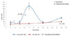
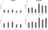
To determine the best time to do the experimental on an old HDF and a young HDF, we compared their proliferation time. Fig. 1 shows that the old HDF had much slower proliferation and lower number of viable fibroblast compared to the young HDF. This is because the young HDF underwent exponential phase, where cell proliferation happened really fast until it reached maximum capacity and then decline, while the old HDF did not show significant exponential phase.
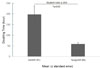
The old HDF had slower doubling time compared to the young HDF, using the same medium without fenugreek. Fig. 2 shows that to double the number of cells, the old HDF needed four times longer compared to the young HDF, which is in accordance with decreased cell growth ability as age increases. This makes the old HDF culture was done until second-third passage, compared to the young HDF which was done until fourth-fifth passage.
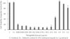
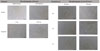
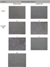
Methanol solvent medium used for fenugreek extract extraction is not toxic and does not influence fibroblasts viability (Fig. 3). Results of fenugreek extract toxicity test at 5 mg/mL to 9.8 µg/mL concentration showed that fenugreek extract was toxic to the old HDF. The toxicity test could also be seen from microphotography pictures showing changes of the old and the young HDF morphology due to fenugreek extract exposure for 24 and 48 hours (Fig. 4 and 5). The best concentration of fenugreek extract for cell viability on the old HDF was 4.9 µg/mL, but HDF morphology examination at 24 and 48 hours showed changes in fibroblast morphology after being exposed to 4.9 µg/mL fenugreek extract for 48 hours. On the young HDF, 4 and 8 µg/mL fenugreek concentration showed changes in morphology examination at 24 hours. Therefore, the concentration of fenugreek extract used to examine COL1A1 and COL3A1 using ELISA test was under 4.9 µg/mL, which was 0.5 – 2 µg/mL.
Fenugreek extract at concentration of 2 µg/mL significantly increased COL1A1 and COL3A1 secretion, both on old HDF (Fig. 6A and 6C) and young HDF (Fig. 6B and 6D). Obtained collagen concentration showed COL1A1 and COL3A1 ratio of 1: 1.5-2 on old HDF and 1:1 on young HDF. Fenugreek extract also seemed to have slightly higher collagen secretion stimulation effect compared to 5 nM estradiol. Fenugreek concentration of 4 µg/mL caused the death of the old HDF so that no collagen secretion data was obtained. However, fenugreek toxicity at concentration of 4 µg/mL was still tolerated by the young HDF. This result showed that fenugreek's effect is not dose-dependent and that 2 µg/mL is the optimal concentration for COL1A1 and COL3A1 on both the old and the young HDF. The statistic test used was paired t-test between control and treatment groups (p<0.05), collagen concentration per 100000 viable cells.
Fenugreek extract at the concentration of 2 µg/mL significantly increased COL1A1 and COL3A1 secretion compared to no treatment and 5 nM estradiol control groups. However, fenugreek extract at the concentration of 1 and 0.5 µg/mL did not show similar results. On the other hand, it tended to inhibit COL1A1 and COL3A1 secretion compared to control (p<0.05). Fenugreek extract at concentration of 0.5 µg/mL produced slightly more COL1A1 and COL3A1 compared to 1 µg/mL concentration. It could be concluded that fenugreek extract effect towards COL1A1 and COL3A1 is not dose-dependent. To find out the cause, further studies are needed to consider specificity of old fibroblast response to low concentration fenugreek extract, and that various active substances content of the extract could have different optimal concentration, therefore giving different cell responses. A study from Agustini12 showed that fenugreek contains a lot of components, such as trigonelline, diosgenin, yamogenin, gitogenin, tiogenin, vitexin, isovitexin, quercetin, luteolin, orientin and isoorientin. Some of those components have agonist or antagonist activity for estrogen receptor α and β. This could explain why different concentrations could produce different results (stimulation or inhibition of collagen secretion).
The results from this study showed an increase of 1:1.5-2 in COL1A1 and COL3A1 ratio of old HDF after fenugreek exposure. Cheng30 showed that COL1A1 and COL3A1 ratio is decreased as age increases. He found that in babies, COL1A1 and COL3A1 levels are both high, but COL3A1 has a higher level compared to COL1A1. As age increases, both COL1A1 and COL3A1 levels are decreased, but the decrease of COL3A1 is much more drastic than COL1A1. In postmenopausal women, COL1A1 level is higher than COL3A1. Based on the results of this study, it is expected that using fenugreek as a postmenopausal skin aging therapy will produce a more significant increase of COL3A1 level compared to COL1A1.
In this study, fenugreek concentration tested to increase secretion of COL1A1 and COL3A1 were 2 µg/mL, 1 µg/mL and 0.5 µg/mL. These concentrations were chosen based on previous toxicity test, the borderline fenugreek concentration was 2 - 10 µg/mL for 48 hours, with which several cultures looked good, while higher concentration caused toxic effect (fibroblast morphology changes). No other study had tested fenugreek using old fibroblast as comparison. Therefore, further studies are still required to investigate about the exact mechanism of action of fenugreek whether it is through specific estrogen binding receptor or other pathways.
In conclusion, fenugreek extract at the concentration of 2 µg/mL could significantly increase COL1A1 and COL3A1 secretion on postmenopausal woman's fibroblast compared to no treatment and 5 nM estradiol controls.
 XML Download