Journal List > Nat Prod Sci > v.22(2) > 1060647

Widowati, Wijaya, Laksmitawati, Widyanto, Erawijantari, Fauziah, Bachtiar, and Sandra: Tea Flavonoids Induced Differentiation of Peripheral Blood-derived Mononuclear Cells into Peripheral Blood-derived Endothelial Progenitor Cells and Suppressed Intracellular Reactive Oxygen Species Level of Peripheral Blood-derived Endothelial Progenitor Cells

Abstract

Endothelial dysfunction in atherosclerosis is associated with increasing oxidative stress that could be reversed by antioxidant. Therefore epigallocatechin gallate (EGCG), epicatechin gallate (ECG), epigallocatechin (EGC) and catechin (C) of tea flavonoids were investigated for their roles in regenerating endothelial cell. Peripheral blood mononuclear cells (PB-MNCs) were isolated, plated and cultured in medium with/without treatment of EGCG, ECG, EGC and C. Results showed that among all EGCG, ECG, EGC and C concentrations tested, 12.5µmol/L was not cytotoxic for peripheral blood-derived endothelial progenitor cells (PB-EPCs). Treatment of EGCG, ECG, EGC or C increased the percentages of CD34, CD133, VEGFR-2 expressions and suppressed hydrogen peroxide-induced percentages of reactive oxygen species (ROS) level in PB-EPCs. Taken together, our current results showed that EGCG, ECG, EGC or C of tea flavonoids could induce differentiation of PB-MNCs into PB-EPCs as well as protect PB-EPCs from oxidative damage by suppresing the intracellular ROS levels.

REFERENCES

(1). Bonetti P. O., Lerman L. O., Lerman A.Arterioscler. Thromb. Vasc. Biol. 2003; 23:168–175.
(2). Higashi Y., Noma K., Yoshizumi M., Kihara Y.Circ. J. 2009; 73:411–418.
(3). Diaz M. N., Frei B., Vita J. A., Keaney J. F. Jr.N. Engl. J. Med. 1997; 337:408–416.
(4). Lin J., Cook N. R., Albert C., Zaharris E., Gaziano J. M., Van Denburgh M., Buring J. E., Manson J. E. J.Natl. Cancer Inst. 2009; 101:14–23.
(5). Yang C. S., Landau J. M. J.Nutr. 2000; 130:2409–2412.
(6). Trevisanato S. I., Kim Y. I.Nutr. Rev. 2000; 58:1–10.
(7). Shenouda S. M., Vita J. A. J.Am. Coll. Nutr. 2007; 26:366S–372S.
(8). Grassi D., Mulder T. P., Draijer R., Desideri G., Molhuizen H. O., Ferri C. J.Hypertens. 2009; 27:774–781.
(9). Hertog M. G., Feskens E. J., Hollman P. C., Katan M. B., Kromhout D.Lancet. 1993; 342:1007–1011.
(10). Mukamal K. J., Maclure M., Muller J. E., Sherwood J. B., Mittleman M. A.Circulation. 2002; 105:2476–2481.
(11). Balentine D. A., Wiseman S. A., Bouwens L. C.Crit. Rev. Food Sci. Nutr. 1997; 37:693–704.
(12). Wolfram S. J.Am. Coll. Nutr. 2007; 26:373S–388S.
(13). Sano M., Tabata M., Suzuki M., Degawa M., Miyase T., Maeda-Yamamoto M.Analyst. 2001; 126:816–820.
(14). Widowati W., Widyanto R. M., Husin W., Ratnawati H., Laksmitawati D. R., Setiawan B., Nugrahenny D., Bachtiar I.Iran J. Basic Med. Sci. 2014; 17:702–709.
(15). Wollert K. C., Drexler H.Circ. Res. 2005; 96:151–163.
(16). Sagara Y., Vanhnasy J., Maher P. J.Neurochem. 2004; 90:1144–1155.
(17). Widowati W., Sardjono C. T., Wijaya L., Laksmitawati D. R., Sandra F. J.US-China Med. Sci. 2012; 9:22–29.
(18). Murphy T. H., Miyamoto M., Sastre A., Schnaar R. L., Coyle J. T.Neuron. 1989; 2:1547–1558.
(19). LeBel C. P., Ischiropoulos H., Bondy S. C.Chem. Res. Toxicol. 1992; 5:227–231.
(20). Jie G., Lin Z., Zhang L., Lv H., He P., Zhao B. J.Agric. Food Chem. 2006; 54:8058–8064.
(21). Valko M., Leibfritz D., Moncol J., Cronin M. T., Mazur M., Telser J.Int. J. Biochem. Cell Biol. 2007; 39:44–84.
(22). Evacuasiany E., Ratnawati H., Liana L. K., Widowati W., Maesaroh M., Mozef T., Risdian C.Oxid. Antioxid. Med. Sci. 2014; 3:141–146.

Fig. 1.
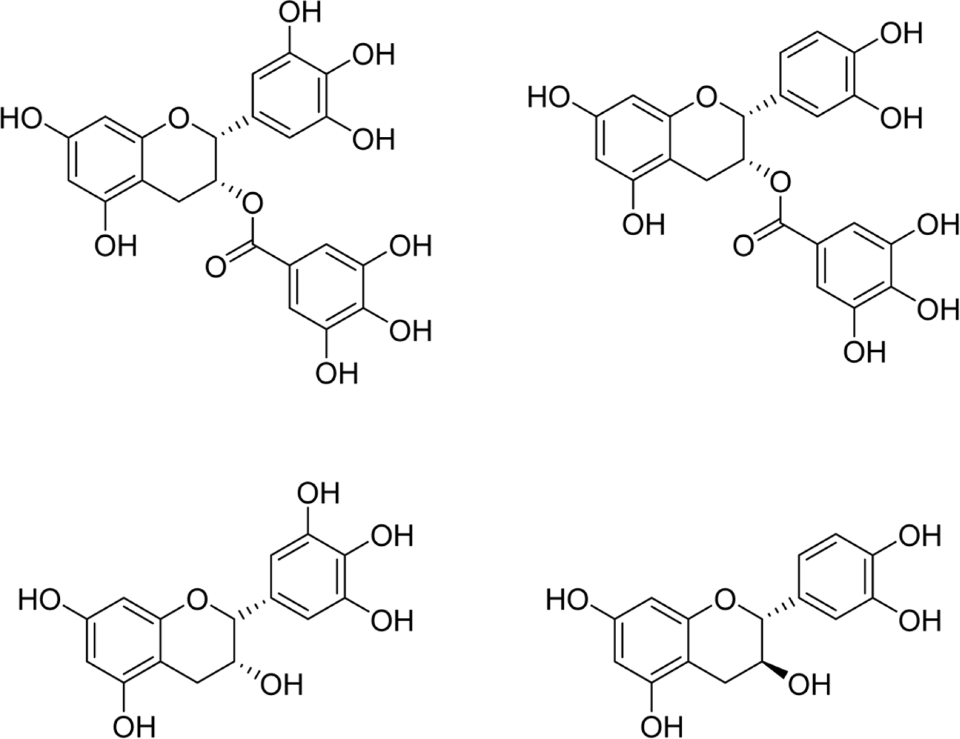
Chemical Structures of catechins. Structures of (−)-epigallocatechin gallate (EGCG) (a), (−)-epicatechin gallate (ECG)(b), (−)-epigallocatechin (EGC) (c), and (+)-catechin (C) (d).
nps-22-87f1.tif
Fig. 2.
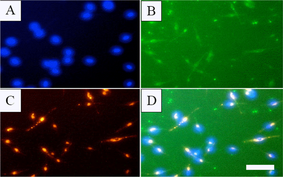
PB-EPCs lectin binding and LDL uptake. PB-EPCs were seeded in 96-well plate and subjected to Fluorescent Staining as described in Experimental. A: DAPI, B: FITC-UEA-I, C: Dil-acLDL, D: Merge of A, B and C. White bar: 50 μ m.
nps-22-87f2.tif
Fig. 3.
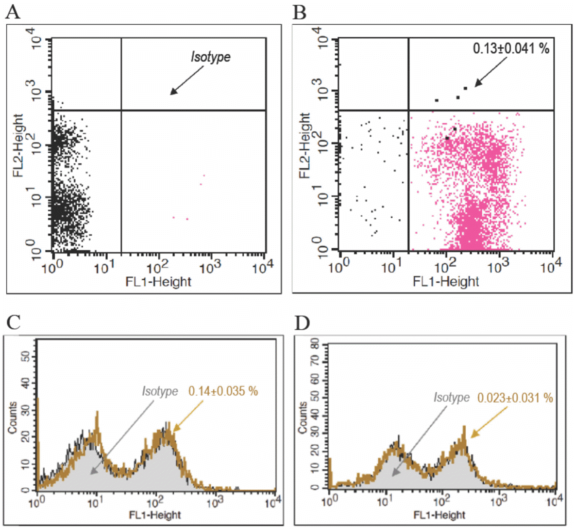
PB-EPCs immunophenotypes. PB-EPCs were detached and subjected to Immunophenotyping as described in Experimental. A: isotype, B: CD34/45, C: CD133 and its isotype, D: VEGF-R2 and its isotype.
nps-22-87f3.tif
Table 1.
Effect of EGCG, ECG, EGC and C on PB-EPCs viability
Treatment Percentage of Viable PB-EPCs (Mean±SD)
EGCG ECG EGC C
Untreated 100.00 ± 8.27cA 100.00 ± 8.27cA 100.00 ± 8.27cA 100.00 ± 8.27cA
0.5% DMSO 98.68 ± 5.04cA 98.68 ± 5.04cA 98.68 ± 5.04cA 98.68 ± 5.04cA
12.5 µmol/L 93.20 ± 5.29cA 94.00 ± 2.27cA 93.73 ± 1.04cA 93.39 ± 0.33bcA
25 µmol/L 78.28 ± 4.02bA 68.45 ± 4.96bA 69.23 ± 4.32cA 93.96 ± 4.84bcB
50 µmol/L 71.29 ± 4.18bA 62.18 ± 2.27bA 63.47 ± 3.55bA 86.21 ± 5.81bB
100 µmol/L 30.59 ± 4.96aA 32.43 ± 4.96aA 54.67 ± 3.06aB 61.76 ± 5.94aB

PB-EPCs were treated with 12.5, 25, 50 or 100 µmol/L of EGCG, ECG, EGC or C for 24 hours. MTS Assay was carried out as described in Experimental. Each treatment was done in triplicate.

a,b,c Means in the same column containing the same superscript are not significant (p ≥ 0.05), while means in the same column containing different superscript in small letter indicate significant differences (p < 0.05).

AB Means in the same row containing the same superscript are not significant (p ≥ 0.05), while means in the same row containing different superscript in capital letter indicate significant differences (p < 0.05). Statistical analysis was performed based on Duncan's post-hoc test. SD: standard deviation.

Table 2.
Effect of EGCG, ECG, EGC and C on apoptosis of PB-EPCs
Treatment Percentage of SubG1
Untreated 11.68 ± 1.20a
0.5% DMSO 14.93 ± 1.60a
EGCG 14.48 ± 6.83a
ECG 14.57 ± 7.67a
EGC 12.31 ± 3.00a
C 15.97 ± 3.62a

Ten thousands PB-EPCs were seeded in each 12-well plate and treated with 12.5 µmol/L EGCG, ECG, EGC or C for 24 hours. After 24 hours, Apoptosis Assay was carried out as described in Experimental. Each treatment was done in triplicate. The apop-totic cells were determined on the basis of the SubG1 area. The data are presented as mean ± standard deviation.

a Means in the same column containing the same superscript are not significant (p ≥ 0.05). Statistical analysis was performed based on Duncan's post-hoc test.

Table 3.
Effect of EGCG, ECG, EGC and C on PB-EPCs immunophenotypes
Treatment CD34 (%) CD133 (%) VEGFR-2 (%)
Untreated 52.86 ± 8.44b 0.63 ± 0.02ab 0.22 ± 0.02a
0.5% DMSO 28.96 ± 9.68a 0.36 ± 0.14a 1.78 ± 0.15c
EGCG 51.09 ± 2.77b 0.83 ± 0.41ab 1.22 ± 0.19b
ECG 60.48 ± 2.60b 0.61 ± 0.08ab 2.42 ± 0.13c
EGC 54.70 ± 3.74b 1.13 ± 0.71b 1.21 ± 0.22b
C 62.24 ± 9.22b 1.01 ± 0.13ab 1.47 ± 0.33bc

Isolated-PB-MNCs were cultured with addition of 12.5 µmol/L EGCG, ECG, EGC or C. PB-EPCs Culture was then carried out as described in Experimental. PB-EPCs were then detached for Immunophenotyping as described in Experimental. Each treatment was done in triplicate. The data are presented as mean ± standard deviation.

a,b,c Means in the same column containing the same superscript are not significant (p ≥ 0.05), while means in the same column containing different superscript in small letter indicate significant differences (p < 0.05). Statistical analysis was performed based on Duncan's post-hoc test.

Table 4.
Effect of EGCG, ECG, EGC and C on ROS of H2 O2-induced PB-EPCs
Treatment ROS Level (%) Ratio of All to Negative Control (%) Ratio of All to Positive Control (%)
Untreated (negative control) 7.20 ± 1.65a 599.95 ± 22.97a 22.81 ± 5.24a
H2 O2 (positive control) 31.55 ± 1.10e 438.15 ± 15.22e 99.99 ± 3.47e
EGCG + H2 O2 12.92 ± 0.70c 179.49 ± 9.67c 40.96 ± 2.21c
ECG + H2 O2 10.83 ± 2.35bc 150.46 ± 32.62bc 34.34 ± 7.44bc
EGC + H2 O2 18.66 ± 2.81d 259.21 ± 39.09d 59.15 ± 8.92d
C + H2 O2 8.85 ± 1.13ab 122.96 ± 15.73ab 28.06 ± 3.59ab

PB-EPCs were treated with 12.5 µmol/L of EGCG, ECG, EGC or C for 30 minutes. PB-EPCs were then treated with H2 O2 with final concentration of 200 µmol/L for 1 hour. Treated-PB-EPCs were subjected to Intracellular ROS Assay as described in Experimental. Each treatment was done in triplicate. The data are presented as mean±standard deviation.

a,b,c,d Means in the same column containing the same superscript are not significant (p ≥ 0.05), while means in the same column containing different superscript in small letter indicate significant differences (p < 0.05). Statistical analysis was performed based on Duncan's post-hoc test.

TOOLS
Similar articles