

 PDF
PDF ePub
ePub Citation
 Print
Print


Abstract
Cerebrospinal fluid (CSF) rhinorrhea can be caused by head trauma, brain or sinus surgery, or neoplastic sinonasal disease. There are many diverse techniques for repairing skull base defects, and recently there has been a shift from using external approaches to endoscopic approaches. The reported success rate after endoscopic repair is 97%, but CSF rhinorrhea may recur in some cases. Recently, we witnessed one case of recurrent CSF rhinorrhea from the posterior wall of the frontal sinus after a traffic accident. The patient was a 48-year-old male and had recurrent CSF rhinorrhea, severe pneumocephalus and mental change even after a CSF leakage repair operation was performed by the neurosurgeon using the periosteal flap. We successfully treated recurrent frontal CSF rhinorrhea with fat graft and mucosal graft, using the above and below approach with the guidance of a navigation system.
References
1). Loew F, Pertuiset B, Chaumier EE, Jaksche H. Traumatic, spontaneous and postoperative CSF rhinorrhea. Adv Tech Stand Neurosurg. 1984; 11:169–207.

2). 대한이비인후과학회 편. 이비인후과학: 두경부외과학 세트(개정판). 일조각;. 2009. 1403–5.
3). Abuabara A. Cerebrospinal fluid rhinorrhoea: diagnosis and management. Med Oral Patol Oral Cir Bucal. 2007; 12:E397–400.
4). Das PT, Balasubramanian D. External frontal sinusotomy and endoscopic repair of cerebrospinal fluid fistula in the posterior wall: preliminary report of a new technique. J Laryngol Otol. 2011; 125:802–6.
5). Heo SJ, Kim JS. Endoscopic Management of Cerebrospinal Fluid Rhinorrhea. Journal of Rhinology. 2014; 1:15–21.
6). Hegazy HM, Carrau RL, Snyderman CH, Kassam A, Zweig J. Transnasal endoscopic repair of cerebrospinal fluid rhinorrhea: a metaanalysis. Laryngoscope. 2000; 110:1166–72.
7). Woodworth BA, Schlosser RJ, Palmer JN. Endoscopic repair of frontal sinus cerebrospinal fluid leaks. J Laryngol Otol. 2005; 119:709–13.
8). Yoon BN, Lee JE, Lee HS, Cho KS, Roh HJ. The Surgical Approach for Removal of Inverted Papilloma Originating from the Frontal Sinus. Korean Journal of Otorhinolaryngology-Head and Neck Surgery. 2008; 9:800–4.
9). Chaaban MR, Conger B, Riley KO, Woodworth BA. Transnasal endoscopic repair of posterior table fractures. Otolaryngol Head Neck Surg. 2012; 147:1142–7.
10). Haegen T, Rehl RM, Vaughan WC. “Above and Below” Techniques in Revision Sinus Surgery: Revision Sinus Surgery. Springer;2008. p. 281–7.
11). Cho SH, Lee YS, Jeong JH, Kim KR. Endoscopic above and below approach with frontal septotomy in a patient with frontal mucocele: a contralateral bypass drainage procedure through the frontal septum. Am J Otolaryngol. 2010; 31:141–3.
12). Maeso PA, Deal RT, Kountakis SE. Combined endoscopic and minitrephination techniques in the surgical management of frontal sinus type IV cell disease. Am J Otolaryngol. 2009; 30:337–9.
13). Lee DH, Lim SC, Joo YE. Treatment outcomes of endoscopic repairs of sinonasal cerebrospinal fluid leaks. J Craniofac Surg. 2011; 22:1266–70.
14). Gassner HG, Ponikau JU, Sherris DA, Kern EB. CSF rhinorrhea: 95 consecutive surgical cases with long term follow-up at the Mayo Clinic. Am J Rhinol. 1999; 13:439–47.
15). Gruss CL, Al Komser M, Aghi MK, Pletcher SD, Goldberg AN, McDermott M, et al. Risk factors for cerebrospinal leak after endoscopic skull base reconstruction with nasoseptal flap. Otolaryngol Head Neck Surg. 2014; 151:516–21.
Fig. 1.
At presentation after trauma, brain CT scan showed multiple craniofacial fractures including anterior wall of the frontal sinus (A). One week after reconstructive surgery, CT scan showed good results of internal fixation of displaced bone fragment without evidences of pneumocephalus (B). However 1 month after surgery, CT scan showed a communication between the frontal sinus and cerebrum, and multiple foci of pneumocephalus (C).
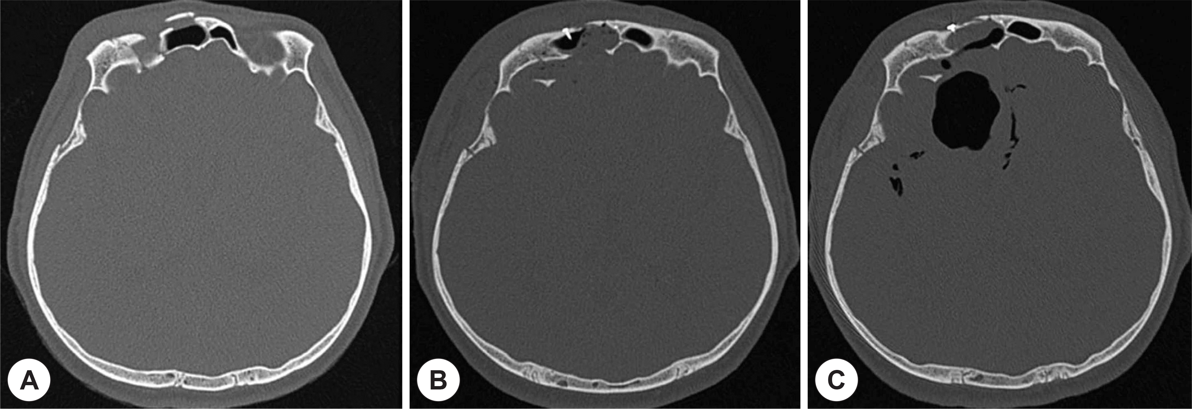
Fig. 2.
At the time of ENT consultation, CT scan showed a clear defect of posterior wall of the frontal sinus and bilateral big-sized pneumocephalus involving frontal lobe and frontal horns of lateral ventricles (A and B). Fortyfive days after revision surgery, CT scan showed a clear boundary between the frontal sinus and cerebrum, and pneumocephalus was replaced by hydrocephalus at right side. However, brain parenchyma was irreversibly changed to encepha-lomalacia.
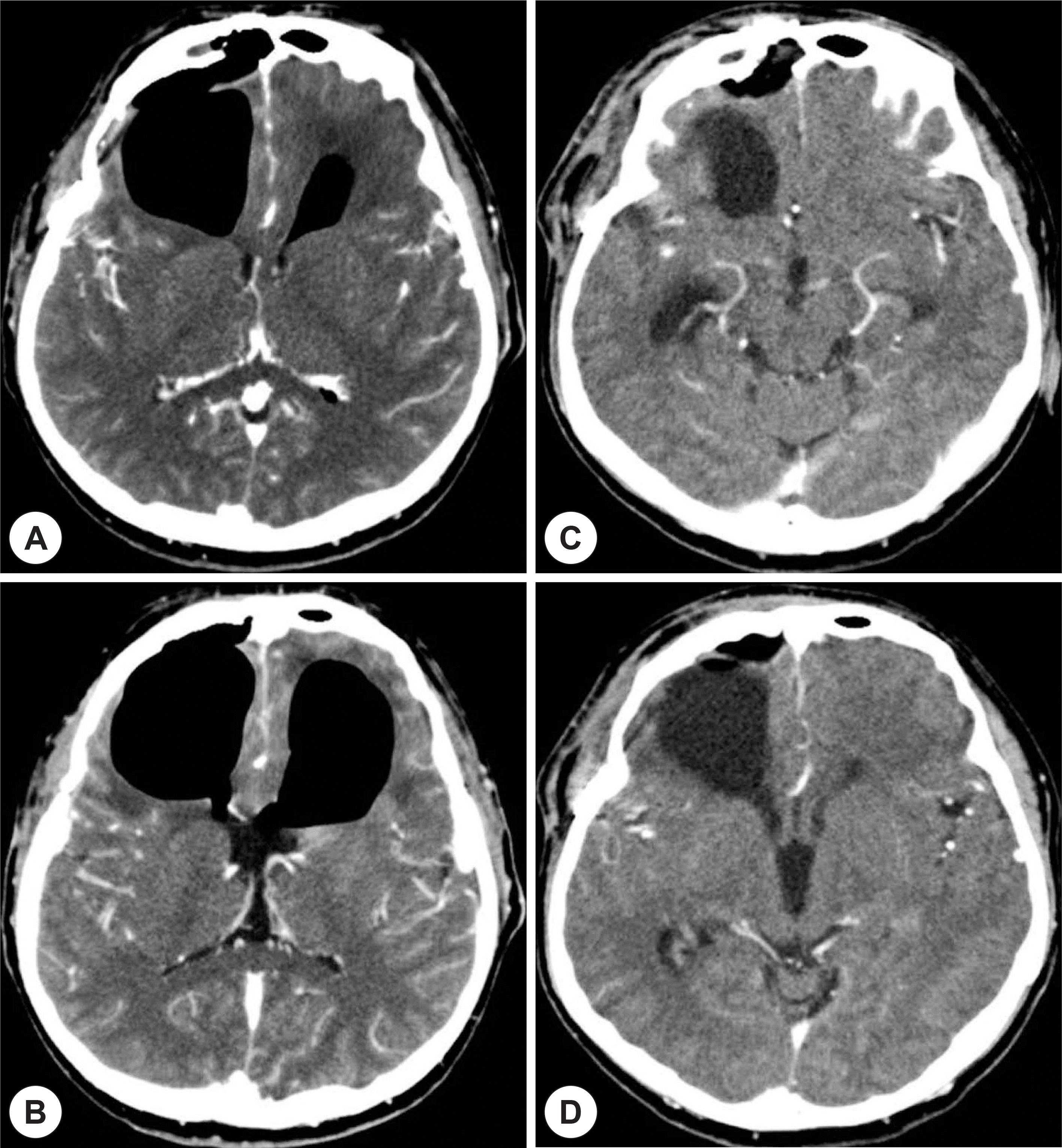
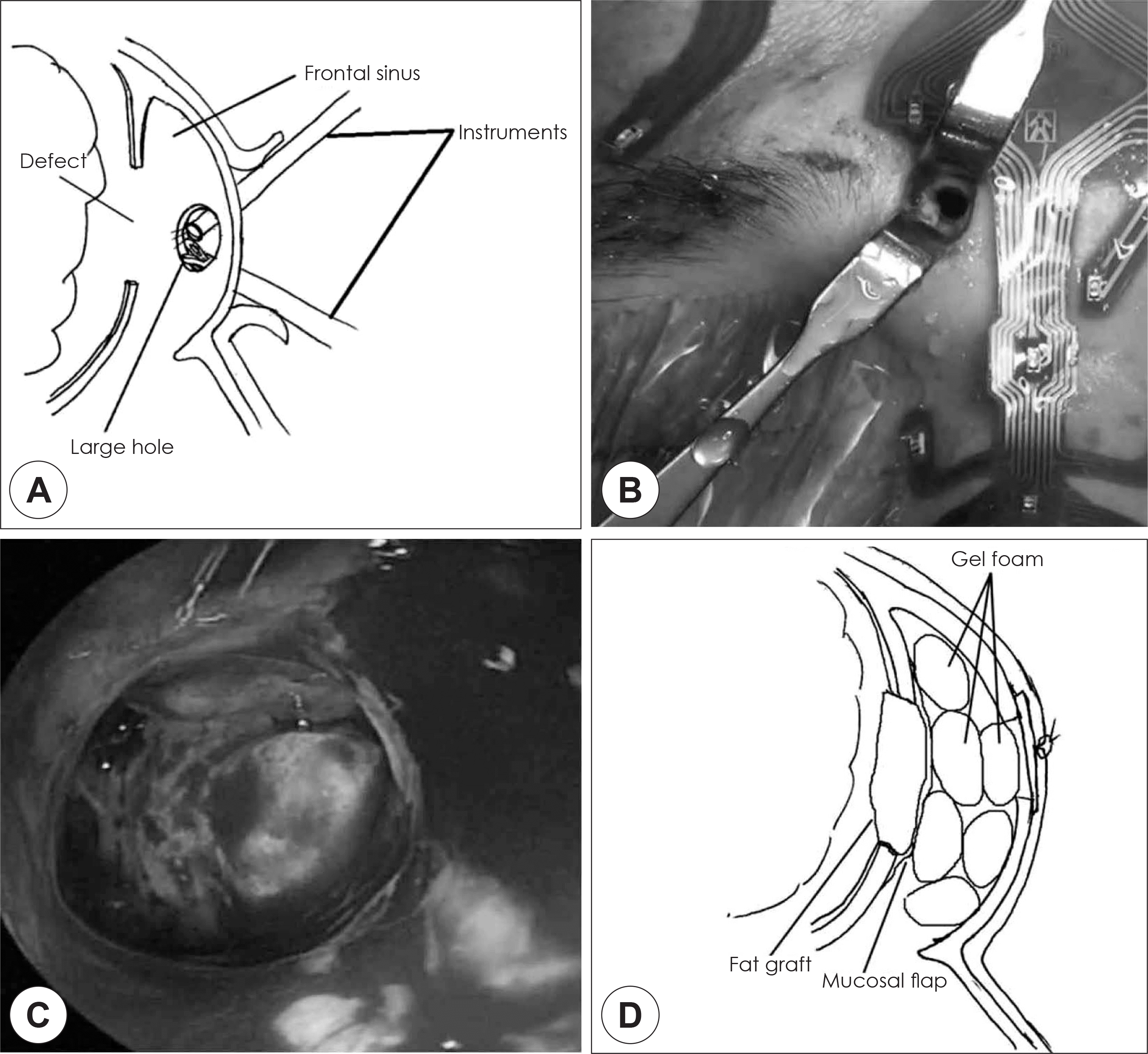
Fig. 3.
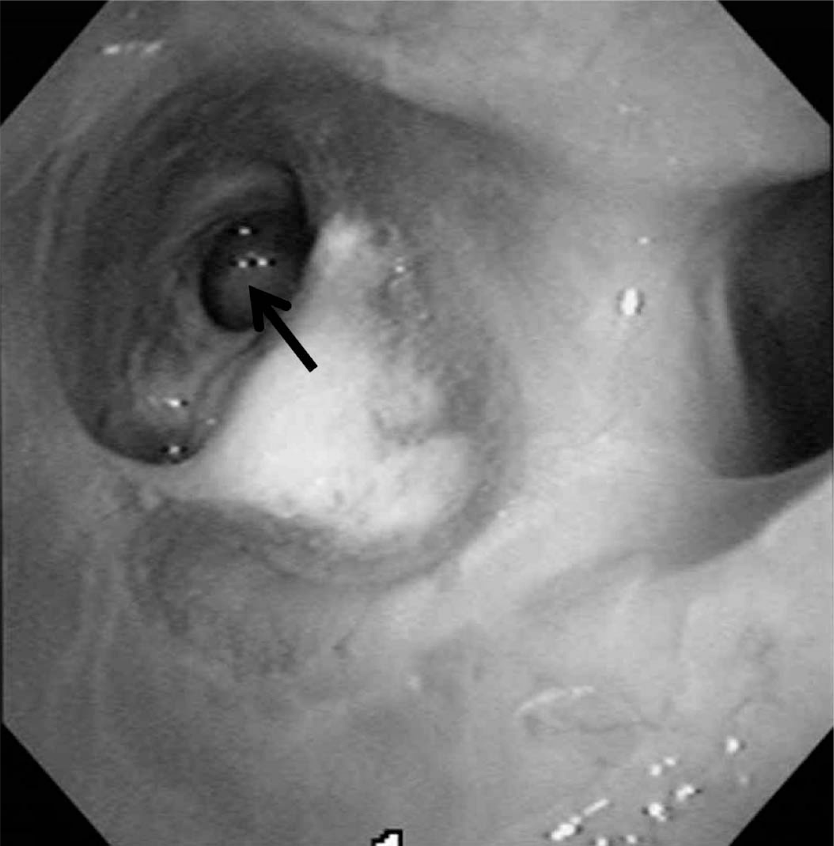
Endoscopic above and below approach for revision reconstructive surgery of posterior wall defect of the frontal sinus. After making one large trephination of the frontal sinus (A and B), loose fat tissues and CSF leaks were noted at the posterior wall (C). After reinforcing with additional fat graft, mucosal graft was overlay on top, and then, frontal sinus was packed with Gelfoam to provide graft stability (D).
 XML Download