

 PDF
PDF ePub
ePub Citation
 Print
Print


INTRODUCTION
Nasotracheal intubation is often preferable to oral intubation in maxillofacial surgery [1]. It provides unrestricted access to the mouth, which facilitates the insertion of instruments. Fiberoptic intubation is a very useful technique for patients with an anticipated difficult airway, such as those with reduced mouth opening due to infection, temporomandibular joint problems, or jaw fracture [23].
With difficult airways, the anatomy is often deviated from normal, and comorbid conditions may lead to complete loss of the airways. Thus, close attention must be devoted to the anesthetic drugs and dosages used to achieve sedation and analgesia for nasal intubation [1]. The ideal sedation technique enables patients to maintain spontaneous ventilation, to be cooperative, and to tolerate passage of a fiberscope to facilitate nasotracheal intubation. It is important for patients undergoing sedated—but awake—fiberoptic intubation to have decreased anxiety, discomfort, and hemodynamic disturbances.
During awake intubation, laryngospasm and coughing in response to intubation can be troublesome. Thus, effective topical airway anesthesia is mandatory for the comfort of the awake patient and subsequent successful airway instrumentation. Profound topical anesthesia of the airway also reduces the need for higher doses of sedatives and analgesics such as midazolam and fentanyl [456].
However, the optimal dose of topical anesthetic drug required before fiberoptic-assisted nasotracheal intubation under sedation remains unclear. We report 2 cases of awake fiberoptic nasotracheal intubation under sedation using a low dose of midazolam and fentanyl in combination with topical lidocaine airway anesthesia.
CASES
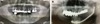
The cases of two patients (1 male, 1 female) who underwent oral maxillofacial surgery under general anesthesia are described. The clinical characteristics of these patients are summarized in Table. 1. Because of physical findings of increased risk for airway compromise, the anesthetic strategy included fiberoptic nasotracheal intubation under sedation and topical anesthesia of the airway (Fig. 1). No premedication was administered to the patients on the day of surgery. After placement of a nasal catheter, oxygen at 6 L/min was initiated. Noninvasive monitoring, including blood pressure and bispectral index (BIS) measurement, electrocardiography, and pulse oximetry, were initiated. After peripheral venous access was obtained, the patients received intravenous fentanyl and midazolam. First, 50 µg of fentanyl was administered two or three times (total 2.2–2.3 µg/kg) at intervals of approximately 2 min. On commencement of 6 L/min of oxygen via mask, 0.5 mg of midazolam was administered one to four times (total 0.02–0.05 mg/kg) at 2-min intervals based on hemodynamics, BIS, and Observer Assessment of Alertness/Sedation Scale (OAA/S) score.
After adequate sedation was achieved with anxiolysis/sedation, as defined by OAA/S level 3 or 4, a wooden applicator stick with a cotton swab, impregnated with 2 mL of 2% lidocaine containing 1:200,000 epinephrine, was inserted through the nasal meatus to verify the nasal passage to the pharyngeal space. A nasopharyngeal airway of either 8.0 mm or 9.5 mm (outside diameter) was inserted to confirm the patency of the nasal passage. After removal of the nasal airway, a nasotracheal tube was then advanced into the pharynx. A fiberoptic scope (Pentax FB-15, HOYA, Tokyo, Japan) was advanced through the nasotracheal tube and 2 mL of 1% lidocaine (20 mg) with 3 mL of air in a 5 mL syringe was first sprayed onto the epiglottis via the working channel of the fiberscope. The fiberscope was then advanced toward the glottis. The patient was instructed to take a deep breath, which lifted the epiglottis anteriorly. The fiberscope was then passed behind the epiglottis to visualize the vocal cords. A second dose of 2 ml of 1% lidocaine (20 mg) was sprayed via the previously described method toward the vocal cords, and the scope was then advanced through the glottis opening into the trachea during spontaneous inspiration. The tip of the fiberscope was then positioned immediately proximal to the carina. A third dose of 1% lidocaine (20 mg [2 mL]) was sprayed into the trachea using the previous method into the trachea. If visualization or orientation was lost at any time, the fiberscope was withdrawn until an appropriate landmark was identified. Finally, with profound topical anesthesia of the entire airway, the nasotracheal tube was then smoothly advanced into its correct final position as confirmed by the presence of positive end-tidal carbon dioxide and auscultation of bilateral breath sounds. Rocuronium (0.6 mg/kg) and propofol (1–1.5 mg/kg) were then intravenously administered for induction of general anesthesia.
Anesthesia was maintained using inhalational anesthetic agents, air and oxygen, in addition to an intravenous infusion of remifentanil. The surgeries proceeded uneventfully and both patients were fully awake after anesthesia for extubation with no complications.
DISCUSSION
Management of the difficult airway is a challenge to anesthesiologists, and may lead to life-threatening complications. It has been reported that 1–18% of patients have a difficult airway [5]. We presented two cases of awake fiberoptic nasotracheal intubation, in which topical anesthetic (lidocaine 60 mg) was sprayed into the airway, combined with a low dose of midazolam (0.02–0.05 mg/kg) and fentanyl (2.2–2.3 µg/kg).
In these patients, we anticipated difficult ventilation or intubation because of possible airway obstruction. It was essential that the sedatives and opioids be administrated slowly and carefully, and titrated in low dosages to prevent any airway complications and to suppress stimulation of the airway with sufficient topical anesthetic.
We previously reported an in vitro study describing the most effective method for spreading the anesthetic spray via the working channel of a fiberscope to mimic the spraying of topical anesthesia in the airway [7]. Spraying involving a fiberoptic scope and a 5 ml syringe containing 2 ml of liquid and 3 ml of air was determined to be most effective method for topical anesthesia of the epiglottis, vocal cord, and trachea. As a topical anesthetic, lidocaine is most frequently used because it has a good safety and efficacy profile, and major adverse effects manifest only if the total dose of injected lidocaine exceeds 5–7 mg/kg [46]. In clinical practice, we have successfully performed sufficient airway anesthesia with a total dose of lidocaine of 60 mg. Therefore, we could perform awake nasotracheal intubation using lower doses of midazolam and fentanyl compared with previous reports [278].
Midazolam is a classic sedative, and is a short-acting benzodiazepine derivative. It has anxiolytic and sedative properties [678]. While midazolam is believed to cause minimal hemodynamic effects, it does have the potential to cause loss of airway reflexes. Thus, careful titration to the desired sedative level is necessary for achieving safe conscious sedation.
Awake intubation could be associated with painful stimulation during passage of the tracheal tube through the nose and the larynx. Opioids are potent analgesics and can help attenuate coughing and hemodynamic changes. Fentanyl is a short-acting opioid that quickly provides analgesia, mild sedation and analgesia with hemodynamic stability, all of which are beneficial for awake intubation; however, there is a risk for respiratory depression [78]. Because the use of fentanyl alone fails to provide sufficient sedation, midazolam is added in clinical practice to improve the quality of sedation. This step-by-step administration of low-dose fentanyl and midazolam, in combination with topical anesthesia, is a useful and safe method for sedated fiberoptic nasotracheal intubation.
In previous studies, it was reported that midazolam (0.05–2.7 mg/kg), propofol (1.0–2.7 mg/kg) and dexmedetomidine (0.5–1.0 µg/kg) as the primary sedative agent generally describe in combination with opioid such as fentanyl (1.0–3.0 µg/kg), sufentanil (the target plasma concentration 0.3 ng/kg) or remifentanil (0.25–0.75 µg/kg) [56789]. However, appropriate levels of sedation for safe awake intubation are very difficult to standardize because the required combination of anxiolysis and analgesia varies widely from case to case [610].

 XML Download