

 PDF
PDF ePub
ePub Citation
 Print
Print


Patients undergoing esophagectomy are at a high risk of pulmonary aspiration before and after surgery due to the loss or alteration of the lower esophageal sphincter [1]. Although no comprehensive study of such a high-risk group has been performed, various methods have been recommended to prevent pulmonary aspiration [23]. Nevertheless, it is not possible to completely prevent pulmonary aspiration during general anesthesia. After pulmonary aspiration, clinical syndromes such as pneumonia and pneumonitis can develop. Due to their high morbidity and mortality, these conditions are clinically important [4].
We would like to present a video clip showing pulmonary aspiration during induction of anesthesia in a patient who is status post esophagectomy, as the case is valuable to elucidate the risk of pulmonary aspiration in patients who have undergone esophageal surgery.
Written consent for publication has been obtained from the patient.
CASE REPORT
A 74-year-old man with mild chronic obstructive pulmonary disease, hepatitis B virus–positive hepatocellular carcinoma, and liver cirrhosis, with suspected hypopharyngeal carcinoma, was scheduled to undergo laser-assisted partial hypopharyngectomy. He had a history of Ivor-Lewis esophagectomy for esophageal cancer performed at 59 years of age. Video-assisted mediastinoscopic lymphadenectomy for lymph node biopsy and neck dissection was planned. No premedication was given, and the fasting period was over 13 hours. The patient entered the operating room with a nasogastric tube inserted. Standard monitoring devices were connected, and general anesthesia induction was planned to be performed by rapid sequence induction.
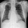
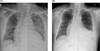
Denitrogenation with 100% oxygen was performed for 3 minutes, followed by intravenous injection of 2 mg/kg of propofol, 1.5 mg/kg of succinylcholine, and 40 mg of lidocaine. During Mallinckrodt laser-tube intubation using a video laryngoscope (McGrath™, Aircraft Medical, United Kingdom), reflux of yellowish-green gastric fluid into the oral cavity was observed (Video 1). As the laryngoscopic view Cormack and Lehane grade was 1, endotracheal intubation was not considered to be difficult, and we decided to suction after intubation. However, intubation was not successful at the first attempt. As the endotracheal tube did not pass beyond the vocal cords, further manipulation of the endotracheal tube near the vocal cord was necessary. The refluxed fluid aspirated into the lungs while manipulating the tube. After suctioning the oral cavity, without removing the tube the video laryngoscope was used to confirm the success of the intubation and the tube was ballooned. Immediately after the intubation, fluid of similar color was aspirated on endotracheal suctioning, and 50 to 100 mL of gastric fluid was drained from the nasogastric tube. During induction, the nasogastric tube was naturally drained. Since the vital signs including the peripheral capillary oxygen saturation (SpO2), as measured by pulse oximetry, were stable, the operation was performed as planned. During laser partial hypopharyngectomy, the fraction of inspired oxygen (FiO2) was maintained at 0.35 to 0.40 and the SpO2 at 95% to 96%. Blood gas analysis showed pH of 7.48, PCO2 of 35 mm Hg, and PO2 of 55 mmHg at FiO2 0.4. After 20 minutes, blood gas analysis reperformed at FiO2 0.5 showed a pH of 7.43, PCO2 of 39 mmHg, and PO2 of 81 mmHg. No fluid or particular reflux was observed in the airway using the laryngoscope when the anode tube was replaced an hour later. During the 4 hours of operation, the vital signs were stable, and the SpO2 remained at 100% at FiO2 0.5. Blood gas analysis performed just before the end of surgery showed pH of 7.41, PCO2 of 41 mm Hg, and PO2 of 119 mm Hg at FiO2 0.5. After the operation, the patient was extubated and transferred to the post-anesthetic care unit without dyspnea. Postoperative chest radiography showed diffuse haziness in the left upper lobe compared with the preoperative image (Figure 1, Figure 2A). The SpO2 decreased to 88% in room air in the post-anesthetic care unit, and the patient was transferred to the ward with a simple mask delivering O2 at 6 L/min, following which the SpO2 was well maintained. The patient received conservative care without antibiotic therapy for the pulmonary aspiration. The patient had mild chronic obstructive pulmonary disease and was diagnosed with cancer-associated segmental pulmonary thromboembolism after surgery. The SpO2 was maintained with a simple mask delivering O2 at 3 L/min until the day before discharge. The patient was discharged on postoperative day 12 without complications related to pulmonary aspiration (Figure 2B).
DISCUSSION
The incidence of perioperative pulmonary aspiration in adults has been reported to be approximately 1 in 4000 to 8000 with a morbidity of 1 in 16,573 and mortality of 1 in 99,441 [56]. A retrospective study from the Mayo Clinic, including cases from between the years 1985 and 1991, reported that the incidence of perioperative pulmonary aspiration after general anesthesia for elective surgery was 1 in 3886 cases [7]. Since the incidence of perioperative pulmonary aspiration is low, precautions to prevent pulmonary aspiration tend to be overlooked. In esophagectomy, the lower esophageal sphincter is excised, the stomach vagal innervation is lost, and a new gastroesophageal anastomosis is made in the midesophagus or cervical esophagus [8]. As in the postesophagectomy state, the flaccid stomach is drained only by gravity, is in the thoracic cavity, and is without the sphincter, the patient becomes vulnerable to aspiration [8]. In such patients, even if there is no oral intake of any food and rapid sequence induction is performed, the possibility of aspiration cannot be completely eliminated. Nonetheless, additional strategies for stomach decompression such as nasogastric tube insertion, head-up position induction, rapid sequence induction with cricoid pressure, and awake tracheal intubation can be considered [8]. Inserting the nasogastric tube does involve risk of anastomotic disruption in the post-esophagectomy state, however, it is possible to safely insert the tube 2 to 3 weeks after the operation [8]. Additionally, the use of premedication such as pro-kinetics, and H2-blockers might assist in preventing pulmonary aspiration [9].
Whether or not to use rapid sequence induction could not be clearly determined through literature review [10], however, patients with moderate to high risk of aspiration should be considered for rapid sequence induction. It has been reported that cricoid pressure does not definitively reduce aspiration [1112], especially in patients with cervical anastomosis where the esophagus is likely to be lateral to the cricoid cartilage, making cricoid pressure ineffective [8]. In our case, cricoid pressure was not applied during induction of anesthesia; even if it was performed, it might not have prevented aspiration completely.
Patients who are status post esophagectomy are at risk of aspiration even if they have never aspirated during induction of anesthesia in the past. In the current case, Levin-tube aspiration before induction and induction at head-up position might have helped reduce the possibility of aspiration. As succinylcholine can increase intraabdominal pressure [13], a high dose of rocuronium might be considered as an alternative. In this case, the risk of pulmonary aspiration was overlooked due to its relatively low incidence. We regret that more meticulous precautions were not taken to prevent pulmonary aspiration during the induction of anesthesia.
Patients who undergo esophago-thoracic procedures are at a higher risk of perioperative pulmonary aspiration [3]. There is still little evidence supporting the role of various precautionary methods of preventing aspiration in this high-risk group. However, as pulmonary aspiration is closely related to postoperative mortality and pulmonary morbidity, anesthesiologists should always pay particular attention to patients at risk of pulmonary aspiration.

 XML Download