

 PDF
PDF ePub
ePub Citation
 Print
Print


The goal of sedation or general anesthesia for dental treatment is to provide high-quality care for patients who have difficulty tolerating typical methods [1]. When normal dental treatment is impossible in a patient who exhibits a negative behavioral reaction from anxiety or fear or has involuntary muscle movement, forceful methods, such as using physical restraint, have often been used [2]. However, physical restraint can impose tremendous stress on the patient, the patient's guardian, and the medical staff, and may also have a negative effect on future treatment [3]. Therefore, sedation, and even general anesthesia in some cases, may be considered for disabled patients who are difficult to treat. However, the choice of method for general anesthesia or sedation may vary depending on the patient's degree of cooperation, type of procedure, invasiveness, treatment time, equipment available in the treatment room, and the availability and capability of the medical staff.
In general, deep sedation can be performed for short procedures that are not highly invasive, in which the patient feels virtually no pain with local anesthesia alone [4]. In such cases, supplying oxygen during the procedure is absolutely necessary, and all available efforts must be made to monitor the patient to prevent airway obstruction or respiratory distress, including the use of carbon dioxide tension monitoring, while appropriate skills are also required to maintain the airway [5]. One of the drugs commonly used to perform deep sedation is propofol, which is known to require a lower dose in disabled patients than in normal adults [6]. Therefore, performing anesthesia depth monitoring using EEG, as with a Bispectral Index (BIS) monitor, can prevent complications associated with overdose and can be helpful in maintaining proper sedation depth [7]. ADMS™ (Anesthetic Depth Monitor for Sedation, Unimedics CO., LTD., Seoul, Korea) is a newly developed anesthetic depth measurement monitor, which displays the patient's arousal state as a score of 0-99 points. Scores of 60-80 indicate sedation state.
In the present case, deep sedation for dental treatment was performed for a total of 30 min on a 27-year-old female patient with intellectual disability and severe dental phobia. For sedation depth monitoring, BIS and the ADMS device were used; the effectiveness of ADMS in sedation depth monitoring is reported here, along with a literature review.
CASE
A 27-year-old female patient with intellectual disability visited the Seoul National University Dental Hospital Special Care Clinic. The patient exhibited several missing teeth (#14, #24, #46, and #47) and malocclusion (anterior open bite). Overall gingival condition was healthy in spite of insufficient oral hygiene maintenance. The patient and her parents wanted to improve her masticatory function by dental restoration.
The first visit for dental treatment was performed under general anesthesia. For restoration of a missing tooth (#14), a three-unit porcelain-fused-to-gold fixed prosthesis was planned. Teeth #13 and #15 were prepared and an impression was obtained. At the second visit, deep sedation was performed. The fixed prosthesis was cemented with resin-modified glass ionomer luting cement (FujiCem, GC, Tokyo, Japan).
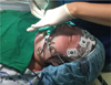
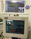
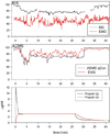
The patient had a history of Tetralogy of Fallot corrective surgery at the age of 1 year, which required administration of antibiotics during dental treatment for prevention of endocarditis, but there were no other specific findings in the pre-anesthesia examination. Although no cervical extension or abnormal mouth opening was found on the airway examination, the patient did show persistent involuntary movements. On the day prior to sedation being performed, the patient was required to fast for 8 h and a 22-gauge intravenous (IV) line was secured. After an IV injection of 30 mg lidocaine, IV propofol was administered using a target concentration infusion (TCI) device (Fresenius Orchestra, Fresenius Kabi, Germany), with effect-site target concentration set to 3 µg/ml. After the patient became unconscious, a pulse oximeter, an electrocardiograph, and a blood pressure monitor were attached to the patient, while oxygen was supplied at a rate of 5 L/min through a nasal cannula, and end-tidal carbon dioxide tension (EtCO2) and respiratory pattern were monitored using the carbon dioxide suction tube on the nasal cannula (Fig. 1). After a few minutes, the patient reached a state of deep sedation, at which time, respiration stabilized to a satisfactory level, oxygen saturation level was stable, and a state of deep sedation in which the patient would not move in response to stimuli from dental treatment was maintained. Throughout the treatment, respiratory rate of 10 breaths/min and EtCO2 of 40-50 mmHg were maintained, while the vital signs were maintained at blood pressure 100-90/60-50 mmHg, heart rate 70-80 beats/min, and body temperature 36.5℃. The BIS scores and qCON scores of ADMS were maintained at around 70 (Fig. 2). Although the overall electromyographic (EMG) value was high, acceptable ADMS values were shown. When the patient regained consciousness, ADMS qCON values increased immediately to > 90 (Fig. 3). The total time required in performing sedation was approximately 30 min; after 30 min in the recovery room, the patient was discharged after confirming that her consciousness and movements had returned to normal. A parent was contacted by telephone after discharge, and no specific complications were reported.
DISCUSSION
Dental treatment of disabled patients has become increasingly common in Korea because of recent economic growth and increased interest among dentists, with many dental hospitals performing treatment with general anesthesia and sedation. Sedation must be performed by medical personnel capable of maintaining the airway and performing emergency treatment, and the patient and guardian must be provided sufficient explanation of the risks associated with sedation; prior written consent must be obtained. Continued patient monitoring must be performed during sedation, and it is also necessary to have the infrastructure necessary to allow the patient to recover safely until discharge [8].
The BIS monitor simplifies the measurement of the degree of sedation and hypnosis from anesthetics or sedatives and is displayed as a number [9]. The BIS value displays the arousal and unconscious state as a score from 0 to 100 by using a specific algorithm to analyze and process the degree of changes in electrical activities of the cerebral cortex in response to anesthesia. Using BIS monitoring can prevent arousal during anesthesia, as well as formation of implicit or explicit memory. Moreover, controlling the drug dose according to BIS can help maintain proper anesthesia depth [9,10]. The US Food and Drug Administration (FDA) approved the use of BIS monitors in 1996, and stated that BIS monitoring during anesthesia can have positive effects, such as helping to expedite arousal after anesthesia and reduce the duration of recovery room stay, along with reducing the total cost of anesthesia [11].
Using BIS during sedation is known to enable earlier discharge [12]. However, it is impossible to use BIS values calculated by processed electroencephalogram (EEG) monitoring to assess the state of sedation, since there are large differences between individuals [13]. Nonetheless, it can be helpful in assessing whether the patient is conscious or unconscious under deep sedation. It is also known that BIS monitoring during dental treatment under IV sedation can be helpful when using midazolam [14].
ADMS™ is a sedation depth-monitoring device that analyzes EEG waveforms to calculate the "Hypnotic Effect" with "Prediction Probability," and displays the result as a value between 0-99 to assist patient monitoring during a procedure. This device was developed by Unimedics CO., LTD of Korea and was introduced to the market after receiving approval from the Korea Ministry of Food and Drug Safety in 2015.
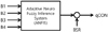
ADMS™ measures the EEG with three electrodes attached to the forehead and shows the sedation depth as qCON value. The algorithm is based on the combination of the energy of four frequency ratios, which generates an index, and is then offset by the value of the EEG suppression rate called the burst suppression ratio (BSR) (Fig. 4). The EEG sampling range is 0.5-45 Hz and the EMG sampling range is 30-75 Hz; the anesthesia depth is calculated using an adaptive neuro fuzzy inference system by power spectral analysis [15].
The BIS was obtained from an algorithm that was derived from numerous EEG analyses by administration of various sedatives and anesthetics to volunteers and patients with no brain diseases [16]. While the BIS algorithm has been updated several times for improved performance, there may still be parts that have not been improved [17]. In all currently available anesthesia depth monitors, a certain amount of delay in calculating and displaying the changes in anesthesia depth is unavoidable. The time it takes to update a BIS value can be between 14 and 155 s [18], meaning the value represents a prior state. ADMS™ has a time delay of 10 s, and calculates and displays the value with a data smoothing ratio of 15 s.
BIS values may be affected by EMG activities and muscle relaxant administration. When EMG activities increase, BIS values increase; when activities decrease with muscle relaxant administration, BIS values decrease [19]. The frequency range of EEG signals used, based on the algorithm for BIS measurement, is 0.5-30 Hz, and the frequency range of the EMG is 30-300 Hz; however, when a low-frequency EMG signal is falsely recognized as a BIS signal, it can be misinterpreted as a high-frequency EEG signal [20]. Since improvements to the BIS algorithm have been made to eliminate such errors, a correlation between the false increase in EMG and BIS no longer exists. As muscle relaxants are not used during sedation and the patient can move during dental treatment, it is important to effectively treat the noise from the EMG signal; with the use of the ADMS, acceptable values were shown despite high EMG values (Fig. 3).
There are several reports of BIS values being inconsistent with the clinical hypnotic state due to brain dysfunction or limitations in the monitor. It may be that because the BIS algorithm was created for individuals with typical EEGs, abnormal EEG patterns may affect BIS monitoring, but a recent report indicated high correlation between patients with neurological disability and sedative effect [21]. However, there are other reports of BIS values being abnormal in patients with neurological disability, as well as reports of BIS values being low in patients with Alzheimer's dementia or multi-infract dementia, even when measured in an awake state [22]; pediatric patients with cerebral palsy showed lower BIS values than normal pediatric patients when anesthetized with sevoflurane [23]. Another study reported that pediatric patients with intellectual disability showed lower BIS values than did normal children during anesthesia maintenance and arousal [24]. When measured in a patient in a persistent vegetative state who had undergone general anesthesia for a dental procedure, pre-anesthesia BIS had decreased to 74-85 points due to neurological injury, and the values decreased even further following sevoflurane administration [25].
In conclusion, successful dental treatment under sedation was possible in the present case using IV propofol and ADMS to monitor the state of sedation. Use of ADMS for dental treatment of disabled patients can provide a level of sedation depth monitoring comparable to that of BIS.
 XML Download