Journal List > Brain Tumor Res Treat > v.5(2) > 1059722

Duong, Nguyen, Sheppard, Ong, Chung, Nagasawa, and Yang: Genomic and Molecular Characterization of Brain Tumors in Asian and Non-Asian Patients of Los Angeles: A Single Institution Analysis

Abstract

Background

Worldwide, approximately 2% of new cancers are of the brain. Five-year survival rates among brain cancer patients have been reported as a little over a third. Differences in clinical outcomes between brain tumor patients of different races remain poorly understood.

Methods

A retrospective chart review was performed on brain tumor resection patients≥18 years old. Demographics, treatment variables, and survival outcomes were collected. Primary outcomes were length of stay, recurrence rate, progression-free survival (PFS), and overall survival (OS).

Results

A total of 452 patients were included in analysis. Females and males had nearly a 1:1 ratio (n=242 and n=220, respectively). Mean age was 54.8 years (SD: 14.5 range: 18–90). Females composed 69% (n=48) of Asian patients; males constituted 31% (n=22). Mean age of the Asian patients was 55.9 years (SD: 14.6 range: 26–89). Asian-only cohort tumor pathologies included glioblastoma (GBM) (n=14), high-grade glioma (n=7), low-grade glioma (n=4), meningioma (n=38), and metastases (n=7). Of the 185 meningioma patients, non-Asian patients comprised 79% of the group (n=146). Of the 65 GBM patients in total, non-Asian patients made up 89% of the GBM cohort (n=58). There were no statistically significant differences between these groups of both cohorts in recurrence (p=0.1580 and p=0.6294, respectively), PFS (p=0.9662 and p=0.4048, respectively), or OS (p=0.3711 and p=0.8183, respectively).

Conclusion

Studies evaluating the survival between patients of different racial backgrounds against several tumor varieties are rare. Patients of certain racial backgrounds may need additional consideration when being attended to despite the same mutational composition as their counterparts. Repeated studies using national databases may yield more conclusive results.

INTRODUCTION

Worldwide, less than 2% of new cancer diagnoses are of the brain and central nervous system [12]. Five-year survival rates among brain cancer patients have been reported as a little over a third, a mortality rate twenty-five times that of its incidence [2]. Comparatively, breast cancer's 5-year survival rates have been reported as over 80% despite having a higher incidence rate than brain cancer [23].
Brain tumor epidemiology in specific races has been documented, and studies into the mechanisms of mutation by race and pathology are well-established [145678910111213141516171819202122232425262728293031]. However, differences in clinical outcomes between brain tumor patients of different races are still being elucidated [253233]. A prior study showed a higher probability of worse outcomes in Asian/Pacific Islanders than in their white counterparts [25]. While these reports evaluated outcomes in a single tumor pathology, studies evaluating the survival between patients of different racial backgrounds against several tumor types are rare.
To optimize treatment for patients with brain tumors, racial differences in progression-free survival (PFS) and overall survival (OS) should be considered. Patients of certain racial backgrounds may need additional consideration despite identical mutational composition as their counterparts because of different underlying genotypic variations [33]. Compared to the white population, the Asian cohort had a higher incidence of isocitrate dehydrogenase (IDH), suggesting additional consideration for glioblastoma (GBM) treatment in Asian patients [33]. The majority of existing studies focus on Asian populations, who compose 5.6% (n=17,320,856) of the total United States population [525343536]. At our institution, Asians comprise over 10% of the brain tumor population, nearly double the national percentage [37]. To further explore differences within this population group, we assessed differences in genetic markers and survival outcomes in patients of either Asian or non-Asian descent diagnosed with brain tumors.

MATERIALS AND METHODS

Inclusion criteria

A retrospective chart review was performed. Patients who had undergone brain tumor resection from March 2013 (year in which the electronic chart system was established at our institution) through January 2017 were included. Patients with “unknown” race were excluded. This study was approved by our center's Medical Institutional Review Board.

Data collection

Patient demographics (age, sex, race, and ethnicity), treatment variables, and survival outcomes were collected. The entire population was stratified by race, with the subgroups as “white,” “black,” “Hispanic or Latino,” and “Asian.” Patients in the Asian category were separately grouped by ethnicity. Categorization was chosen based on examples from the most recent United States Census Bureau data [3637]. Treatment variables included operation duration, extent of resection (gathered from operative report), chemotherapy, and radiation modality and dosage. Primary outcomes were length of stay, discharge disposition, recurrence rate, PFS, and OS.
Mutational statuses and diagnoses were gathered from pathology reports. For patients diagnosed with GBM specifically, statuses were gathered for IDH, phosphate and tensin homologue (PTEN), tumor protein p53 (p53), chromosome 1p and chromosome 19q (1p/19q), epidermal growth factor receptor (EGFR), type III epidermal growth factor receptor (EGFRvIII), overall antigen Ki-67 (Ki67) percentages, and methylation mutational statuses. World Health Organization (WHO) grade, glial fibrillary acidic protein (GFAP), epithelial membrane antigen (EMA), and S100 mutational statuses were accumulated for meningioma patients. Patients were then organized by tumor histological subtype.

Statistical analysis

Wilcoxon signed-rank and Pearson's tests were performed. Correlation analyses were performed to identify relationships between race and all variables of interest for GBM and meningioma patients. Statistical analyses were performed using SAS, version 9.3 (SAS Institute, Cary, NC, USA). A p-value less than 0.05 was considered statistically significant.

RESULTS

General population

A total of 452 patients were included in the study. Demographics of both cohorts are summarized in Table 1. Whites comprised 71% of the cohort (n=323), Asian 16% (n=70), Hispanic or Latino 9% (n=40), black 4% (n=18), and unspecified 0.2% (n=1). Pathologies stratified by race are summarized in Table 2. The Asian-only population consisted of: Filipino 17% (n=12), Chinese 14% (n=10), Korean 10% (n=7), Vietnamese 4% (n=3), Pacific Islander 3% (n=2), Japanese 3% (n=2), Burmese 1% (n=1), and unspecified 47% (n=33). Tumor pathologies in the Asian-only cohorts included are summarized in Table 3.

Glioblastoma

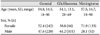
We identified 65 GBM patients in total across all races. Of patients diagnosed with GBM, non-Asian patients comprised 89% of the cohort (n=58) with the remaining 11% (n=7) being Asian patients. There were no statistically significant differences between the groups in PTEN loss (p=0.705), p53 (p=0.086), 1p/19q loss (p=0.282), EGFR amplification (p=0.709), EGFRvIII (p=0.118), overall Ki67 (p=0.695), O6-methylguanine methyltransferase (MGMT) gene promoter methylation status (p=0.090), or other gathered variables (Table 4).

Meningioma

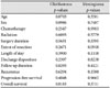
A total of 185 meningioma patients were included. Non-Asian patients comprised 79% of the group (n=146) while Asian patients composed the last 21% of meningioma patients (n=39). There were no statistically significant differences between these groups in WHO grade (p=0.643), histological subtype (p=0.783), GFAP (p=0.197), EMA (p=0.057), S100 (p=0.549), Ki67 (p=0.592), or other collected variables (Table 4).

DISCUSSION

Differences in races, specifically Asians in contrast to other groups, in respect to brain tumor epidemiology has already been well studied [145678910111213141516171819202122232425262728293031]. Due to the considerable Asian patient population at our institution, we endeavored to compare races within the cohort. We performed a retrospective chart analysis of patients, who underwent brain tumor resection at our institution. Race and all collected variables were tested in both GBM and meningioma patients. Survival outcomes were measured in those two cohorts against our collected variables.
Though several studies have reported significant proclivities between tumor pathologies and certain races, there were no statistically significant differences between race and tumor pathologies in our population cohort [1456789101112131415161718192021222324252627282930313839404142]. Maile et al. [8] studied 35,663 patients in England and found that the general white population had the highest occurrence of GBM among other races. They also reported Pakistanis had two times the incidence of brain neoplasms compared to that of Bangladeshis (p<0.001) [8]. However Brown et al. [4] found that in a cohort of 2,096 pediatric patients, age-specific incidence of tumor pathology was not statistically different between races. In that report, even when stratified by age, there were no significant differences or correlations between race and tumor pathology [4].
In our patients with GBM, there was no significant difference between Asians and non-Asians for any of our variables. PFS and OS between the two groups were not statistically distinct from one another. There are several studies that document the mutational statuses of GBM, with some demonstrating disparities in PFS and OS between races [232526283233354344454647484950515253545556575859606162636465666768]. Dai et al. [33] performed a meta-analysis of GBM patients which included studies from Europe, North America, and Asia. In their study of 3,464 patients, IDH mutation associated mortality rate decreased nearly two times more in European populations than it did in Asian populations [33]. Additional multivariate analysis between race, treatment modalities, and mutational variation is one approach that warrants further study to understand PFS and OS. A study by Wu et al. [28] examined MGMT methylation in GBM patients of different ethnic groups and found no statistically significant differences between these groups with mutational statuses and OS. Because MGMT methylation results in higher sensitivity to chemotherapeutic agents, the lack of differences between methylation scores of various ethnic groups should contribute to similar PFS or OS among races [69707172]. This is consistent with our analysis of races in the GBM population. Emerging racial disparities already published in GBM patients could be used in large metaanalysis studies to continue this line of research.
In addition, we examined correlates between race and other prognostic markers in patients with meningioma. While we found no significant correlations, several studies show a proclivity of meningiomas with certain ethnicities [5737475]. Das et al. [5] published an article on 48,001 patients from a Singaporean hospital and found that Chinese patients had the highest rate of meningioma occurrence. According to their study, over 90% of their malignant meningiomas were expressed in Chinese patients [5]. Despite the ethnic propensity for meningioma they found, PFS and OS in patients are comparable between ethnicities [5]. Analyses of mutational statuses and their effects as confounding factors in the survival outcomes of these patients propose a valid area of research that should be explored more in depth. This model of analysis has already been well-applied to GBM research.
Our study was a retrospective review, which incurs several limitations. Data was gathered in a limited setting; therefore, the tested variables are not as homogenous as a prospective study. Patients with unspecified race or unknown mutational statuses, although still included here, could not be further distinguished. Additionally, we reported from a single institution, while several similar studies acquired their cohort from a large, national database [67813253234386176]. Therefore, our sample population may be limited in both size and location. Socio-economic status of patients has been acknowledged to affect rates of brain tumor occurrence and may be a confounding variable [132021222832427677787980818283]. While our analysis only used univariate statistics, future studies analyzing confounding factors, such as age, in survival rates are the next approaches in identifying racial disparities.
Our single institution study facilitated the acquisition of socio-economic status, which in our future studies could elucidate the role of socio-economic status on OS and PFS. Additional multivariate analysis includes investigating the role of mutational statuses as confounding factors in race and survival outcome correlations. Racial disparities have been documented in a few studies similar to the one performed by the authors. Continued evaluation of those disparities is necessary to assure standardized treatment across all races.
In conclusion, while there are few studies assessing survival outcomes of different racial cohorts with various tumor pathologies, patients with the same mutational configuration may not have the same treatment response between varying racial backgrounds. Additional studies using larger cohorts, such as the Surveillance, Epidemiology, and End Results database, are necessary for more decisive results.

Figures and Tables

Table 1

Demographics of all populations

btrt-5-64-i001
General Glioblastoma Meningioma
Age (years, SD, range) 54.8, 14.5, 18–90 54.1, 13.1, 28–69 57.5, 14.7, 18–90
Sex, % (n)
 Female 52.4 (242) 38.8 (64) 71.9 (133)
 Male 47.6 (220) 61.2 (101) 28.1 (52)

SD, standard deviation

Table 2

Summary of pathologies by race

btrt-5-64-i002
Pathology Race
White or Caucasian Black or African American Hispanic or Latino Asian
GBM/gliosarcoma 30.5 (141) 0.2 (1) 3.1 (14) 3.1 (14)
Low-grade glioma 5.8 (27) 0 (0) 0 (0) 0.9 (4)
High-grade glioma 8.2 (38) 0.2 (1) 0 (0) 1.5 (7)
Meningioma 22.9 (106) 3.1 (14) 5.6 (26) 8.4 (38)
Metastases 2.4 (11) 0.4 (2) 0 (0) 1.5 (7)

All values given in percentage and (n). GBM, glioblastoma

Table 3

Summary of pathologies by Asian ethnicity

btrt-5-64-i003
Pathology Race
Pacific Islander Burmese Korean Filipino Vietnamese Chinese Japanese Unspecified
GBM/gliosarcoma 0 (0) 0 (0) 2.86 (2) 2.86 (2) 1.43 (1) 2.86 (2) 0 (0) 10.00 (7)
Low-grade glioma 1.43 (1) 0 (0) 0 (0) 0 (0) 0 (0) 0 (0) 0 (0) 4.29 (3)
High-grade glioma 1.43 (1) 0 (0) 0 (0) 1.43 (1) 0 (0) 0 (0) 0 (0) 7.14 (5)
Meningioma 0 (0) 1.43 (1) 7.14 (5) 11.83 (8) 1.43 (1) 10.00 (7) 2.86 (2) 20.00 (14)
Metastases 0 (0) 0 (0) 0 (0) 1.43 (1) 1.43 (1) 1.43 (1) 0 (0) 5.71 (4)

All values given in percentage and (n). GBM, glioblastoma

Table 4

Elements investigated in pathology cohorts

btrt-5-64-i004
Glioblastoma
p-values
Meningioma
p-values
Age 0.0703 0.3581
Sex 0.0986 0.7487
Chemotherapy 0.2547 0.9903
Radiation 0.6093 0.5779
Surgery duration 0.5631 0.3395
Extent of resection 0.2671 0.0918
Length of stay 0.3900 0.1118
Discharge disposition 0.2397 0.8238
Follow up duration 0.8293 0.4121
Recurrence 0.6294 0.1580
Progression-free survival 0.4048 0.9662
Overall survival 0.8183 0.3711

Acknowledgments

Lawrance K. Chung is supported by an American Medical Association 27 (AMA) Foundation Seed Grant and an AΩA Carolyn L. Kuckein Student Research Fellowship. Isaac Yang was partially supported by a Visionary Fund Grant, an Eli and Edythe Broad Center of Regenerative Medicine and Stem Cell Research UCLA Scholars in Translational Medicine Program Award, the Jason Dessel Memorial Seed grant, the UCLA Honberger Endowment Brain Tumor Research Seed Grant, and the STOP CANCER Research Career Development Award.

Notes

Conflicts of Interest The authors have no financial conflicts of interest.

References

1. Ferlay J, Soerjomataram I, Dikshit R, et al. Cancer incidence and mortality worldwide: sources, methods and major patterns in GLOBOCAN 2012. Int J Cancer. 2015; 136:E359–E386.
crossref
2. Howlader N, Noone AM, Krapcho M, et al. SEER Cancer Statistics Review (CSR) 1975-2014. Bethesda, MD: National Cancer Institute;Accessed July 28, 2017. https://seer.cancer.gov/csr/1975_2014.
3. Chen JG, Zhu J, Zhang YH, et al. Cancer survival in Qidong between 1972 and 2011: a population-based analysis. Mol Clin Oncol. 2017; 6:944–954.
crossref
4. Brown M, Schrot R, Bauer K, Dodge J. Incidence of first primary central nervous system tumors in California, 2001-2005: children, adolescents and teens. J Neurooncol. 2009; 94:263–273.
crossref
5. Das A, Tang WY, Smith DR. Meningiomas in Singapore: demographic and biological characteristics. J Neurooncol. 2000; 47:153–160.
6. Dubrow R, Darefsky AS. Demographic variation in incidence of adult glioma by subtype, United States, 1992-2007. BMC Cancer. 2011; 11:325.
crossref
7. Gittleman H, Ostrom QT, Farah PD, et al. Descriptive epidemiology of pituitary tumors in the United States, 2004-2009. J Neurosurg. 2014; 121:527–535.
crossref
8. Maile EJ, Barnes I, Finlayson AE, Sayeed S, Ali R. Nervous system and intracranial tumour incidence by ethnicity in England, 2001-2007: a descriptive epidemiological study. PLoS One. 2016; 11:e0154347.
crossref
9. Kohler BA, Ward E, McCarthy BJ, et al. Annual report to the nation on the status of cancer, 1975-2007, featuring tumors of the brain and other nervous system. J Natl Cancer Inst. 2011; 103:714–736.
crossref
10. Linabery AM, Ross JA. Trends in childhood cancer incidence in the U.S. (1992-2004). Cancer. 2008; 112:416–432.
crossref
11. Howard SC, Metzger ML, Wilimas JA, et al. Childhood cancer epidemiology in low-income countries. Cancer. 2008; 112:461–472.
crossref
12. Heshmat MY, Kovi J, Simpson C, Kennedy J, Fan KJ. Neoplasms of the central nervous system. Incidence and population selectivity in the Washington DC, metropolitan area. Cancer. 1976; 38:2135–2142.
crossref
13. Ohgaki H, Kleihues P. Epidemiology and etiology of gliomas. Acta Neuropathol. 2005; 109:93–108.
crossref
14. Davis FG, McCarthy B, Jukich P. The descriptive epidemiology of brain tumors. Neuroimaging Clin N Am. 1999; 9:581–594.
15. Fan KJ, Pezeshkpour GH. Ethnic distribution of primary central nervous system tumors in Washington, DC, 1971 to 1985. J Natl Med Assoc. 1992; 84:858–863.
16. Jukich PJ, McCarthy BJ, Surawicz TS, Freels S, Davis FG. Trends in incidence of primary brain tumors in the United States, 1985-1994. Neuro Oncol. 2001; 3:141–151.
crossref
17. Kuratsu J, Takeshima H, Ushio Y. Trends in the incidence of primary intracranial tumors in Kumamoto, Japan. Int J Clin Oncol. 2001; 6:183–191.
18. McLendon RE, Robinson JS Jr, Chambers DB, Grufferman S, Burger PC. The glioblastoma multiforme in Georgia, 1977-1981. Cancer. 1985; 56:894–897.
crossref
19. Stiller CA, Nectoux J. International incidence of childhood brain and spinal tumours. Int J Epidemiol. 1994; 23:458–464.
crossref
20. Deorah S, Lynch CF, Sibenaller ZA, Ryken TC. Trends in brain cancer incidence and survival in the United States: Surveillance, Epidemiology, and End Results Program, 1973 to 2001. Neurosurg Focus. 2006; 20:E1.
crossref
21. Chakrabarti I, Cockburn M, Cozen W, Wang YP, Preston-Martin S. A population-based description of glioblastoma multiforme in Los Angeles County, 1974-1999. Cancer. 2005; 104:2798–2806.
crossref
22. Preston-Martin S. Descriptive epidemiology of primary tumors of the brain, cranial nerves and cranial meninges in Los Angeles County. Neuroepidemiology. 1989; 8:283–295.
crossref
23. Barnholtz-Sloan JS, Sloan AE, Schwartz AG. Racial differences in survival after diagnosis with primary malignant brain tumor. Cancer. 2003; 98:603–609.
crossref
24. Sadetzki S, Modan B, Chetrit A, Freedman L. An iatrogenic epidemic of benign meningioma. Am J Epidemiol. 2000; 151:266–272.
25. Thumma SR, Fairbanks RK, Lamoreaux WT, et al. Effect of pretreatment clinical factors on overall survival in glioblastoma multiforme: a Surveillance Epidemiology and End Results (SEER) population analysis. World J Surg Oncol. 2012; 10:75.
crossref
26. Barnholtz-Sloan JS, Maldonado JL, Williams VL, et al. Racial/ethnic differences in survival among elderly patients with a primary glioblastoma. J Neurooncol. 2007; 85:171–180.
crossref
27. Robertson JT, Gunter BC, Somes GW. Racial differences in the incidence of gliomas: a retrospective study from Memphis, Tennessee. Br J Neurosurg. 2002; 16:562–566.
crossref
28. Wu CC, Wang TJ, Jani A, et al. A modern radiotherapy series of survival in Hispanic patients with glioblastoma. World Neurosurg. 2016; 88:260–269.
crossref
29. Sze M, Butow P, Bell M, et al. Migrant health in cancer: outcome disparities and the determinant role of migrant-specific variables. Oncologist. 2015; 20:523–531.
crossref
30. Grenade C, Phelps MA, Villalona-Calero MA. Race and ethnicity in cancer therapy: what have we learned? Clin Pharmacol Ther. 2014; 95:403–412.
crossref
31. Allard JE, Maxwell GL. Race disparities between black and white women in the incidence, treatment, and prognosis of endometrial cancer. Cancer Control. 2009; 16:53–56.
crossref
32. Aizer AA, Ancukiewicz M, Nguyen PL, Shih HA, Loeffler JS, Oh KS. Underutilization of radiation therapy in patients with glioblastoma: predictive factors and outcomes. Cancer. 2014; 120:238–243.
crossref
33. Dai Y, Ning X, Han G, Li W. Assessment of the association between isocitrate dehydrogenase 1 mutation and mortality risk of glioblastoma patients. Mol Neurobiol. 2016; 53:1501–1508.
crossref
34. Jung KW, Yoo H, Kong HJ, Won YJ, Park S, Lee SH. Population-based survival data for brain tumors in Korea. J Neurooncol. 2012; 109:301–307.
crossref
35. Mukasa A, Takayanagi S, Saito K, et al. Significance of IDH mutations varies with tumor histology, grade, and genetics in Japanese glioma patients. Cancer Sci. 2012; 103:587–592.
crossref
36. Hoeffel EM, Rastogi S, Kim MO, Shahid H. The Asian population: 2010. US census bureau;2010. Accessed March 23, 2016. https://www.census.gov/prod/cen2010/briefs/c2010br-11.pdf.
37. Humes KR, Jones NA, Ramirez RR. Overview of Race and Hispanic Origin: 2010. US census bureau;2010. Accessed March 23, 2016. https://www.census.gov/prod/cen2010/briefs/c2010br-02.pdf.
38. Nomura K. Epidemiology of germ cell tumors in Asia of pineal region tumor. J Neurooncol. 2001; 54:211–217.
39. Araki C, Matsumoto S. Statistical reevaluation of pinealoma and related tumors in Japan. J Neurosurg. 1969; 30:146–149.
crossref
40. Oi S, Matsuzawa K, Choi JU, Kim DS, Kang JK, Cho BK. Identical characteristics of the patient populations with pineal region tumors in Japan and in Korea and therapeutic modalities. Childs Nerv Syst. 1998; 14:36–40.
crossref
41. Surawicz TS, Davis F, Freels S, Laws ER Jr, Menck HR. Brain tumor survival: results from the National Cancer Data Base. J Neurooncol. 1998; 40:151–160.
42. Ostrom QT, Gittleman H, Xu J, et al. CBTRUS statistical report: primary brain and other central nervous system tumors diagnosed in the United States in 2009-2013. Neuro Oncol. 2016; 18:suppl_5. v1–v75.
crossref
43. Sanson M, Marie Y, Paris S, et al. Isocitrate dehydrogenase 1 codon 132 mutation is an important prognostic biomarker in gliomas. J Clin Oncol. 20091; 27:4150–4154.
crossref
44. Weller M, Felsberg J, Hartmann C, et al. Molecular predictors of progression-free and overall survival in patients with newly diagnosed glioblastoma: a prospective translational study of the German Glioma Network. J Clin Oncol. 2009; 27:5743–5750.
crossref
45. Hartmann C, Hentschel B, Wick W, et al. Patients with IDH1 wild type anaplastic astrocytomas exhibit worse prognosis than IDH1-mutated glioblastomas, and IDH1 mutation status accounts for the unfavorable prognostic effect of higher age: implications for classification of gliomas. Acta Neuropathol. 2010; 120:707–718.
crossref
46. Parsons DW, Jones S, Zhang X, et al. An integrated genomic analysis of human glioblastoma multiforme. Science. 2008; 321:1807–1812.
crossref
47. Carrillo JA, Lai A, Nghiemphu PL, et al. Relationship between tumor enhancement, edema, IDH1 mutational status, MGMT promoter methylation, and survival in glioblastoma. Am J Neuroradiol. 2012; 33:1349–1355.
crossref
48. Juratli TA, Kirsch M, Geiger K, et al. The prognostic value of IDH mutations and MGMT promoter status in secondary high-grade gliomas. J Neurooncol. 2012; 110:325–333.
crossref
49. Phillips JJ, Aranda D, Ellison DW, et al. PDGFRA amplification is common in pediatric and adult high-grade astrocytomas and identifies a poor prognostic group in IDH1 mutant glioblastoma. Brain Pathol. 2013; 23:565–573.
crossref
50. Shin JH, Lee YS, Hong YK, Kang CS. Correlation between the prognostic value and the expression of the stem cell marker CD133 and isocitrate dehydrogenase1 in glioblastomas. J Neurooncol. 2013; 115:333–341.
crossref
51. Labussière M, Boisselier B, Mokhtari K, et al. Combined analysis of TERT, EGFR, and IDH status defines distinct prognostic glioblastoma classes. Neurology. 2014; 83:1200–1206.
crossref
52. Molenaar RJ, Verbaan D, Lamba S, et al. The combination of IDH1 mutations and MGMT methylation status predicts survival in glioblastoma better than either IDH1 or MGMT alone. Neuro Oncol. 2014; 16:1263–1273.
crossref
53. Bleeker FE, Atai NA, Lamba S, et al. The prognostic IDH1( R132 ) mutation is associated with reduced NADP+-dependent IDH activity in glioblastoma. Acta Neuropathol. 2010; 119:487–494.
crossref
54. Killela PJ, Pirozzi CJ, Healy P, et al. Mutations in IDH1, IDH2, and in the TERT promoter define clinically distinct subgroups of adult malignant gliomas. Oncotarget. 2014; 5:1515–1525.
crossref
55. Castells X, Acebes JJ, Majós C, et al. Development of robust discriminant equations for assessing subtypes of glioblastoma biopsies. Br J Cancer. 2012; 106:1816–1825.
crossref
56. Stancheva G, Goranova T, Laleva M, et al. IDH1/IDH2 but not TP53 mutations predict prognosis in Bulgarian glioblastoma patients. Biomed Res Int. 2014; 2014:654727.
57. Takahashi Y, Nakamura H, Makino K, et al. Prognostic value of isocitrate dehydrogenase 1, O6-methylguanine-DNA methyltransferase promoter methylation, and 1p19q co-deletion in Japanese malignant glioma patients. World J Surg Oncol. 2013; 11:284.
crossref
58. Wang XW, Boisselier B, Rossetto M, et al. Prognostic impact of the isocitrate dehydrogenase 1 single-nucleotide polymorphism rs11554137 in malignant gliomas. Cancer. 2013; 119:806–813.
crossref
59. Yan W, Zhang W, You G, et al. Correlation of IDH1 mutation with clinicopathologic factors and prognosis in primary glioblastoma: a report of 118 patients from China. PLoS One. 2012; 7:e30339.
crossref
60. Zhang W, Zhang J, Yan W, et al. Whole-genome microRNA expression profiling identifies a 5-microRNA signature as a prognostic biomarker in Chinese patients with primary glioblastoma multiforme. Cancer. 2013; 119:814–824.
crossref
61. Das A, Tan WL, Teo J, Smith DR. Glioblastoma multiforme in an Asian population: evidence for a distinct genetic pathway. J Neurooncol. 2002; 60:117–125.
62. Watanabe K, Tachibana O, Sata K, Yonekawa Y, Kleihues P, Ohgaki H. Overexpression of the EGF receptor and p53 mutations are mutually exclusive in the evolution of primary and secondary glioblastomas. Brain Pathol. 1996; 6:217–223. discussion 223-4.
crossref
63. Ng HK, Lo SY, Huang DP, Poon WS. Paraffin section p53 protein immunohistochemistry in neuroectodermal tumors. Pathology. 1994; 26:1–5.
crossref
64. Wang Y, Pan L, Sheng XF, Chen S, Dai JZ. Nimotuzumab, a humanized monoclonal antibody specific for the EGFR, in combination with temozolomide and radiation therapy for newly diagnosed glioblastoma multiforme: first results in Chinese patients. Asia Pac J Clin Oncol. 2016; 12:e23–e29.
crossref
65. Yang H, Wei D, Yang K, Tang W, Luo Y, Zhang J. The prognosis of MGMT promoter methylation in glioblastoma patients of different race: a meta-analysis. Neurochem Res. 2014; 39:2277–2287.
crossref
66. Lehrer S, Green S, Ramanathan L, Rosenzweig K, Labombardi V. No consistent relationship of glioblastoma incidence and cytomegalovirus seropositivity in whites, blacks, and Hispanics. Anticancer Res. 2012; 32:1113–1115.
67. Krishnamachari B, Il'yasova D, Scheurer ME, et al. A pooled multisite analysis of the effects of atopic medical conditions in glioma risk in different ethnic groups. Ann Epidemiol. 2015; 25:270–274.
crossref
68. Tang J, Shao W, Dorak MT, et al. Positive and negative associations of human leukocyte antigen variants with the onset and prognosis of adult glioblastoma multiforme. Cancer Epidemiol Biomarkers Prev. 2005; 14:2040–2044.
crossref
69. Esteller M, Hamilton SR, Burger PC, Baylin SB, Herman JG. Inactivation of the DNA repair gene O6-methylguanine-DNA methyltransferase by promoter hypermethylation is a common event in primary human neoplasia. Cancer Res. 1999; 59:793–797.
70. Hegi ME, Liu L, Herman JG, et al. Correlation of O6-methylguanine methyltransferase (MGMT) promoter methylation with clinical outcomes in glioblastoma and clinical strategies to modulate MGMT activity. J Clin Oncol. 2008; 26:4189–4199.
crossref
71. Kitange GJ, Carlson BL, Mladek AC, et al. Evaluation of MGMT promoter methylation status and correlation with temozolomide response in orthotopic glioblastoma xenograft model. J Neurooncol. 2009; 92:23–31.
crossref
72. Amatu A, Sartore-Bianchi A, Moutinho C, et al. Promoter CpG island hypermethylation of the DNA repair enzyme MGMT predicts clinical response to dacarbazine in a phase II study for metastatic colorectal cancer. Clin Cancer Res. 2013; 19:2265–2272.
crossref
73. Kepes J. Meningiomas. Biology, Pathology and Differential Diagnosis. New York: Masson;1982.
74. Bondy M, Ligon BL. Epidemiology and etiology of intracranial meningiomas: a review. J Neurooncol. 1996; 29:197–205.
crossref
75. Rachlin JR, Rosenblum ML. Etiology and biology of meningiomas. In : Al-Mefty O, editor. Meningiomas. NY: Raven Press;1991. p. 27–35.
76. Sadetzki S, Zach L, Chetrit A, et al. Epidemiology of gliomas in Israel: a nationwide study. Neuroepidemiology. 2008; 31:264–269.
crossref
77. Pashaki AS, Hamed EA, Mohamadian K, Abassi M, Safaei AM, Torkaman T. Efficacy of high dose radiotherapy in post-operative treatment of glioblastoma multiform--a single institution report. Asian Pac J Cancer Prev. 2014; 15:2793–2796.
crossref
78. Gurney JG, Kadan-Lottick N. Brain and other central nervous system tumors: rates, trends, and epidemiology. Curr Opin Oncol. 2001; 13:160–166.
crossref
79. Ohgaki H, Kleihues P. Population-based studies on incidence, survival rates, and genetic alterations in astrocytic and oligodendroglial gliomas. J Neuropathol Exp Neurol. 2005; 64:479–489.
crossref
80. Elia-Pasquet S, Provost D, Jaffré A, et al. Incidence of central nervous system tumors in Gironde, France. Neuroepidemiology. 2004; 23:110–117.
crossref
81. Fleury A, Menegoz F, Grosclaude P, et al. Descriptive epidemiology of cerebral gliomas in France. Cancer. 1997; 79:1195–1202.
crossref
82. Iwamoto FM, Reiner AS, Nayak L, Panageas KS, Elkin EB, Abrey LE. Prognosis and patterns of care in elderly patients with glioma. Cancer. 2009; 115:5534–5540.
crossref
83. Formenti SC, Meyerowitz BE, Ell K, et al. Inadequate adherence to radiotherapy in Latina immigrants with carcinoma of the cervix. Potential impact on disease free survival. Cancer. 1995; 75:1135–1140.
crossref
TOOLS
Similar articles