

 PDF
PDF ePub
ePub Citation
 Print
Print


INTRODUCTION
Esophageal carcinoma frequently metastasizes to the lungs and liver, but carcinoma of the esophagus has been considered an uncommon source of metastatic brain tumor [12]. Three studies from Japan reported the incidence of brain metastasis in patients diagnosed with esophageal cancer in the 1980s to early 2000s as ranging from 1.4–1.5% [34]. Several other studies reported that the incidence of brain metastasis in a contemporary series of patients with carcinoma of the esophagus ranged from 0.5% to 4.8% [345]. Although some studies have mentioned esophageal cancer with solitary brain metastasis [678] or with meningocerebral metastasis [129] or with skull metastasis [110], multiple meningocerebral metastasis and extensive skull metastasis from squamous cell carcinoma of esophagus has not been reported in the literature. We report a patient with an extensive osteolytic change of the skull and multiple meningocerebral metastases from esophageal carcinoma. In this report, we emphasized the clinical and radiological findings of the patient with skull and multiple meningocerebral metastases from an esophageal carcinoma, and reviewed the pertinent literatures.
CASE REPORT
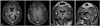
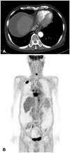
A 71-year-old man presented with a 3-month history of headache and enlarging painless scalp mass on both high parietal convexities. He was undertaken esophagectomy, Ivor-Lewis operation, for esophageal cancer and confirmed as a squamous cell carcinoma of the esophagus 3 years ago. He had received chemotherapy and radiotherapy for esophageal carcinoma. The physical examination showed a subcutaneous mass over both parietal convexities. There were several small and non-tender lymph nodes in the right supraclavicular region, but no axillary lymphadenopathy. There were no significant deficits on the neurological examination. The brain computed tomography (CT) scans demonstrated brain metastasis with peritumoral edema in the right temporal lobe and the left parietal lobe. In addition, expansile osteolytic change was observed in the skull vault and both parietal convexities (Fig. 1). The soft tissue tumor was measured about 8.9×5.5×4.3 cm in size with intra- and extra-cranial compression of the brain. Brain magnetic resonance imaging (MRI) scans showed two brain metastatic lesions with peritumoral edema in the right temporal lobe and the left parietal lobe. Diffuse meningeal thickening and enhancement was also noted in the fronto-parietal area (Fig. 2). A CT scan of his chest showed diffuse wall thickening of the lower esophagus from subcarina to epiphrenic portion (Fig. 3A). Positron emission tomography showed multiple metastatic foci in the brain, bones, right lung, liver, anastomotic site of esophagus, right adrenal gland, and mesenteric lymph nodes (Fig. 3B). The patient underwent operation by a bicoronal scalp incision with bifrontal craniectomy, removal of the bony tumor, and cranioplasty. Pathological findings revealed the metastatic squamous cell carcinoma. The final diagnosis was squamous cell carcinoma of the esophagus with skull metastasis. The patient was referred for concurrent chemotherapy and radiation therapy. Two month later, the patient complained chest discomfort and underwent fine needle aspiration biopsy, diagnosed as a small cell lung cancer. Then, he could not take chemotherapy due to his age and poor general condition.
DISCUSSION
The incidence of esophageal cancer has been increasing over the past thirty years [11], with an expected 16,470 cases and 14,530 deaths in the United States in 2009 [12]. Nevertheless, esophageal carcinoma is among the most challenging oncologic problems. It is well known that esophageal cancer may cause distant metastasis, most frequently in the lungs, pleura, liver, peritoneum, and the adrenal gland. However, metastasis to the bones is infrequent, and skull metastasis from esophageal cancer may be extremely rare [12]. Brain metastasis secondary to esophageal carcinoma is considered to be a rare event, with a reported incidence of between 0.5 and 4.8% [345]. Several studies in the United States also reported low incidence of brain metastasis in patients with esophageal cancer [51113]. Although some studies have reported esophageal cancer with brain metastasis [678], dural metastasis has been reported only 3 case in the past literature [129]. Akhavan and Navabii [1] reported a case of leptomeningeal carcinomatosis from squamous cell carcinoma of esophagus presented by hoarseness. Chen and Huang [2] reported the case of esophageal squamous cell carcinoma with dural and bone marrow metastases. This report also discussed the pathogenesis of unusual metastatic diseases and differential diagnosis of pachymeningeal thickening. Irie et al. [9] reported the case of solitary meningocerebral metastasis from squamous cell carcinoma of the esophagus. Stark et al. [14] reported the characteristic clinical features of metastatic skull tumor, which are distinctive from those of primary skull tumor. Metastatic skull tumor typically appear as expansile, osteolytic, hypervascular lesions and MRI scans showed iso- or hypointensity on T1 and T2 weighted images with moderate enhancement. And can cause local swelling that is usually painless, but they rarely lead to neurologic dysfunction [14]. However, Ellis and McDonald [15] reported a case of acute epidural hematoma secondary to skull metastasis from esophageal carcinoma, which proved that skull metastasis can also lead to severe neurologic dysfunction. For the metastatic skull tumor, surgical treatment should be considered in patients who have neurologic consequences, for example, high risk of extradural or intratumoral hemorrhage or brain compression. Surgical resection might not cure the underlying disease, but it is a relatively safe and satisfactory treatment for relief of neurologic symptoms in patients with cranial metastasis.
This is an uncommon case of squamous cell carcinoma of the esophagus including leptomenigeal dissemination, extensive osteolytic skull lesions, and multiple brain metastases.

 XML Download