Abstract
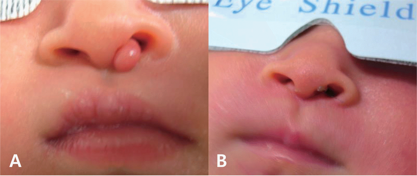
Pai syndrome is a rare disorder, first described in 1987. Diagnostic criteria is the presence of the nasal polyp and one of the following: midline cleft lip, congenital polyp of mid-anterior alveolar process, and pericallosal lipoma. Thirty-six cases of Pai syndrome have been described so far. We report 1 case of Pai syndrome accompanied by congenital nasal polyp and callosal lipoma with partial agenesis of corpus callosum, the first time in Korea.
REFERENCES
1.Lederer D., Wilson B., Lefesvre P., Poorten VV., Kirkham N., Mitra D, et al. Atypical findings in three patients with Pai syndrome and literature review. Am J Med Genet A. 2012. 158A:2899–904.

2.Pai GS., Levkoff AH., Leithiser RE Jr. Median cleft of the upper lip associated with lipomas of the central nervous system and cutaneous polyps. Am J Med Genet. 1987. 26:921–4.

3.Castori M., Rinaldi R., Bianchi A., Caponetti A., Assumma M., Grammatico P. Pai syndrome: first patient with agenesis of the corpus callosum and literature review. Birth Defects Res A Clin Mol Teratol. 2007. 79:673–9.

4.Al-Mazrou KA., Al-Rekabi A., Alorainy IA., Al-Kharfi T., Al-Serhani AM. Pai syndrome: a report of a case and review of the literature. Int J Pediatr Otorhinolaryngol. 2011. 61:149–53.

5.Vaccarella F., Pini Prato A., Fasciolo A., Pisano M., Carlini C., Seymandi PL. Phenotypic variability of Pai syndrome: report of two patients and review of the literature. Int J Oral Max-illofac Surg. 2008. 37:1059–64.

6.Chousta A., Ville D., James I., Foray P., Bisch C., Depardon P, et al. Pericallosal lipoma associated with Pai syndrome: prenatal imaging findings. Ultrasound Obstet Gynecol. 2008. 32:708–10.

7.Savasta S., Chiapedi S., Perrini S., Tognato E., Corsano L., Chiara A. Pai syndrome: a further report of a case with bifid nose, lipoma, and agenesis of the corpus callosum. Childs Nerv Syst. 2008. 24:773–6.

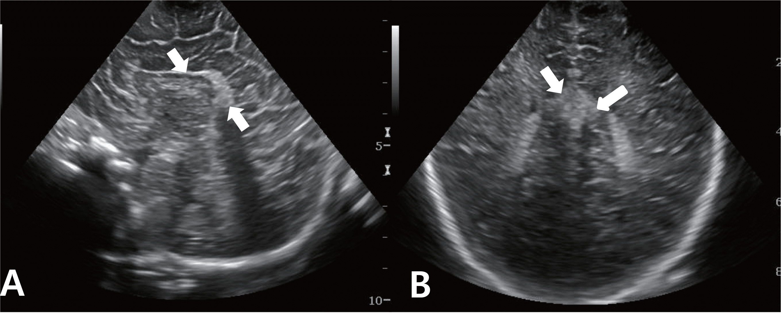
Fig. 2.
Brain ultrasonogrphy: sagittal (A) and coronal (B) image showing lipoma (arrow) at body to tail portion of corpus callosum.

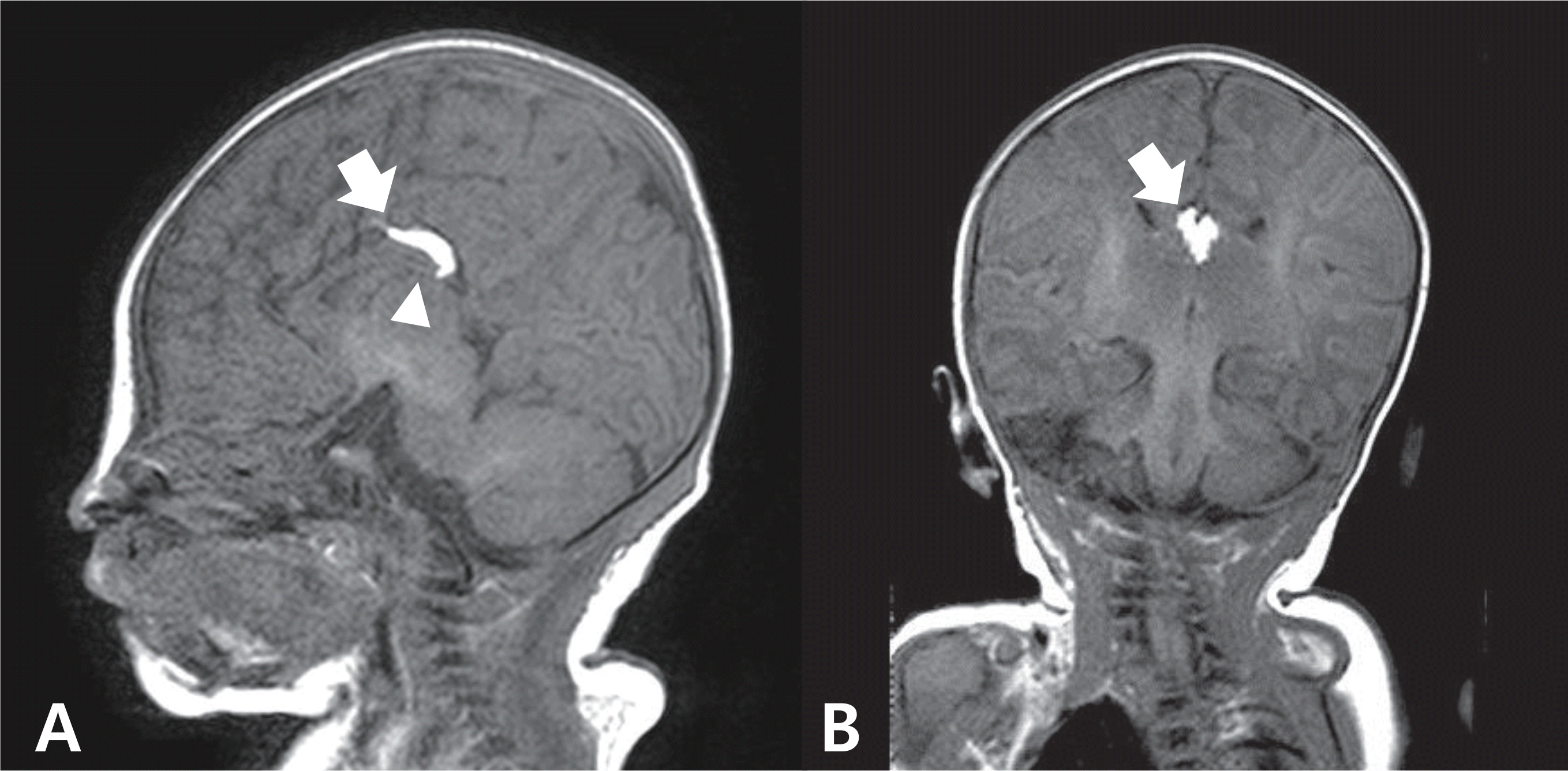
Fig. 3.
Brain magnetic resonance imaging: sagittal (A) and coronal (B) T-1 weighted images showing lipoma in the body and tail portion of corpus callosum (arrow) and tail portion of the corpus callosum was replaced by the mass (arrow head).

Table 1.
Clinical Summary of Pai syndrome Patients Described in the Literature1-9 and Our Patient


 PDF
PDF ePub
ePub Citation
 Print
Print


 XML Download