

 PDF
PDF ePub
ePub Citation
 Print
Print


Abstract
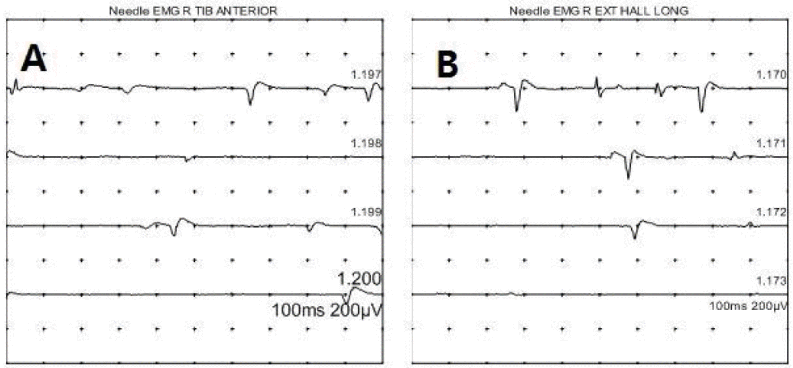
Postpartum common peroneal neuropathy is rare. Causes of peroneal neuropathy include prolonged external knee compression like lithotomy position or knee hyperflexion and epidural analgesia. We report a case of right common peroneal neuropathy after vaginal delivery. A 20-year-old nulliparous woman complained of weakness and abnormal sensation of right foot after vaginal delivery. Lumbar magnetic resonance imaging results are normal, but needle electromyography reveals abnormal spontaneous activities in right tibialis anterior and right extensor hallucis longus. These findings are suggestive of right common peroneal neuropathy with mild axonotmesis. The patient had fully recovered at 6 weeks after the delivery with conservative management include stretching and range of motion exercise. Peroneal neuropathy is rare, but understanding of the risk factor and preventive measure for peroneal neuropathy is important for delivery team.
REFERENCES
1). Wong CA., Scavone BM., Dugan S., Smith JC., Prather H., Ganchiff JN, et al. Incidence of postpartum lumbosacral spine and lower extremity nerve injuries. Obstet Gynecol. 2003. 101:279–88.

2). Wong CA. Nerve injuries after neuraxial anaesthesia and their medicolegal implications. Best Pract Res Clin Obstet Gynaecol. 2010. 24:367–81.
3). Holdcroft A., Gibberd FB., Hargrove RL., Hawkins DF., Dellaportas CI. Neurological complications associated with pregnancy. Br J Anaesth. 1995. 75:522–6.
4). Radawski MM., Strakowski JA., Johnson EW. Acute common peroneal neuropathy due to hand positioning in normal labor and delivery. Obstet Gynecol. 2011. 118(2 Pt 2):421–3.
5). Indusuyi OB., Morrey BF. Peroneal nerve palsy after total knee arthroplasty. Assessment of predisposing and prognostic factors. J Bone Joint Surg Am. 1996. 78:177–84.
6). Marciniak C. Fibular(peroneal) neuropathy: electrodiagnostic features and clinical correlates. Phys Med Rehabil Clin N Am. 2013. 24:121–37.
7). Bsteh G., Wanschitz JV., Gruber H., Seppi K., Löscher WN. Prognosis and prognostic factors in non-traumatic acute-onset compressive mono-neuropathies—radial and peroneal mononeuropathies. Eur J Neurol. 2013. 20:981–5.
8). Bunch K., Hope E. An uncommon case of bilateral peroneal nerve palsy following delivery: a case report and review of the literature. Case Rep Obstet Gynecol. 2014. 2014:746480.
 XML Download