

 PDF
PDF ePub
ePub Citation
 Print
Print


INTRODUCTION
Despite improvements in perioperative outcomes, pancreatic cancer has a poor prognosis, with a 5-year survival rate of only 8%–10% among patients.12 Most patients are diagnosed in the advanced stages, and effective systemic therapies are lacking. According to the annual report of cancer statistics in Korea, the 5-year survival rate of overall pancreatic cancer patients was 10% in 2014, with the higher rate due to increased early diagnoses following the introduction of a routine cancer screening program for all citizens conducted by the National Health Insurance, and increased detection of less aggressive pancreatic malignancies.3 In addition, effective systemic treatments including neoadjuvant therapies (NATs) have been introduced in an attempt to overcome the limitations of prior treatments and to improve patient outcomes, especially in patients with borderline resectable, locally advanced, and even metastatic pancreatic cancer; therefore, the outcomes of pancreatic cancer patients have been improved by additional surgical resection after systemic treatment.456 However, because the definitions of borderline resectable and locally advanced pancreatic cancer differ among institutions, it is impossible to compare survival rates according to clinical stage in pancreatic cancer patients. In addition, the data published by most institutions do not include patients with metastatic or locally advanced pancreatic cancer.4789 SEER (Surveillance, Epidemiology, and End Results program, USA) data cover a large number of patients but are limited by the high variation among the involved hospitals and a lack of accurate clinical staging.10 Furthermore, as there are no recent data on clinical stage based on accurate imaging criteria in overall patients, it is difficult to grasp the actual status of pancreatic cancer treatment in Korea. To date, very few studies have identified trends in the treatment of pancreatic cancers, including locally advanced and metastatic cancers, in a single-center study.
Therefore, the objective of this study was to categorize all patients according to clinical stage using uniform imaging diagnostic criteria and to evaluate the impact of treatment modality on the survival of pancreatic cancer patients included in a large-scale prospective database at Seoul National University Hospital.
MATERIALS AND METHODS
Database
We conducted a retrospective cohort study using prospectively recorded information of patients registered in a clinical oncology database at Seoul National University Hospital between January 1, 2007 and December 31, 2014. The study was approved by the Institutional Review Board at Seoul National University (1706-159-863).
Patients and groups
We included patients who were diagnosed with stage I–IV pancreatic cancer between January 1, 2007 and December 31, 2014 and aged ≥19 years at diagnosis. The diagnoses were made using computed tomography with a pancreas-specific protocol and/or magnetic resonance imaging, positron emission tomography, endoscopic ultrasonography, with or without cytological or histological confirmation of conventional ductal adenocarcinoma of the pancreas. Patients were excluded if surgery was performed at another hospital or the diagnosis was made outside the index period. The National Comprehensive Cancer Network 2017 guidelines were used for pancreatic cancer staging, based on computed tomography performed at the time of diagnosis (Table 1). The patients were first categorized into four groups according to the clinical stage, as follows: resectable (RPC), borderline resectable (BRPC), locally advanced (LAPC), or metastatic pancreatic cancer (MPC).4 Patients in each stage group were further categorized according to the treatment modality, as follows: NAT followed by surgery (NAT+surgery group), surgery±adjuvant chemoradiotherapy (surgery group), chemotherapy alone (CTx group), and palliative care (palliative care group). NAT was defined as the first course of chemotherapy, radiation, or concurrent chemoradiotherapy (CCRT) prior to surgery.
Data extraction
The primary outcome was survival time, which was defined as the time from the date of diagnosis to the date of death or the last follow-up. The patients' characteristics, date of diagnosis, type of surgery, tumor location, tumor size, serum level of carbohydrate antigen (CA) 19-9 (a tumor marker; normal range <37 U/ml), and pathologic reports were extracted from the medical records.11 We re-reviewed the images of computed tomography and magnetic resonance examinations performed at the time of diagnosis. The patients were categorized according to clinical stage (according to the National Cancer Center Network 2017 guidelines) and again by treatment modality. The tumor response was defined according to the Response Evaluation Criteria in Solid Tumors using computed tomography performed after NAT, and was categorized as complete response (disappearance of the target lesion), partial response (≥30% decrease in tumor diameter), stable disease (neither sufficient shrinkage to qualify as partial response nor sufficient increase to qualify as progressive disease), or progressive disease (≥20% increase in diameter).12
The TNM residual tumor classification was recorded in patients who underwent pancreatectomy with curative intent.13 We used the tumor regression grading system of the College of American Pathologists, which comprises the following four grades according to the extent of residual carcinoma in post-NAT pancreatectomy specimens: grade 0, no viable residual tumor (pathologic complete response); grade 1, marked response (minimal residual cancer with single cells or small groups of cancer cells); grade 2, moderate response (residual cancer outgrown by fibrosis); and grade 3, poor or no response (extensive residual cancer).14
Neoadjuvant chemotherapy and/or radiotherapy
Neoadjuvant chemotherapy regimens included gemcitabine, conventional 5-fluouracil (5-FU), or FOLFIRINOX. Gemcitabine chemotherapy consisted of 400 mg/m2 body surface area (BSA) intravenous gemcitabine administered weekly for 6 weeks. Three-dimensional conformal radiotherapy consisted of a total dose of 45 Gy (1.8 Gy daily fraction, 5 fractions per week for 5 weeks) with a boost dose of 9 Gy (1.8 Gy daily fraction, 5 fractions). 5-FU based CCRT consisted of 20-Gy dose to the tumor given in 10 daily fractions over a 2-week period plus an intravenous bolus of 5-FU (500 mg/m2 of BSA on each of the first 3 days of radiotherapy and again after a planned break of 2 weeks). FOLPIRINOX consisted of oxaliplatin at a dose of 85 mg/m2 followed by leucovorin at a dose of 400 mg/m2 both administered as a 2-hour intravenous infusion with the addition of 180 mg/m2 irinotecan after 30 minutes given over 90 minutes as an intravenous infusion. This treatment was followed by 5-FU at a dose of 400 mg/m2 administered as an intravenous bolus followed by a continuous infusion of 2,400 mg/m2 for a 46-hour period (one cycle) every 2 weeks. The choice of chemotherapy and/or radiotherapy was determined by each patient's general performance and ease of access to the hospital. Dose were reduced depending on the patient's status or when adverse events were noted.
Adjuvant chemotherapy and/or radiotherapy
Adjuvant chemotherapy and/or radiotherapy was recommended to patients after operation. Adjuvant treatment regimen included gemcitabine, conventional 5-FU, or FOLFIRINOX. Similar to neoadjuvant treatment, the regimen was determined considering each patient's general performance and ease of access to the hospital.
Statistical analysis
Patient demographics, treatment modalities, and tumor characteristics were compared among the four clinical stages and four treatment groups using analysis of variance or t tests for continuous variables and χ2 tests for categorical variables. Kaplan-Meier analyses were conducted to compare survival rates among treatment groups according to clinical stage, and log rank tests were used to compare differences in survival. The median survival time was estimated from the Kaplan-Meier curves. Cox proportional hazards models were used to evaluate risk factors affecting survival according to clinical stage. All analyses were performed using PASW software, version 18.0 (SPSS Inc., Chicago, IL, USA). Differences were considered significant at p-value <0.05.
RESULTS
Patient characteristics
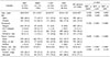
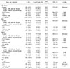
Overall, 1,646 patients were included in this retrospective study. The patients were divided into four groups according to their clinical stage: RPC (n=475, 28.9%), BRPC (n=129, 7.8%), LAPC (n=384, 23.3%), and MPC (n=658, 40.0%). There were significant differences among the four groups in terms of clinical stage, tumor location, tumor size, and serum CA 19-9 level, but not sex (Table 2). Patients in the RPC group was older than those in the MPC group (64.9 years vs. 63.0 years, respectively, p=0.020). The proportions of males and females were similar in all four groups (approximately 60% males). Pancreatic head cancer was dominant in the RPC group compared to the LAPC and MPC groups (Both p<0.001). The rate of pancreatic body or tail cancer was greater in the LAPC (47.9%) and MPC (66.6%) than in the RPC (30.5%) and BRPC (20.0%) groups. Tumor size was greater in the BRPC (32.0 mm), LAPC (38.9 mm) and MPC (44.0 mm) groups than in the RPC (26.5 mm) group (All p<0.001).
Treatment groups
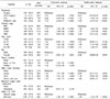
The patients in each clinical stage group were further classified according to treatment modality: surgery (n=495, 30.1%), NAT+surgery (n=35, 2.1%), CTx (n=794, 48.2%), and palliative care (n=322, 19.6%). Surgery without NAT was the dominant treatment modality in the RPC group (434/495, 87.6%), NAT+surgery in the BRPC group (28/35, 80.0%), and CTx or palliative care in the LAPC (285/794, 35.9%) and MPC groups (449/794, 56.5%). The surgical procedures performed in the NAT+surgery and surgery groups are summarized in Table 3. Overall, 30 patients (85.7%) in the NAT+surgery group and 428 patients (86.4%) in the surgery group underwent surgical resection with curative intent. There were significant differences between these two groups in terms of the type of surgery (p=0.019) and main vessel resection (p<0.001), but not the reason for palliative surgery (p=0.793). Of 495 patients in surgery group, 353 patients underwent adjuvant treatment after surgical resection and 142 patients did not undergo adjuvant treatment after surgery.
Median survival time according to clinical stage
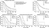
We analyzed the median survival time of the patients according to clinical stage and treatment modality (Table 4). Kaplan-Meier plots for survival according to clinical stage are presented in Fig. 1. In the RPC patients, the median survival time in the surgery group (22 months) was significantly longer than those in the CTx (8 months, p<0.001) and palliative care (11 months, p<0.001) groups. In the BRPC patients, the median survival time in the surgery group (16 months) was significantly different from those in the NAT+surgery (24 months, p=0.049) and palliative care group (7 months, p<0.001) but was not significantly longer than that in the CTx group (12 months, p=0.091). Similarly, in LAPC patients, the median survival time was not significantly different between the surgery and CTx groups (10 and 13 months, respectively, p=0.142). In the MPC patients, the median survival time in the CTx group (7 months) was not significantly different from that in the NAT+surgery group (12 months, p=0.138) but was longer than that in the palliative care group (3 months, p<0.001). In RPC and BRPC groups, the median survival time in the surgery with adjuvant therapy group (25 and 17 months, respectively) was significantly different from those in the surgery without adjuvant therapy (15 and 8 months, respectively, both p<0.001).
Pathologic profiles and types of NAT
The stage of pancreatic cancer in the 35 patients who received NAT+surgery was borderline resectable in 24, locally advanced in 4 (11.4%), and metastatic in 3 (8.6%). Two patients had single liver metastasis, and one had para-aortic lymph node metastasis; these three patients showed improvements following NAT and then underwent surgical resection. Twenty patients (57.2%) underwent concurrent chemoradiotherapy and 15 patients (42.8%) underwent CTx only. The CTx regimens were gemcitabine in 24 patients (68.6%), FOLFIRINOX (a combination of 5-fluorouracil, oxaliplatin, irinotecan, and leucovorin) in 5 patients (14.3%), gemcitabine plus erlotinib in 4 patients (11.4%), and 5-fluorouracil plus cisplatin in 2 patients (5.7%). The tumor response after NAT was classified as partial response in 11 patients (31.4%), stable disease in 20 patients (57.2%), and progressive disease in 4 patients (11.4%). The CA 19-9 level after NAT was normalized in 15 patients (42.9%), stable in 16 patients (45.7%), and elevated in 4 patients (11.4%).
In the NAT+surgery and surgery groups in particular, there was no difference in terms of TNM residual tumor classification, which was classified as R0 in 26 patients (86.7%) in the NAT+surgery group and 362 patients (84.6%) in the surgery group. However, there were significant differences in terms of the pathologic T stage (p<0.001), pathologic N stage (p=0.016), and AJCC pathologic stage (p=0.004) between these two groups. The mean number of lymph nodes removed was 15.42 in the NAT+surgery group and 17.32 in the surgery group (p=0.387). The mean number of metastatic lymph nodes was 0.71 in the NAT+surgery group and 1.87 in the surgery group (p<0.001).
Prognostic factors for survival according to clinical stage
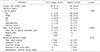
Table 5 lists the results of the Cox proportional hazards regression analyses. The NAT+surgery group conferred a 35% lower hazard of mortality compared with the surgery group (hazard ratio [HR]=0.65, p=0.045). In contrast, the CTx (HR=1.35, p=0.032) and palliative care (HR=3.95, p<0.001) groups were associated with greater risk compared with the surgery group. The risk of mortality was greater in patients aged 71–80 years (HR=1.39, p<0.001) or 81–100 years (HR=1.51, p=0.002) at diagnosis than in patients aged 18–60 years at diagnosis. The risk of mortality was lower in females than in males (HR=0.89, p=0.035) and was higher in patients with elevated CA 19-9 levels than in patients with normal CA 19-9 levels (HR=1.39, p<0.001). Main vessel invasion, regional lymph node metastasis at the time of diagnosis, and tumor location showed no significant effects on survival rate.
Among the RPC patients, compared with the surgery group, regional lymph node metastasis (HR=1.42, p=0.004) and an elevated initial CA 19-9 level (HR=2.09, p<0.001) were associated with higher risk in terms of survival. In BRPC patients, compared with the surgery group, there were no significant differences in risk in the NAT+surgery or CTx group but a higher risk in the palliative care group (HR=3.08, p<0.001). In LAPC patients, the NAT+surgery, CTx, and palliative care groups did not show significant differences in risk compared with the surgery group. In MPC patients, the NAT+surgery group did not show a significant difference in risk compared with CTx group, but the palliative care (HR=3.23, p<0.001) group was associated with a higher risk compared with the CTx group.
DISCUSSION
This retrospective cohort study aimed to categorize patients according to clinical stage using uniform imaging diagnostic criteria based on high-resolution imaging and to evaluate the impact of treatment modalities on the survival of pancreatic cancer patients. Of the 1,646 total patients, 475 (28.9%) were diagnosed with RPC, 129 (7.8%) with BRPC, 384 (23.3%) with LAPC, and 658 (40.0%) with MPC. There were 530 (32.2%) surgically treatable pancreatic cancer patients, which is more than reported previously, and 35 (2.1%) patients underwent surgery after NAT.3 Of the 475 RPC patients, 438 (91.4%) underwent surgery, and R0 resection was achieved in 388 (81.0%).
According to the annually reported summary staging of pancreatic cancer by the Korea National Cancer Registry, there were 2,576 (10.9%) localized, 7,492 (31.8%) regional, and 10,735 (45.6%) distant cases of metastasis, and 2,715 (11.7%) unknown cases in 2014, 5-year prevalence (5,948 were newly diagnosed with pancreatic cancer in 2014; however, this report did not include the incidence of pancreatic cancer according to clinical stage).3 Summary staging is the most basic method of categorizing how far a cancer has spread from its point of origin, but it does not reflect the clinical stage, which has been essential for determining resectability the suitability for NAT.15
A large retrospective cohort study using the 2003–2011 dataset from the National Cancer Database based in the U.S. reported that of 18,332 pancreatic cancer patients, 7,095 (38.7%) were clinical stage I, 9,760 (53.2%) clinical stage II, and 1,477 (53.2%) clinical stage III.10 However, that study has several limitations in that patients with clinical stage IV or unknown stage pancreatic cancer or those who did not undergo surgical resection were excluded. In addition, BRPC and LAPC were not distinguished in clinical stage III patients, and the multicenter data were heterogenous because of patient collection from over 1500 facilities. Most of the classification systems focus on anatomical findings and there is still debate whether these are a solid classification, due to a lack of prospective studies in this regard. Guidelines that also incorporate biological features are needed to help predict early recurrence, even in resectable pancreatic cancer, and to support indications for NAT in select patients with resectable pancreatic cancer.791617
Another recent meta-analysis showed that of 4,394 (from 111 studies) pancreatic cancer patients, 46.9% who were initially staged as unresectable underwent surgical exploration.18 Of these patients, 69.9% were resected successfully, leading to a resectability rate after NAT of 33.2% (comparable R0 resection rate to that in the initially resectable group), suggesting that patients with pancreatic cancer who can undergo surgical resection are increasing more than one out of five in the past by applying NAT at an advanced stage.18 Furthermore, an increase in the early diagnosis rate, detection of various low-malignancy-risk pancreatic cancers, and development of surgical techniques have accelerated the surgical resection rate of pancreatic cancer.310
Katz et al. conducted a prospective, multicenter, single- arm pilot study in 2013 of 22 patients with borderline resectable pancreatic cancer who were treated with a modified FOLFIRINOX regimen and chemoradiotherapy as NAT. The median survival was 21.7 months, and R0 resection was achieved in 20 (93%) patients.19 In 2014, Tzeng et al. published the results of a retrospective study, in which 84 patients received gemcitabine or 5-fluorouracil- based NAT followed by surgery, and 57 patients underwent NAT without surgery. The R0 resection rate was 92% in the NAT+surgery group. The median survival was 30.9 months in the NAT+surgery group versus 12 months in the NAT only group.20 In our study, among the patients with BRPC, the median survival was 24 months in the NAT+surgery group, 16 months in the surgery group, 12 months in the CTx group, and 7 months in the palliative care group. Only 32 (6.2%) patients with BRPC or LAPC underwent surgery after NAT, and 61 (12.0%) underwent surgery without NAT. Few studies have assessed the overall median survival in patients with pancreatic cancer stratified by treatment modality and disease stage in the same period of time.
Among patients who underwent curative resection, the rate of R0 resection was comparable between the NAT+surgery and surgery groups, even though the proportion of patients with advanced cancer was greater in the former group. We recognize that the NAT+surgery group tended to undergo more aggressive surgical procedures than did the surgery group; for example, the main vessel resection rate was greater in the former group (22.9% vs. 9.7%; p<0.001). Since the dense stroma associated with pancreatic cancer may result in little change on computed tomographic imaging, despite an excellent cellular response, there may be some discrepancy between the Response Evaluation Criteria in Solid Tumors criteria and tumor regression grade; hence, adjuvant surgery may be necessary to identify a pathological response.21
Tumor response after neoadjuvant treatment were assessed radiologically with pancreatobiliary protocol CT and were categorized according to the new response evaluation criteria in RECIST guidelines in this study. Also, serum concentrations of CA 19-9 were measured at the first visit to the hospital, after neoadjuvant treatment and after surgery. Kim et al. reported a retrospective study in 2017 of 40 BRPC patients who were treated with NAT followed by resection that the RECIST criteria and reduced serum CA 19-9 concentration were associated with biological response.22 Several recent studies were conducted that Positron Emission Tomography/Computer tomography (PET/CT) could increase the chance of detecting patients with progressive pancreatic cancer after neoadjuvant therapy compared to the conventional anatomical- based assessment of RECEIST criteria.2324
We also conducted multivariate analysis using the Cox proportional hazards model to determine prognostic factors for survival in patients with pancreatic cancer. The results showed surgery after NAT compared with surgery or CTx alone, female sex, resectable stage, a CA 19-9 level within the normal range at diagnosis, and tumor location in the pancreatic body or tail to be prognostic factors for improved survival in patients with pancreatic cancer. Tzeng et al. also reported that serum CA 19-9 is a dynamic preoperative marker of tumor biology and the response to NAT, and provides prognostic information in patients with unresected or resected BRPC.20 In addition, another study revealed that patients with advanced pancreatic cancer whose serum tumor marker levels had normalized after NAT may be appropriate candidates for tumor resection.25
Some strengths and limitations of this study also warrant discussion. First, because this was a retrospective analysis of patients treated at a single institution, the results are subject to the biases and limitations inherent to retrospective studies. Nevertheless, this is one of the largest reported cohorts of patients with resectable, borderline resectable, locally advanced, or metastatic pancreatic cancer in which the cancer was staged using objective radiographic criteria and classified using consensus guidelines. Second, we divided the patients into four groups based on their treatment modalities. Unfortunately, the numbers of patients varied among the groups, with some groups being too small for detailed analyses. As a result of ethical issues and difficulty in recruiting patients, no randomized clinical trials of prior surgery compared with NAT have been performed. Nevertheless, several phase I/II trials and on-going phase III clinical trials, such as NEOPAC (NCT01521702) and Prep-02/JSAP-05 (UMIN-No. 000009634) are underway and will hopefully yield further insight into the benefits of NAT.19262728 Third, this study included heterogenous regimens of NAT, which showed different oncologic outcomes.
In conclusion, based on the results of this retrospective, single-center, large cohort study, categorizing all pancreatic cancer patients according to clinical stage using uniform imaging diagnostic criteria with high-resolution images, the rates of resectable and surgically treatable pancreatic cancer were 29.1% and 32.2%, which are higher than those reported previously. In the RPC group, 434 (91.4%) patients underwent surgery, and R0 resection was achieved in 388 (81.0%). Although our findings support the use of NAT in eligible patients with borderline resectable pancreatic cancer, the survival rate of advanced-stage patients is still low; thus, more effective systemic therapies are required. In addition, further studies are needed to evaluate the effects of specific NAT regimens and to update consensus guidelines with biological features.
 XML Download