

 PDF
PDF ePub
ePub Citation
 Print
Print


Fibroma of tendon sheath (FTS) was initially described in 1936 by Geschickter and Copeland1) as benign firmed soft tissue tumor which is rare, less common than another soft tissue tumor especially giant cell tumor of the tendon sheath (GCTTS). Chung and Enzinger2) in 1979 described the clinical and pathological features of 138 cases of the FTS. In cases of 138 patients, 98.6% of FTS occurred in the extremities: 86.2% in the upper extremities and 12.3% in the lower extremities; of the latter 5 were in the foot and 3 at the ankle. Kirby et al.3) in 1989 described the 83 cases of the soft tissue tumor of foot. They reported only one case of fibroma in the foot, which is the same result that very few were found in the foot. This article describes a 51-year-old patient of FTS that developed in the extensor tendon of foot, the only known FTS to form in this area. We report this case with a review of the lietrature because of the satisfactory results of surgical treatment of FTS. This study was approved by the Medical Research Ethics Review Committee of Kangbuk Samsung Hospital.
CASE REPORT
A 51-year-old female came in an orthopedic surgery clinic with a 2-month experience of an enlarging, painless mass along the dorsal side of second metatarsophalangeal (MTP) joint. She didn't suffer pain when she walk or sleep. She denied weight loss and reported no associated fever, chills, or night sweats. Medical history and family history of her were not cause and there wasn't a history of trauma.
Physical examination revealed 3.5 cm×2 cm oval-shaped subcutaneous, soft and non-tender mass at the dorsal side of second MTP joint. The mass appeared to slightly move with the extensor digitorum longus (EDL) and extensor digitorum brevis (EDB) with dorsiflexion of ankle. There was no restriction of foot motions and not associated heating sense, erythema, edema. Muscle atrophy, and neurovascular examination were unremarkable. Routine laboratory data including white blood cell count, erythrocyte sedimentation rate, and C-reactive protein level were normal.
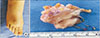
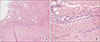
During general anesthesia of the patient, longitudinal incision were subsequently performed to show a well circumscribed 3.3 cm×2 cm×1.5 cm mass which was sturdily adherent to the dorsal side of second MTP joint capsule and conjoined tendon of EDL and EDB (Fig. 2). But no neurologic structure and vascular peduncle was found. And then excisional biopsy of the mass was subsequently performed. The gross specimen was shown as white-tan, multilobular, and rubbery mass. The histopathologic examination of the mass test showed fibrous lobulated tumor organized of dense fibrous tissue with some spindle cells (Fig. 3). The tumor was reported as FTS.
The patient left the hospital the 2 days after the operation with a simple compression dressing. After one month she returned to her normal foot activities with full range of motion. Perioperative complication wasn't occurred. At the 3 months follow-up, there was no evidence of clinical or radiological recurrence.
DISCUSSION
FTS of plantar side is associated with trauma history commonly. Even if some of them did not have trauma history, the tumor occurring resulted from surface of plantar area was rubbed frequently.4) Because the extensor tendons of foot have a shorter excursion than the flexor tendons of foot,5) there is less rub in the extensor tendons. Thus, FTS of extensor tendon is thought to be rare than flexor tendon.
Clinically, the most common presentation is slow-growing least painful rare entity arising from tendon or tendon sheath. The median age of 138 cases was 31 years, and male patients were affected about twice as many, with a ratio of 3.1:1.5.2) Five cases were found around foot. In the feet, 4 of the 5 cases were removed from the plantar region.2) Several other authors also described FTS around the foot, one each in the peroneus longus tendon, the flexor hallucis longus tendon, the plantar area.46) Involvement of the conjoined tendon of EDL and EDB was first reported in our patient.
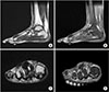
Plain radiographies of the foot are usually normal, except when large tumor mass protrudes from surrounding structures, or erosive bony changes were occurred which is greatly unusual. MRI features of fibroma are variable. T1-weighted images showed low signal intensity and T2-weighted images showed various signal intensities and enhancement after contrast images.7) This variation depends on the amount of hyalinization and sclerosis of the tumor.7) Proliferation fibroblasts numbers can change T2-weighted images. Our patient's MRI also showed features alike; heterogeneous low signal intensity in both T1- and T2-weighted images. GCTTS, fibromatosis, and nodular fasciitis was considered as alterative diagnosis because they are usual in foot and also show signal intensities alike on MRI.7) Differential diagnosis from fibromatosis is distinctly circumscribed margins of the fibroma. And the nodular fasciitis can be distinguished by character that relatively straight forward because fibroma is lobulated and not nodular.
Histologically, FTS are composed of fibroblasts' benign proliferation which are surrounded by collagen fibers, commonly including scattered axis shaped fibroblasts and narrow slit-like vascular spaces but sometimes stellate shaped fibroblast cells.12) Scattered fibroblasts which shape is star like can exist in collagen bundles rarely and are frequently concentrated around the vascular spaces.8) In histologically we can distinguish it and the GCTTS through fibroblastic spread. Due to similarities between some characteristic, some articles have described that FTS could have progressed from GCTTS, because of the progressive vascular damage.
The treatment of choice is local excision, with a reported recurrence rate of 18% to 24%. Most recurrences are owing to troubles of complete excision because of adherence to tendons and tendon sheaths.910) So we should carefully and completely resect.
In our case we are presenting a patient who has tendon sheath fibroma which occurred in the EDL and EDB combined with tendon sheath which is unusual location. We think this is first reported case of this tumors developing in these tendon sheath, we report our case with a literature review.
 XML Download