Abstract
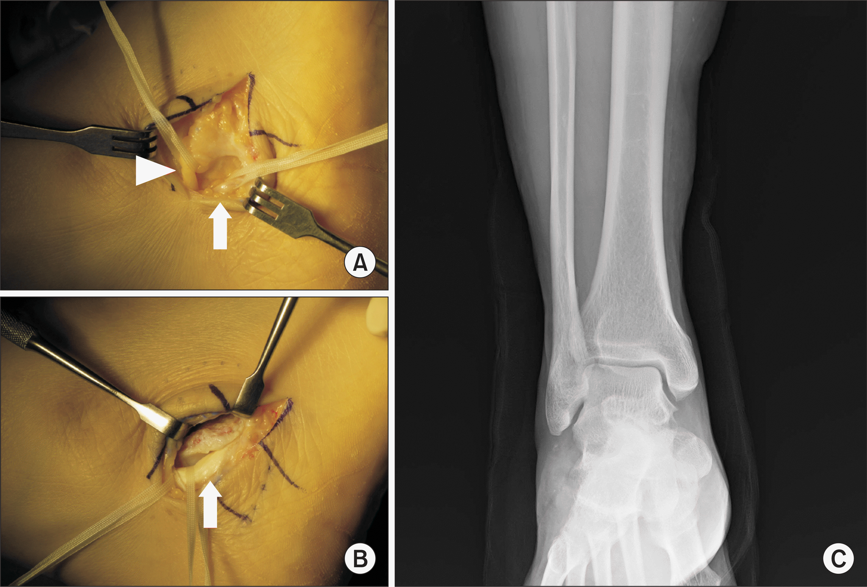
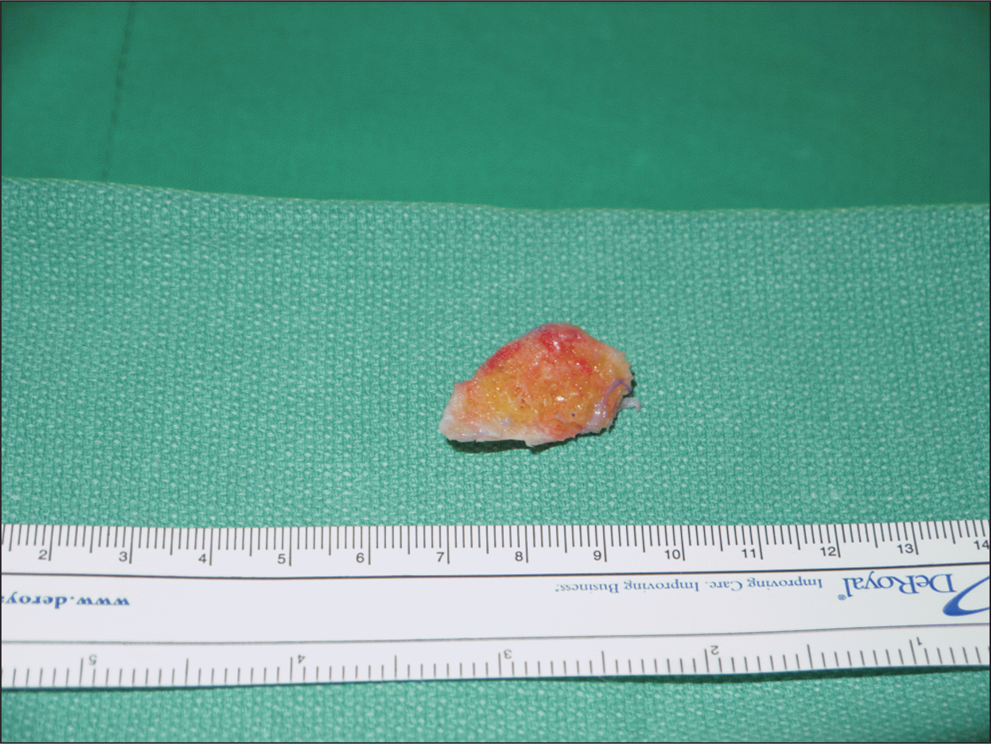
A hypertrophied peroneal tubercle can present as a bony prominence at the lateral aspect of the foot and a peroneal tenosynovitis or tear. We report a case of a 52-year-old man complaining of lateral foot tingling pain and numbness. The sural nerve entrapment and peroneus longus tenosynovitis by hypertrophied peroneal tubercle were confirmed. Good results were obtained after excision of the hypertrophied peroneal tubercle and sural nerve release.
References
1. Bruce WD, Christofersen MR, Phillips DL. Stenosing tenosynovitis and impingement of the peroneal tendons associated with hypertrophy of the peroneal tubercle. Foot Ankle Int. 1999; 20:464–7.
2. Burman M. Stenosing tendovaginitis of the foot and ankle; studies with special reference to the stenosing tendovaginitis of the peroneal tendons of the peroneal tubercle. AMA Arch Surg. 1953; 67:686–98.
3. Chen YJ, Hsu RW, Huang TJ. Hypertrophic peroneal tubercle with stenosing tenosynovitis: the results of surgical treatment. Changgeng Yi Xue Za Zhi. 1998; 21:442–6.
4. Ochoa LM, Banerjee R. Recurrent hypertrophic peroneal tubercle associated with peroneus brevis tendon tear. J Foot Ankle Surg. 2007; 46:403–8.

5. Hyer CF, Dawson JM, Philbin TM, Berlet GC, Lee TH. The peroneal tubercle: description, classification, and relevance to peroneus longus tendon pathology. Foot Ankle Int. 2005; 26:947–50.

6. Shibata Y, Sakuma E, Yoshida Y, Wakabayashi K, Iguchi H, Sekiya I, et al. Morphometric analysis of the peroneal tubercle using a three-dimensional computed tomography model. Foot (Edinb). 2014; 24:200–2.

7. Pomeroy G, Wilton J, Anthony S. Entrapment neuropathy about the foot and ankle: an update. J Am Acad Orthop Surg. 2015; 23:58–66.
8. Sugimoto K, Takakura Y, Okahashi K, Tanaka Y, Ohshima M, Kasanami R. Enlarged peroneal tubercle with peroneus longus tenosynovitis. J Orthop Sci. 2009; 14:330–5.

10. Park SJ, Jeong HJ, Kim E, Lee JW. Operative treatment of stenosing tenosynovitis of the peroneus longus tendon associated with hypertrophy of the peroneal tubercle: a case report–1 case. J Korean Foot Ankle Soc. 2013; 17:150–3.
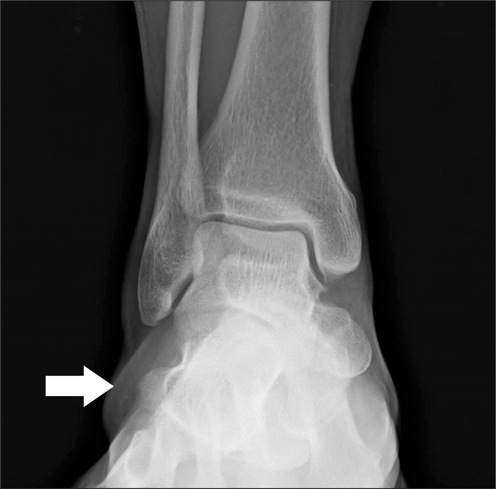
Figure 1.
A 52-year-old man with right foot lateral pain anteroposterior radiographs of right ankle show hypertrophied peroneal tubercle of calcaneus (arrow).

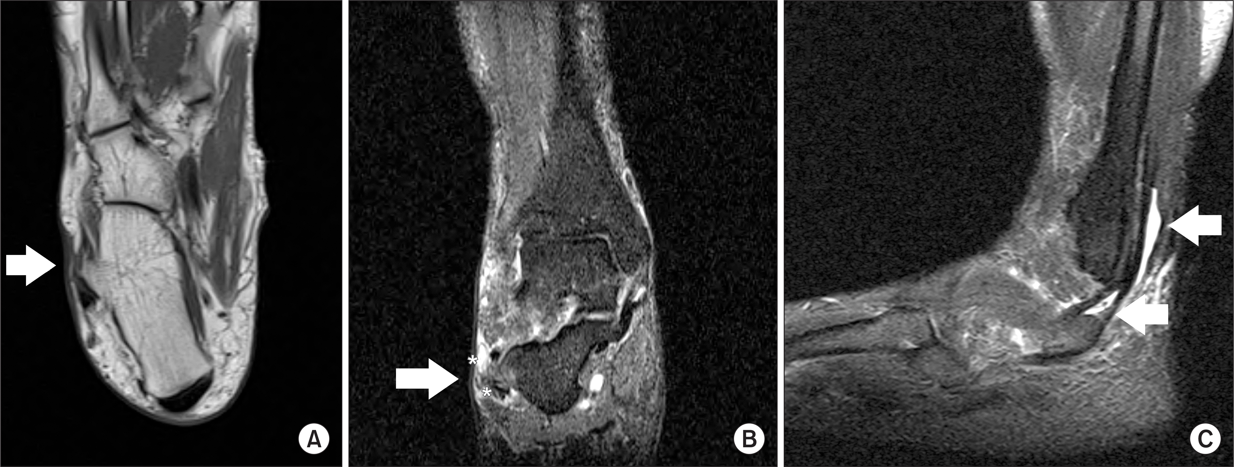
Figure 2.
Using magnetic resonance imaging. (A) T1-weighted axial image shows hypertrophied peroneal tubercle of calcaneus (arrow). (B) T2-weighted coronal image shows focal edema (arrow) and thickening of branch of sural nerve (asterisks). (C) T2-weighted sagittal image shows tenosynovitis of peroneus longus and fluid collection at the proximal level (arrows).


 PDF
PDF ePub
ePub Citation
 Print
Print


 XML Download