Abstract
Purpose:
The purpose of this study was to compare the clinical and radiologic results of arthrodesis between anterior approach and transfibular approach arthrodesis in ankle arthritis.
Materials and Methods:
There were 61 cases of ankle arthritis treated by anterior or transfibular ankle arthrodesis in our hospital from April 2008 to March 2012. We investigated 29 cases (27 patients) who underwent ankle arthrodesis with an anterior approach (15 cases) and transfibular approach (14 cases), and were followed for over two years. Clinically, American Orthopaedic Foot and Ankle Society (AOFAS) ankle-hindfoot score, pain visual analogue scale (VAS), and subjective satisfaction degrees were evaluated. In addition, ankle coronal and sagittal alignments were evaluated using plain radiographs at 6 and 24 months, postoperatively.
Results:
Clinically, preoperative mean AOFAS score and VAS was 41.3 and 6.4, and were changed to 58.9 and 3.3 postoperatively in the anterior approach group. In the transfibular approach group, preoperative mean AOFAS score was 36.6 and VAS was 7.1, and they were changed to 54.9 and 3.4 postoperatively. However, no significant differences in the clinical results were observed between the two groups (p=0.297). Duration of attaining union was 8.1 weeks in the anterior approach group and 10.4 weeks in the transfibular approach group. Complications were delayed union in one case, nonunion in three cases, cancellous screw breakage in three cases, and complex regional reflex syndrome in one case.
Conclusion:
After transfibular ankle arthrodesis as treatment of ankle osteoarthritis, the tendency for valgus angulation of the ankle at the final follow-up was observed and 6.5 mm cancellous screw breakage occurred frequently. Therefore, in order to achieve better stability, it is necessary to use 6.5 mm cannulated screws rather than 6.5 mm cancellous screws for ankle arthrodesis.
REFERENCES
1.Raikin SM. Arthrodesis of the ankle: arthroscopic, mini-open, and open techniques. Foot Ankle Clin. 2003. 8:347–59.

2.Akra GA., Middleton A., Adedapo AO., Port A., Finn P. Outcome of ankle arthrodesis using a transfibular approach. J Foot Ankle Surg. 2010. 49:508–12.

3.Stranks GJ., Cecil T., Jeffery IT. Anterior ankle arthrodesis with cross-screw fixation. A dowel graft method used in 20 cases. J Bone Joint Surg Br. 1994. 76:943–6.

4.Gordon D., Zicker R., Cullen N., Singh D. Open ankle arthrodeses via an anterior approach. Foot Ankle Int. 2013. 34:386–91.

5.Maurer RC., Cimino WR., Cox CV., Satow GK. Transarticular cross-screw fixation. A technique of ankle arthrodesis. Clin Or-thop Relat Res. 1991. 268:56–64.
6.Chen YJ., Huang TJ., Shih HN., Hsu KY., Hsu RW. Ankle arthrodesis with cross screw fixation. Good results in 36/40 cases followed 3-7 years. Acta Orthop Scand. 1996. 67:473–8.
7.Zwipp H., Rammelt S., Endres T., Heineck J. High union rates and function scores at midterm followup with ankle arthrodesis using a four screw technique. Clin Orthop Relat Res. 2010. 468:958–68.

8.Monroe MT., Beals TC., Manoli A 2nd. Clinical outcome of arthrodesis of the ankle using rigid internal fixation with cancellous screws. Foot Ankle Int. 1999. 20:227–31.

9.Mann RA., Rongstad KM. Arthrodesis of the ankle: a critical analysis. Foot Ankle Int. 1998. 19:3–9.

10.Colman AB., Pomeroy GC. Transfibular ankle arthrodesis with rigid internal fixation: an assessment of outcome. Foot Ankle Int. 2007. 28:303–7.

11.Reilingh ML., Beimers L., Tuijthof GJ., Stufkens SA., Maas M., van Dijk CN. Measuring hindfoot alignment radiographically: the long axial view is more reliable than the hindfoot alignment view. Skeletal Radiol. 2010. 39:1103–8.

12.Holt ES., Hansen ST., Mayo KA., Sangeorzan BJ. Ankle arthrodesis using internal screw fixation. Clin Orthop Relat Res. 1991. 268:21–8.
13.Kennedy JG., Hodgkins CW., Brodsky A., Bohne WH. Outcomes after standardized screw fixation technique of ankle arthrodesis. Clin Orthop Relat Res. 2006. 447:112–8. S.

14.Nielsen KK., Linde F., Jensen NC. The outcome of arthroscopic and open surgery ankle arthrodesis: a comparative retrospective study on 107 patients. Foot Ankle Surg. 2008. 14:153–7.
15.Campbell CJ., Rinehart WT., Kalenak A. Arthrodesis of the ankle. Deep autogenous inlay grafts with maximum cancellous-bone apposition. J Bone Joint Surg Am. 1974. 56:63–70.
16.Adams JC. Arthrodesis of the ankle joint: experiences with the transfibular approach. J Bone Joint Surg Br. 1948. 30B:506–11.
17.Mann RA., Van Manen JW., Wapner K., Martin J. Ankle fusion. Clin Orthop Relat Res. 1991. 268:49–55.
18.Wang GJ., Shen WJ., McLaughlin RE., Stamp WG. Transfibular compression arthrodesis of the ankle joint. Clin Orthop Relat Res. 1993. 289:223–7.

19.Moeckel BH., Patterson BM., Inglis AE., Sculco TP. Ankle arthrodesis. A comparison of internal and external fixation. Clin Orthop Relat Res. 1991. 268:78–83.
20.O’Brien TS., Hart TS., Shereff MJ., Stone J., Johnson J. Open versus arthroscopic ankle arthrodesis: a comparative study. Foot Ankle Int. 1999. 20:368–74.
21.Schutz M., Ruedi TP. Principles of internal fixation. Tornetta P, Rockwood CA, editors. editors.Rockwood & Green fractures in adults. 8th ed.Philadelphia: Lippincott Williams & Wilkins;2014. p. 195–226.
22.Kish G., Eberhart R., King T., Holzaepfel JL., Pollock T. Ankle arthrodesis placement of cannulated screws. Foot Ankle. 1993. 14:223–4.

Figure 1.
(A) Preoperative standing radiograph shows medial compartment ankle osteoarthritis. Ankle arthrodesis was performed with anterior approach using three cannulated screws (B, anteroposterior view; C, lateral view). (D) Last follow-up radiograph shows complete bony union without breakage or loosening.

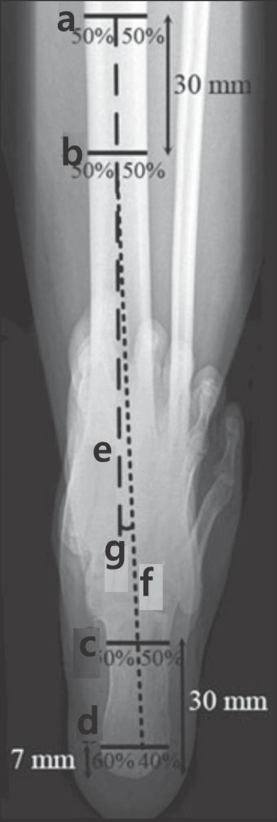
Figure 2.
Measurement method for the long axial view. We defined the mid-diaphyseal axis of the tibia by bisecting the tibia into two mid-diaphyseal points (lines a and b) 30 mm apart and extending the line distally (linee). The mid-diaphyseal axis of the calcaneus is defined by a line through two points in the calcaneus. At a distance of 7 mm from the most distal part of the calcaneus, a horizontal line is drawn (line d). Lined is divided into a 40%:60% ratio, where the length of the 40% line is measured from the lateral side. A second line (linec) is drawn horizontally, 30 mm from the most distal part of the calcaneus. The calcaneus axis (line f) is drawn by connecting the 40% mark at lined and the bisected line c. The hindfoot angle (g) is the angle between lines e and f. Revised from the article of Reilingh, et al. (Skeletal Radiol. 2010;39:1103-8).11)

Figure 3.
(A) Preoperative standing radiograph shows medial compartment ankle osteoarthritis. (B) Ankle arthrodesis was performed with trans-fibular approach using three cancellous screws. (C) Follow-up radiograph shows breakage of cancellous screws and valgus tibiotalar alignment. (D) Revision arthrodesis with cannulated screws was performed and bony union was evident 2 months after surgery.


 PDF
PDF ePub
ePub Citation
 Print
Print


 XML Download