

 PDF
PDF ePub
ePub Citation
 Print
Print


INTRODUCTION
An incidental dural tear resulting in cerebrospinal fluid (CSF) leakage is a complication of spinal surgery, with a reported incidence of between 0.3% and 16% (1, 2). The patient is likely to experience symptoms of CSF hypovolemia, including postural headache plus nausea, vomiting, pain or tightness in the neck or back, dizziness, diplopia, photophobia, tinnitus, and/or blurred vision. The usual treatment of CSF leakage consists of drainage of the CSF through a subarachnoid catheter (3) and surgical repair of the dural tear (4), although an epidural blood patch may also be used (5). To provide appropriate treatment, it is essential to detect the exact site of CSF leakage. Among the diagnostic modalities used to determine the site of CSF leakage are magnetic resonance (MR) imaging, computed tomographic (CT) cisternography, and radioisotope cisternography. Here, we describe two patients with surgery-related CSF leakage in whom the leakage sites were accurately detected on MR myelography.
CASE REPORTS
Patient 1
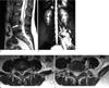
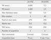
A 28-year-old woman developed severe headache accompanied by nausea and vomiting one day after a lumbar discectomy at L4-5 level for an intervertebral disc herniation. Spinal MR imaging showed a hyperintense T2 signal at the discectomy site, suggesting postoperative changes (Fig. 1a). Axial T2 weighted images of conventional MR showed left paravertebral fluid collection (Fig. 1b, c), but failed to detect exact leakage site. MR myelography was performed to detect possible CSF leakage using a 2-dimensional (D) turbo spin echo (TSE) technique on a 1.5-T scanner (Gyroscan Intera, Philips Medical Systems, Best, the Netherlands). The scanning parameters were summarized in the table. MR myelography showed a small, hyperintense CSF leakage site at the left lateral aspect of the L4 level. We also observed diffuse hyperintensity in the left paravertebral area along the muscle bundles, representing local fluid accumulation secondary to CSF leakage (Fig. 1d). The patient was promptly treated with an epidural blood patch targeted to the leakage site. Her symptoms were relieved by the next day, and she was discharged without further events.
Patient 2
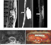
A 64-year-old woman was admitted due to an orthostatic headache that developed after spinal surgery. Six months earlier, she had undergone surgery to remove a spinal intradural extramedullary mass at the T11-12 level, which was pathologically confirmed as a meningioma. After the operation, she developed orthostatic headache, suggesting postoperative CSF leakage. She underwent two additional operations, but her symptoms did not improve. She was therefore admitted to our institution. A physical examination and laboratory tests showed no significant abnormal findings. Spine MR revealed a localized fluid collection at the previous operation site with a compression fracture of the T12 superior aspect. The patient underwent MR myelography to detect the leakage site, with 2D TSE and 3D balanced turbo field echo (BTFE) images obtained on the same 1.5-T scanner as in Patient 1. Detailed scanning parameters are summarized in the table. 2D TSE images and a maximum-intensity projection 3D BTFE rotational view showed the position of the fistula site in relation to the thecal sac and pseudomeningocele. Axial-reconstructed 3D BTFE images showed that the exact site of the fistula was in the left posterolateral aspect of the thecal sac at the T12 level (Figs. 2a-d).
A repeat operation at the T12 level confirmed the dural defect in the left posterolateral aspect of the thecal sac at the T12 level, corresponding to the position of the fistula on MR myelography (Fig. 2e). The defect was sealed using an artificial dura. The patient's headache was relieved after the operation, and her clinical course was uneventful.
DISCUSSION
We performed MR myelography to detect possible CSF leakage in 2 patients with severe headaches after spinal surgeries. We found that MR myelography successfully identified the exact location of the leakage site or communication with a pseudomeningocele.
Postoperative detection of a dural tear may be difficult. The diagnostic techniques used to detect these sites are MRI, CT, CT cisternography, and radionuclide cisternography.
The basic principle of MR myelography is enhancement of the CSF signal by suppression of the adjacent tissue signal. MR myelography eliminates the signal from epidural fatty tissue because of its extremely long echo time and effective fat suppression (6, 7). MR myelography was useful in detecting CSF leakage in patients with spontaneous CSF hypovolemia (8).
We used a TSE sequence for 2D images and a BTFE sequence for 3D images. No MR contrast material was used for both sequences. In Patient 1, thick slab 2D TSE MR myelography clearly demonstrated the fistula site. However, in Patient 2, 2D TSE MR myelography demonstrate the fistula level, but could not clearly visualize its exact location, making 3D images necessary. 3D BTFE revealed better delineation of anatomic structures, with axial-reconstructed images clearly showing that the fistula site was on the left side of the thecal sac.
Several case reports have described the use of MR myelography to detect CSF leakage in patients with spontaneous CSF hypovolemia (9, 10). MR myelography has several advantages compared with RI cisternography and CT myelography. It is a noninvasive technique, there is no radiation hazard, and it can be performed easily within a short examination time. The false negativity of RI cisternography is relatively high than CT myelography (11). Contrast material should be used in CT myelography however, MR myelography is no need to use contrast material. In addition, MR myelography may show specific structural information of ligamentous integrity and spinal cord derangement (12). In cases with CSF leakage after spinal surgery, it is very important to determine the exact site of leakage for proper management. In our patients, MR myelography provided valuable information about the leakage sites, and both of them could be treated successfully.
 XML Download