

 PDF
PDF ePub
ePub Citation
 Print
Print


Abstract
Purpose
The aim of this study was to evaluate the diagnostic performance of a computer-aided detection (CAD) system used with automated breast ultrasonography (ABUS) for suspicious lesions detected on breast MRI, and CAD-false lesions.
Materials and Methods
We included a total of 40 patients diagnosed with breast cancer who underwent ABUS (ACUSON S2000) to evaluate multiple suspicious lesions found on MRI. We used CAD (QVCADTM) in all the ABUS examinations. We evaluated the diagnostic accuracy of CAD and analyzed the characteristics of CAD-detected lesions and the factors underlying false-positive and false-negative cases. We also analyzed false-positive lesions with CAD on ABUS.
Results
Of a total of 122 suspicious lesions detected on MRI in 40 patients, we excluded 51 daughter nodules near the main breast cancer within the same quadrant and included 71 lesions. We also analyzed 23 false-positive lesions using CAD with ABUS. The sensitivity, specificity, positive predictive value, and negative predictive value of CAD (for 94 lesions) with ABUS were 75.5%, 44.4%, 59.7%, and 62.5%, respectively. CAD facilitated the detection of 81.4% (35/43) of the invasive ductal cancer and 84.9% (28/33) of the invasive ductal cancer that showed a mass (excluding non-mass). CAD also revealed 90.3% (28/31) of the invasive ductal cancers measuring larger than 1 cm (excluding non-mass and those less than 1 cm). The mean sizes of the true-positive versus false-negative mass lesions were 2.08 ± 0.85 cm versus 1.6 ± 1.28 cm (P < 0.05). False-positive lesions included sclerosing adenosis and usual ductal hyperplasia. In a total of 23 false cases of CAD, the most common (18/23) cause was marginal or subareolar shadowing, followed by three simple cysts, a hematoma, and a skin wart.
References
1. An YY, Kim SH, Kang BJ. The image quality and lesion characterization of breast using automated whole-breast ultrasound: a comparison with handheld ultrasound. Eur J Radiol. 2015; 84:1232–1235.

2. Jeh SK, Kim SH, Choi JJ, et al. Comparison of automated breast ultrasonography to handheld ultrasonography in detecting and diagnosing breast lesions. Acta Radiol. 2016; 57:162–169.
3. Wang ZL, Xu JH, Li JL, Huang Y, Tang J. Comparison of automated breast volume scanning to handheld ultrasound and mammography. Radiol Med. 2012; 117:1287–1293.
4. van Zelst JCM, Tan T, Platel B, et al. Improved cancer detection in automated breast ultrasound by radiologists using computer aided detection. Eur J Radiol. 2017; 89:54–59.
5. Shin HJ, Kim HH, Cha JH. Current status of automated breast ultrasonography. Ultrasonography. 2015; 34:165–172.
6. Kim Y, Kang BJ, Kim SH, Lee EJ. Comparison and combination of two ultrasound modalities, handheld ultrasound and automated breast volume scanner, with and without knowledge of MRI. Iran J Radiol. 2018; 15:e60176.
7. Wang HY, Jiang YX, Zhu QL, et al. Differentiation of benign and malignant breast lesions: a comparison between automatically generated breast volume scans and handheld ultrasound examinations. Eur J Radiol. 2012; 81:3190–3200.
8. Chang JM, Moon WK, Cho N, Park JS, Kim SJ. Radiologists' performance in the detection of benign and malignant masses with 3D automated breast ultrasound (ABUS). Eur J Radiol. 2011; 78:99–103.
9. Van Zelst JC, Platel B, Karssemeijer N, Mann RM. Multiplanar reconstructions of 3D automated breast ultrasound improve lesion differentiation by radiologists. Acad Radiol. 2015; 22:1489–1496.
10. Kim JH, Cha JH, Kim N, et al. Computer-aided detection system for masses in automated whole breast ultrasonography: development and evaluation of the effectiveness. Ultrasonography. 2014; 33:105–115.
11. Drukker K, Sennett CA, Giger ML. Automated method for improving system performance of computer-aided diagnosis in breast ultrasound. IEEE Trans Med Imaging. 2009; 28:122–128.
12. Berg WA, Gutierrez L, NessAiver MS, et al. Diagnostic accuracy of mammography, clinical examination, US, and MR imaging in preoperative assessment of breast cancer. Radiology. 2004; 233:830–849.
13. Wiratkapun C, Duke D, Nordmann AS, et al. Indeterminate or suspicious breast lesions detected initially with MR imaging: value of MRI-directed breast ultrasound. Acad Radiol. 2008; 15:618–625.
14. Abe H, Schmidt RA, Shah RN, et al. MR-directed ("SecondLook") ultrasound examination for breast lesions detected initially on MRI: MR and sonographic findings. AJR Am J Roentgenol. 2010; 194:370–377.
Fig. 1.
Images from a 32-year-old woman with a suspicious lesion detected on MRI and investigated subsequently with automated breast ultrasound (ABUS). (a) MRI image showed an approximately 1.1 cm enhancing mass at the 10 o'clock position on the left breast (arrow). (b) Mammography showed microcalcifications with suspicious architectural distortion involving the upper left inner quadrant (arrows). (c) Handheld ultrasonography revealed about 1.1-cm irregular mass with microcalcifications in the same direction. (d) 3D ABUS revealed correlating suspicious lesion on CAD in the left AP and medial views, later confirmed as invasive ductal carcinoma.
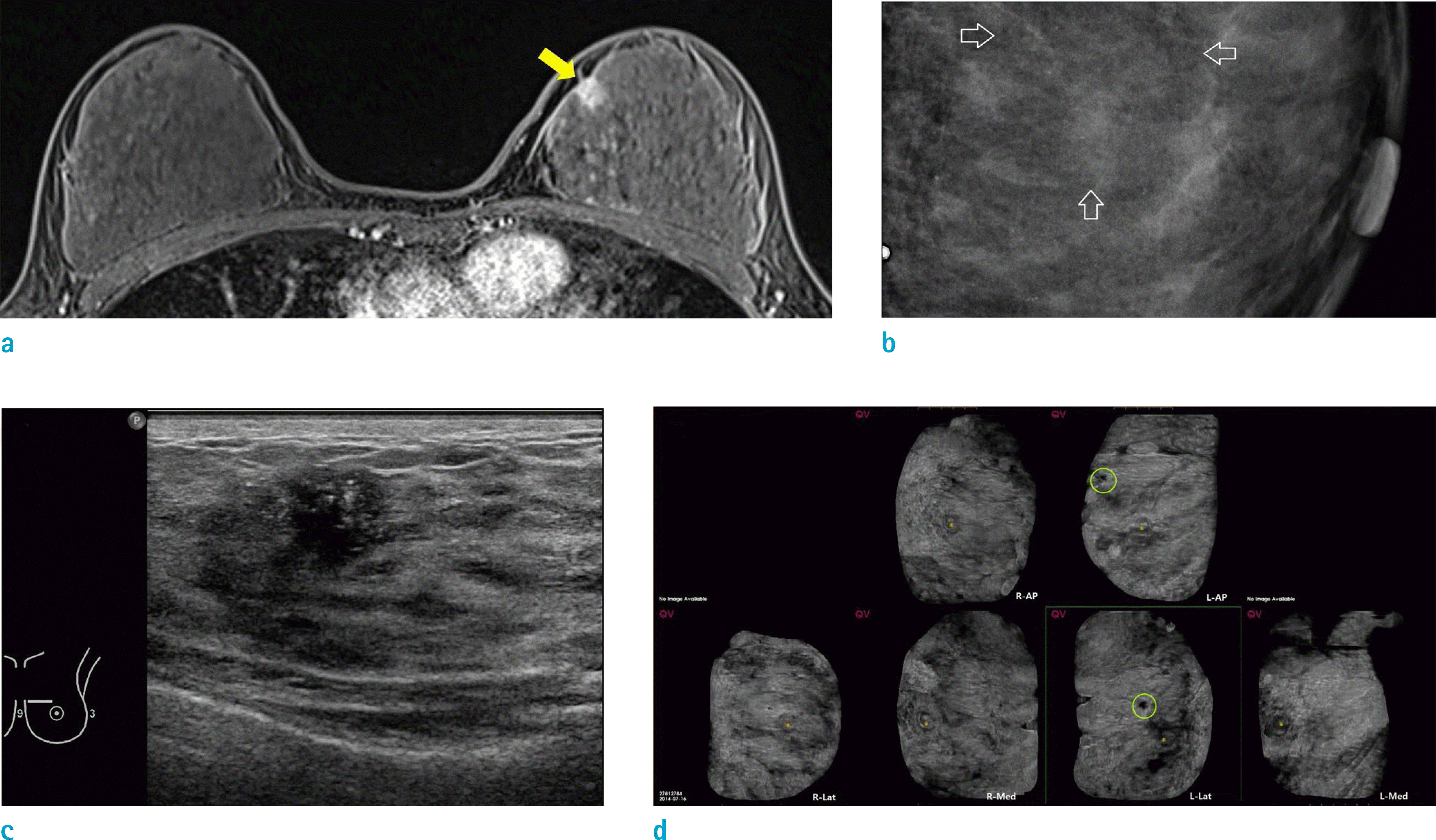
Fig. 2.
A 62-year-old woman who showed three CAD-detected lesions in both breasts. (a) CAD revealed a suspicious lesion in the right breast and only one marked lesson in the right medial view. The other two marked lesions involved the left breast: one was only marked in the left AP view, and the other one was marked in the whole 3D views. (b) Axial (white box) and maximal intensity projection (MIP) reconstruction image (yellow box) shows a right breast lesion that was confirmed as a pseudo lesion based on marginal shadowing. (c) Axial (white box) and MIP reconstruction image (yellow box) of one of the left breast lesions, which was marked by CAD only on AP view; it was also a pseudo lesion due to marginal shadowing. (d) Axial (white box) and MIP reconstruction image (yellow box) of the other left breast lesion, which was entirely marked by CAD in 3D view, and confirmed as IDC. (e) HHUS image of a biopsy-proven IDC lesion involving left breast shows a 1.8-cm marked hypoechoic mass with microlobulation in the left 2-h direction. (f) MRI of biopsy-proven IDC lesion shows 1.7-cm markedly enhanced mass in the left breast at the 1 o'clock position.

Table 1.
Characteristics of Patients and Lesions
Table 2.
Sensitivity and Specificity of Computer-Aided Detection (CAD) with Automated Breast Ultrasonography for Suspicious Lesions Detected on Breast MRI and CAD-False Lesions
Table 3.
Sensitivity and Specificity of Computer-Aided Detection (CAD) for Breast Cancer According to Mass/Non-Mass Lesions and Size in MRI
Table 4.
Correlation between Computer-Aided Detection (CAD)-Positive Lesions Based on MRI and Automated Breast Ultrasonography ABUS)
 XML Download