

 PDF
PDF ePub
ePub Citation
 Print
Print


Abstract
Reduction malarplasty (RMP) to reshape the facial contour is one of the most popular aesthetic surgical procedures in Asia. Here we report a case of intracerebral hematoma (ICH) after RMP. A 31-year-old woman was referred to our hospital following RMP. On arrival at our emergency room, she presented with deep drowsy mentality and right hemiparesis. Computed tomographic scan demonstrated an ICH. We conducted stereotactic aspiration of the blood clots. Because of increased ICH after the operation, the patient underwent craniotomy and hematoma evacuation. After removal of hematoma, intraoperatively a defect of the middle cranial fossa dura, a skull base bony defect, and a bony fragment were noticed. We think that these lesions have been caused by aggressive manipulation of surgical instruments. To our knowledge, ICH after RMP was not reported. The radiologic features of this case and suggested mechanism of the complication are described.
References
1. Baek RM, Kim J, Kim BK. Three-dimensional assessment of zygomatic malunion using computed tomography in patients with cheek ptosis caused by reduction malarplasty. J Plast Reconstr Aesthet Surg. 65:448–455. 2012.

2. Hwang K. Lateral rectus muscle injury, orbital fracture, mouth locking, and facial palsy resulting from reduction malarplasty. J Craniofac Surg. 22:151–154. 2011.
3. Kook MS, Jung S, Park HJ, Ryu SY, Oh HK. Reduction malarplasty using modified L-shaped osteotomy. J Oral Maxillofac Surg. 70:e87–e91. 2012.
4. Lee KC, Ha SU, Park JM, Kim SK, Park SH, Kim JH. Reduction malarplasty by 3-mm percutaneous osteotomy. Aesthetic Plast Surg. 30:333–341. 2006.
5. Ma YQ, Zhu SS, Li JH, Luo E, Feng G, Liu Y, et al. Reduction malarplasty using an L-shaped osteotomy through intraoral and sideburns incisions. Aesthetic Plast Surg. 35:237–241. 2011.
6. Mahatumarat C, Rojvachiranonda N. Reduction malarplasty without external incision: a simple technique. Aesthetic Plast Surg. 27:167–171. 2003.
7. Nakanishi Y, Nagasao T, Shimizu Y, Miyamoto J, Kishi K, Fukuta K. The boomerang osteotomy – a new method of reduction malarplasty. J Plast Reconstr Aesthet Surg. 65:e111–e120. 2012.
8. Onizuka T, Watanabe K, Takasu K, Keyama A. Reduction malar plasty. Aesthetic Plast Surg. 7:121–125. 1983.
9. Powell NB, Riley RW, Laub DR. A new approach to evaluation and surgery of the malar complex. Ann Plast Surg. 20:206–214. 1988.
10. Sumiya N, Kondo S, Ito Y, Ozumi K, Otani K, Wako M. Reduction malarplasty. Plast Reconstr Surg. 100:461–467. 1997.
FIGURE 1.
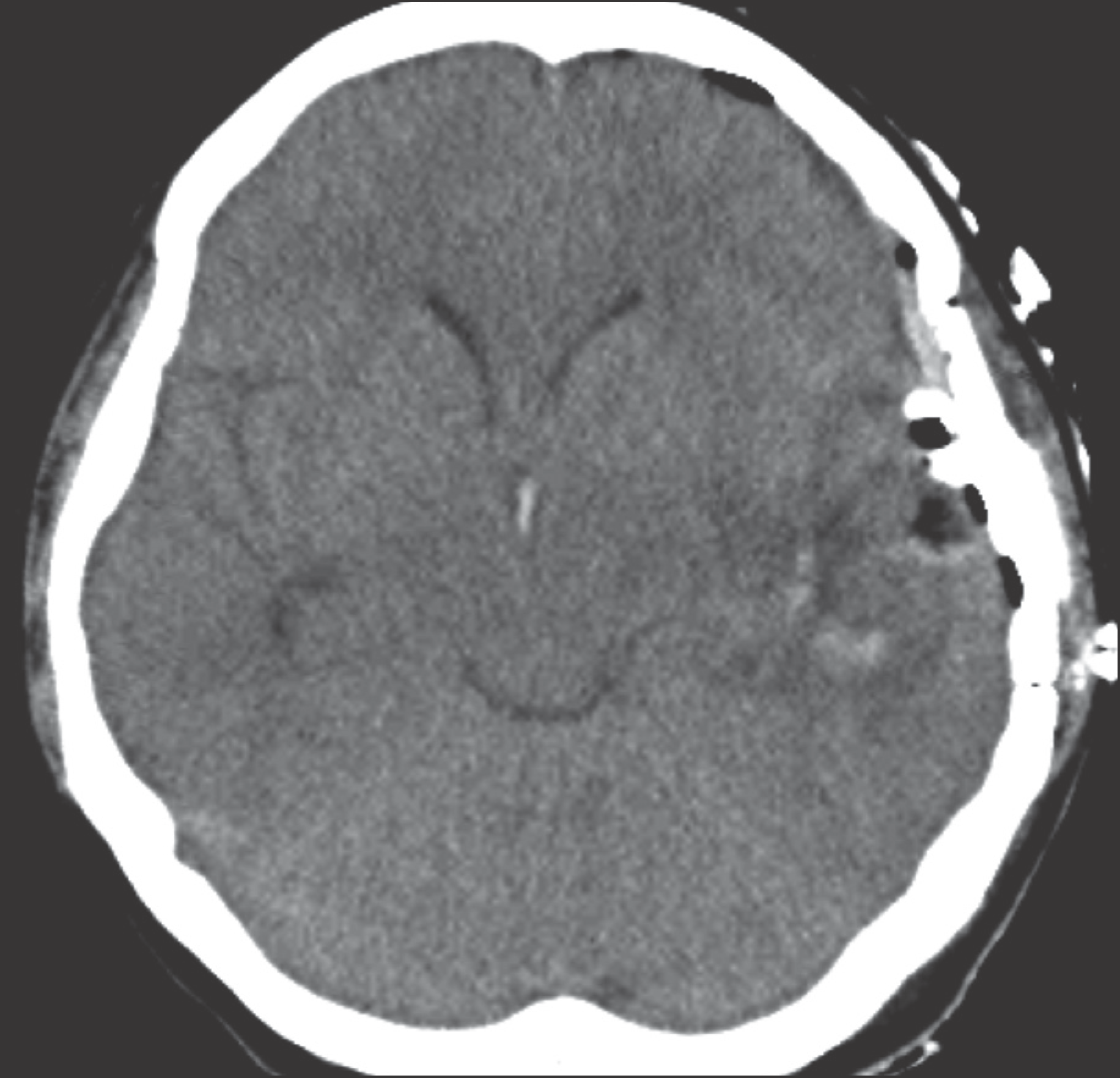
Brain noncontrast computed tomographic scan performed at the emergency room shows intracerebral hematoma located in the left temporal lobe.
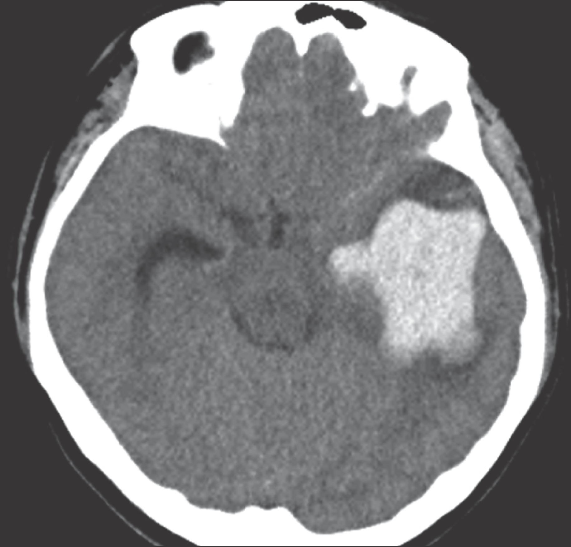
FIGURE 2.
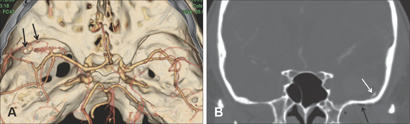
Skull AP (A) and lateral X-ray (B) images demonstrate the postoperative state of reduction malarplasty. AP: antero-posterior.
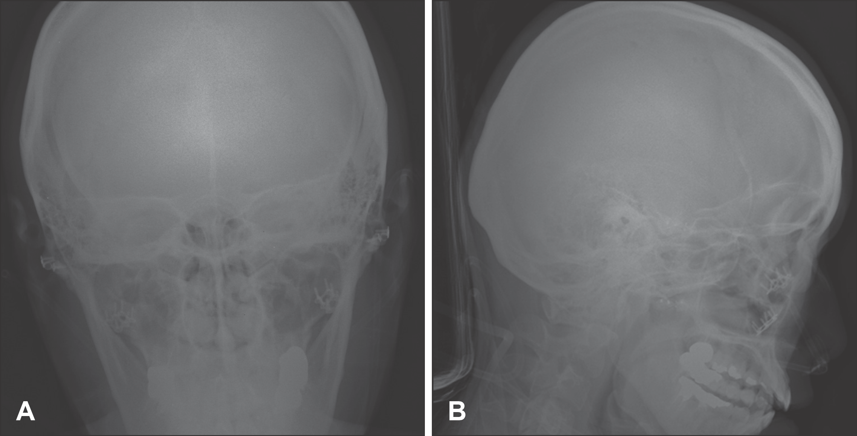
FIGURE 3.
Brain noncontrast computed tomographic scan performed after 1st operation demonstrates intracerebral hematoma located in the left temporal lobe.
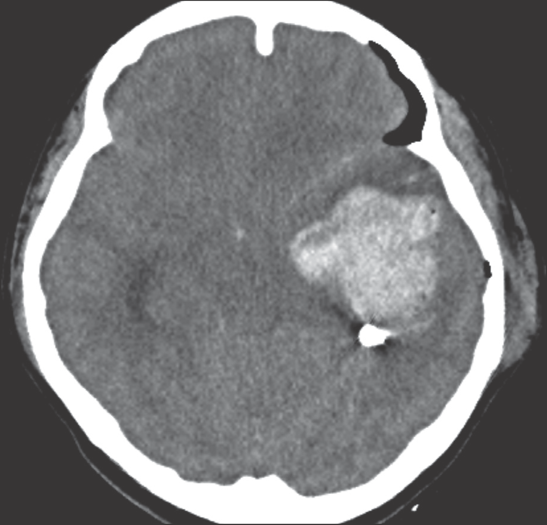
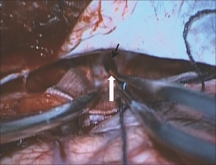
FIGURE 4.
Intraoperative finding of craniotomy and hematoma evacuation shows the dural defect (black arrow) located in the left middle cranial fossa and a bony fragment (white arrow).
 XML Download