

 PDF
PDF ePub
ePub Citation
 Print
Print


INTRODUCTION
Previous studies, including the Familial Atherosclerosis Treatment Study, HDL-Atherosclerosis Treatment Study, Armed Forces Regression Study, Carotid Plaque Composition by MRI during Lipid-Lowering Study, and Arterial Biology for the Investigation of the Treatment Effects of Reducing Cholesterol 6-HDL and LDL Treatment Strategies in Atherosclerosis Trial (ARBITER 6-HALTS), have reported that niacin slows progression of coronary artery stenosis1 and carotid intima-media thickness (IMT).23 However, analyses synthesizing both old and new studies now suggest that niacin is unlikely to be effective in reducing clinical events in people with high cardiovascular risk.45678
Elevated lipoprotein (Lp) (a) is an independent risk factor of cardiovascular disease in patients with diabetes mellitus who also commonly have atherogenic dyslipidemia beyond hyper low-density lipoprotein (LDL)-cholesterolemia.9 Additionally, Lp (a) is known to contribute to residual cardiovascular risk in a subset of patients who are able to achieve optimal level of LDL-cholesterol (LDL-C).10 Furthermore, it has been reported that Lp (a) may contribute to the progression of aortic stenosis.11 A recent study also demonstrated that Lp (a) induces monocyte trafficking in the arterial wall.12
The use of niacin to control Lp (a) is well known, especially in Korea.13 A prior study reported that niacin decreases the production of apolipoprotein (a) and Lp (a)-apoB-100. Likewise, niacin regulates lipid levels by reducing the production of very LDL.14 Nevertheless, no clinical trial has analyzed the effects of niacin pharmacotherapy on Lp (a) level, clinical outcomes, or surrogate markers.15
The aim of this study was to examine the effect of niacin on the progression of carotid intima-media thickening in patients at LDL-C goal but high Lp (a) level. Specifically, we hypothesized that the effect of niacin on Lp (a) could partially inhibit vascular changes in a population with residual cardiovascular risk.
MATERIALS AND METHODS
1. Study population
Written informed consent was obtained from all study participants. Patients who visited the outpatient clinic of the Division of Cardiology, Severance Hospital during the study period were initially screened. The inclusion criteria were age 20–79 years, at LDL-C goal as defined by National Cholesterol Education Program Adult Treatment Panel III guidelines,16 Lp (a) level >25 mg/dL, and mean carotid IMT >0.75 mm. Patients who met all inclusion criteria were enrolled in the study. Individuals were eligible regardless of whether they were taking lipid-lowering medication. The exclusion criteria were history of acute coronary syndrome or cerebrovascular accident within three months prior to study enrollment, uncontrolled hypertension (systolic blood pressure >160 mmHg or diastolic blood pressure >100 mmHg), or diabetes mellitus (hemoglobin A1c >9%).We also excluded patients with a history of an acute or chronic systemic inflammatory disease, malignancy, chronic endocrine-, hepatic-, or renal disease; patients who were pregnant or breast feeding, actively using hormone replacement therapy, or who had a history of niacin intolerance.
2. Study protocol
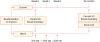
The Institutional Review Board of Severance Hospital, Seoul, Korea, approved this study (4-2008-3530). This was a 24-month, open-label, randomized study. At the screening visit, each patient was interviewed about medical history and underwent physical examination, laboratory assessment, and carotid ultrasound. Patients who met the inclusion criteria were randomized at a 1:2 ratio into one of 2 groups for 24 months: control or niacin extended release 1,500 mg (Exlip; Seoul Pharma, Seoul, Korea). We selected a 2:1 randomization ratio in anticipation of a higher number of adverse events (AEs) and discontinuation in the niacin group. We also used an extended release niacin, which has a better tolerance for flushing than other niacin formulations. Furthermore, extended release niacin was the only formulation of niacin available in Korea at the time the study was performed. In the treatment group, the initial dose of niacin was 500 mg/day, which was sequentially increased at the beginning of weeks 5 (1,000 mg/day) and 9 (1,500 mg/day). The final dose was continued to the end of the study (Fig. 1). Patients were enrolled between March 2012 and November 2014, and a total of 96 patients was randomized.
Blood samples were collected at the time of randomization and at 24 months of treatment. Patients were instructed to fast and avoid alcohol and smoking for at least 12 hours prior to sampling. Samples were analyzed within 4 hours by the local laboratory, which was certified by the Korean Society of Laboratory Medicine. Levels of total cholesterol, triglycerides (TG), and high-density lipoprotein-cholesterol (HDL-C) were measured by enzymatic methods using a Hitachi 7600-120 chemistry analyzer (Hitachi, Tokyo, Japan). LDL-C was calculated using the Friedwald formula. Lp (a) was measured by rate nephelometry (IMMAGE system; Beckman Coulter, Brea, CA, USA). Tolerability for the treatment was assessed by history taking and physical examination.
3. Carotid ultrasound
High-resolution carotid ultrasound was performed at baseline and after 24 months according to a standard protocol by 2 trained sonographers. The exam was performed using commercially available equipment (Acuson X300; Siemens Healthineers, Erlangen, Germany) with a 7.5-MHz linear transducer. B-mode images of the far wall of the distal right and left common carotid arteries were captured at the end-diastolic period. Carotid IMT was measured by a single trained reader in a 1-cm segment from 1 cm proximal to the carotid bifurcation using an automated system. IMT values for each side were averaged and used in all analyses. The intra-observer and inter-observer coefficients of variation were 5.4% and 6.4%, respectively.
4. Statistical analysis
The primary study outcomes were the percentage changes in mean and maximal carotid IMTs from time of randomization to 24 months of treatment. The percentage changes in total cholesterol, TG, HDL-C, LDL-C, non-HDL-C, and Lp (a) were evaluated as secondary outcome variables. Outcome variables were evaluated by per protocol analysis. A minimum of 25 and 50 participants were planned for the control and niacin groups, respectively, assuming a power of 0.80 and a significance level of 0.05 to demonstrate superiority of niacin over control. A 3% difference in the percentage change of mean carotid IMT with a standard deviation of 3.8% between the two groups was predefined as significant. To compensate for an anticipated drop-out rate of 20%, we aimed to enroll at least 30 and 60 participants in the placebo and niacin groups, respectively. Group differences in categorical variables were assessed using Fisher's exact test. Student's t-test was used to compare the mean values of continuous variables between the two groups. Paired t-test was used to assess differences in parameters before and after treatment within each group. Differences were considered significant if the p-value was <0.05. All analyses were performed using SPSS version 17.0 (SPSS Inc., Chicago, IL, USA).
RESULTS
1. Patient characteristics
Among 96 randomized patients, 31 (18 of 33 in the control group and 13 of 63 in the niacin group) completed the study. A total of 65 patients dropped from the study and was excluded from efficacy analysis, with 42 due to AEs, 15 due to withdrawal of consent, and eight due to protocol violation. The clinical characteristics of the patients who were randomized and completed the study are listed in Table 1. The mean age of randomized patients was 65 years, and 44% were male. The rates of diabetes mellitus, coronary artery disease (CAD), and statin-use were 13%, 32%, and 48%, respectively. The mean carotid IMT was 0.822±0.192 mm. Among the 31 patients who completed the study, there was a higher proportion of males, and the mean age and prevalence of CAD were lower in the niacin group. However, none of the clinical or laboratory variables were significantly different between the two groups.
Table 1
Characteristics of study subjects
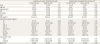
Values are presented as number (%), mean±standard deviation, or median (interquartile range).
DM, diabetes mellitus; CAD, coronary artery disease; BMI, body mass index; TC, total cholesterol; TG, triglycerides; HDL-C, high-density lipoprotein-cholesterol; LDL-C, low-density lipoprotein-cholesterol; Lp (a), lipoprotein (a); HbA1c, hemoglobin A1c; IMT, intima-media thickness.
2. Changes in primary and secondary outcomes
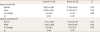
At the 24-month follow-up, percentage change in mean carotid IMT was not significantly different between the 2 groups (−1.4%±15.5% and −1.1%±7.3% in the control and niacin groups, respectively, p=0.95). The maximal carotid IMT did decrease in the niacin group compared to before treatment, but the difference was not significant (p=0.07). Likewise, the overall percentage change was similar for the two groups (0.7%±16.5% and −4.4%±11.6%, respectively, p=0.35) (Table 2).
Table 2
Changes of primary outcome variables (n=31)
At follow-up, HDL-C was significantly elevated in the niacin group (p=0.03), and hemoglobin A1c was decreased in the control group (p=0.02). None of the other parameters were significantly changed between the two groups. Although Lp (a) level decreased by −21.4%±35.6% in the niacin group, the difference was not statistically significant. Elevation of HDL-C tended to be greater in the niacin group (2.8%±13.3% and 13.2%±17.6%, in the control and niacin group, respectively, p=0.07), while the percentage change in hemoglobin A1c was significantly different between the two groups (−1.9%±2.2% and 3.3%±6.7%, respectively, p=0.02) (Table 3).
Table 3
Changes of secondary outcome variables (n=31)
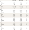
Values are presented as mean±standard deviation, or median (interquartile range).
TC, total cholesterol; TG, triglycerides; HDL-C, high-density lipoprotein-cholesterol; LDL-C, low-density lipoprotein-cholesterol; Lp (a), lipoprotein (a); HbA1c, hemoglobin A1c.
*Comparison before and after treatment; †Comparison between two groups.
3. AEs
Forty-two patients experienced AEs during the study period and discontinued niacin as a result (Table 4). The most common AEs were skin-related and included complaints of pruritus, urticaria, and flushing (n=29, 69%). The second most common AEs were related to gastrointestinal complaints. Other less frequent AEs included chest symptoms and hyperglycemia.
Table 4
Discontinuation of niacin due to adverse events over the study period

DISCUSSION
To the best of our knowledge, this is the first study to examine the effect of niacin specifically in patients with elevated Lp (a) level. We found that treatment with niacin for two years in this patient population had a neutral effect on mean and maximal carotid IMT. Compared to the control group, the group treated with niacin exhibited significant change in glucose metabolism and marginal change in HDL-C level. Lp (a) was decreased to a greater extent after niacin treatment, but the difference was not significant.
Previous studies have shown that niacin does not reduce cardiovascular risk even though it improves multiple lipid parameters including Lp (a). However, existing studies pertaining to associations between niacin, Lp (a), and cardiovascular results did not use pre-determined endpoints.17 Although we hypothesized that niacin would inhibit thickening of carotid IMT in individuals with high Lp (a), we were unable to find evidence for such an effect. Our results are also in contrast to the ARBITER 6-HALTS study, which demonstrated regression of carotid IMT after niacin therapy.3 Although that study included populations of similar age to our study, there were differences in baseline LDL-C and carotid IMT, being higher and slightly lower compared to our patients, respectively. It remains uncertain which of the two study populations had a greater baseline cardiovascular risk and the extent to which that influenced the study outcomes. The reason for the discrepancy between the negative results of our trial and those of the ARBITER 6-HALTS study are not completely understood. The follow-up duration of our study was 24 months, 10 months longer than that of the ARBITER 6-HALTS study, making an insufficient treatment duration an unlikely explanation for the difference in results and lack of efficacy of niacin in our study. Because the inclusion criteria were difficult to meet and the discontinuation rate was high due to drug AEs, we also considered the possibility that the number of subjects who completed the study was insufficient to show a significant difference. Although niacin tended to decrease Lp (a) in our study, the standard deviation of the value was quite large, suggesting that there was considerable individual variation in the response to niacin therapy. Another possibility is that there may be no beneficial effect of niacin on carotid arteries. Indeed, no studies have demonstrated an association between pharmacologically increased level of HDL-C and reduction in cardiovascular outcomes.18 In addition, another study reported that niacin does not improve endothelial function in statins despite further improvement of lipid profiles.19 In recent studies, the effect of niacin on HDL functionality has been variable and not always beneficial.20 Although niacin significantly increases HDL-C level, it does not improve ABCA1-specific cholesterol efflux and has insufficient effects on the atheroprotective population of HDL.21
Our study population was selected on the basis of Lp (a) level, but not TG or HDL-C level. In contrast to our hypothesis, there may be a better candidate population for niacin therapy than individuals with high Lp (a) level. For example, patients with residual cardiovascular risk with high TG and low HDL-C levels might be better suited for niacin therapy. “Atherogenic dyslipidemia” includes a wide spectrum of lipid profiles, from hypertriglyceridemia, low HDL-C, qualitative changes of LDL, and elevated remnant lipoprotein levels. Individuals with these conditions may have high residual risk even if they have achieved the LDL-C target, and they may also gain benefit from additive lipid modifying therapy.22 In support of this possibility, a prior study demonstrated greater beneficial effects of niacin on endothelial cells in patients with low HDL-C.23
There were several potential limitations to the present study that should be considered when interpreting our results. First, as mentioned above, the drop-out rate was relatively high, and the number of patients who completed the study may have been insufficient to derive solid conclusions. Second, we used carotid IMTs as the primary study outcome. However, more recent international guidelines have suggested against routine measurement of carotid IMT for the purposes of risk stratification.24 Therefore, the decreasing clinical relevance of target variables in our study is another limitation of our study. Finally, the beneficial effect of niacin tends to be smaller in Chinese subjects compared to Europeans, according to the Heart Protection Study 2-Treatment of HDL to Reduce the Incidence of Vascular Events (HPS2-THRIVE) study.5 Therefore, caution should be used when applying our results to other ethnicities.
In conclusion, a two-year treatment with niacin 1,500 mg/day did not block progression of carotid intimal-medial thickening in patients at LDL-C goal and high Lp (a). Although niacin induced metabolic changes to lipids and glucose, it did not have a significant effect on the carotid arteries. However, due to suboptimal sample size, these results may have been underpowered to provide a solid conclusion.
 XML Download