Abstract
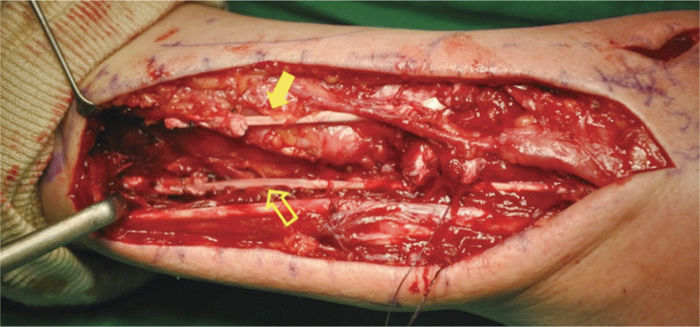
Extensor tendon rupture is well known complication following distal radius fracture after either conservative treatment or volar plating. However, there are not many reports in literature about concomitant ruptures of other extensor tendons. We report a case of delayed rupture of extensor pollicis longus (EPL), second extensor digitorum communis (EDC II), and extensor indicis proprius (EIP) tendons 4 weeks after volar plating for distal radius fracture. Due to the absence of EIP, EIP transfer was discouraged for EPL reconstruction. Thumb and index finger extension was restored by palmaris longus tendon graft for EPL and EDC II.
References
1. Roth KM, Blazar PE, Earp BE, Han R, Leung A. Incidence of extensor pollicis longus tendon rupture after nondisplaced distal radius fractures. J Hand Surg Am. 2012; 37:942–7.

2. Tarallo L, Mugnai R, Zambianchi F, Adani R, Catani F. Volar plate fixation for the treatment of distal radius fractures: analysis of adverse events. J Orthop Trauma. 2013; 27:740–5.
3. Ward JP, Kim LT, Rettig ME. Extensor indicis proprius and extensor digitorum communis rupture after volar locked plating of the distal radius: a case report. Bull NYU Hosp Jt Dis. 2012; 70:273–5.
4. Hattori Y, Doi K, Sakamoto S, Yukata K. Delayed rupture of extensor digitorum communis tendon following volar plating of distal radius fracture. Hand Surg. 2008; 13:183–5.

5. de Boer SW, van Kooten EO, Ritt MJ. Extensor pollicis longus tendon rupture with concomitant rupture of the extensor digitorum communis II tendon after distal radius fracture. J Hand Surg Eur Vol. 2010; 35:679–81.

6. Caruso G, Vitali A. del Prete F. Multiple ruptures of the extensor tendons after volar fixation for distal radius fracture: a case report. Injury. 2015; 46(Suppl 7):S23–7.
7. Gelb RI. Tendon transfer for rupture of the extensor pollicis longus. Hand Clin. 1995; 11:411–22.

8. Hamlin C, Littler JW. Restoration of the extensor pollicis longus tendon by an intercalated graft. J Bone Joint Surg Am. 1977; 59:412–4.

9. Thoma A, Quttainah A. Extensor indicis proprius tendon transfer for extensor pollicis longus rupture. Can J Plast Surg. 2001; 9:139–42.

10. Chung US, Kim JH, Seo WS, Lee KH. Tendon transfer or tendon graft for ruptured finger extensor tendons in rheumatoid hands. J Hand Surg Eur Vol. 2010; 35:279–82.
Fig. 1.
Axial and three-dimensional computed tomography images demonstrating a fracture of Lister’s tubercle on the right side (arrows). A bony spur created a the gap between the fragment and the main radius part and might be the cause for tear of the tendon.
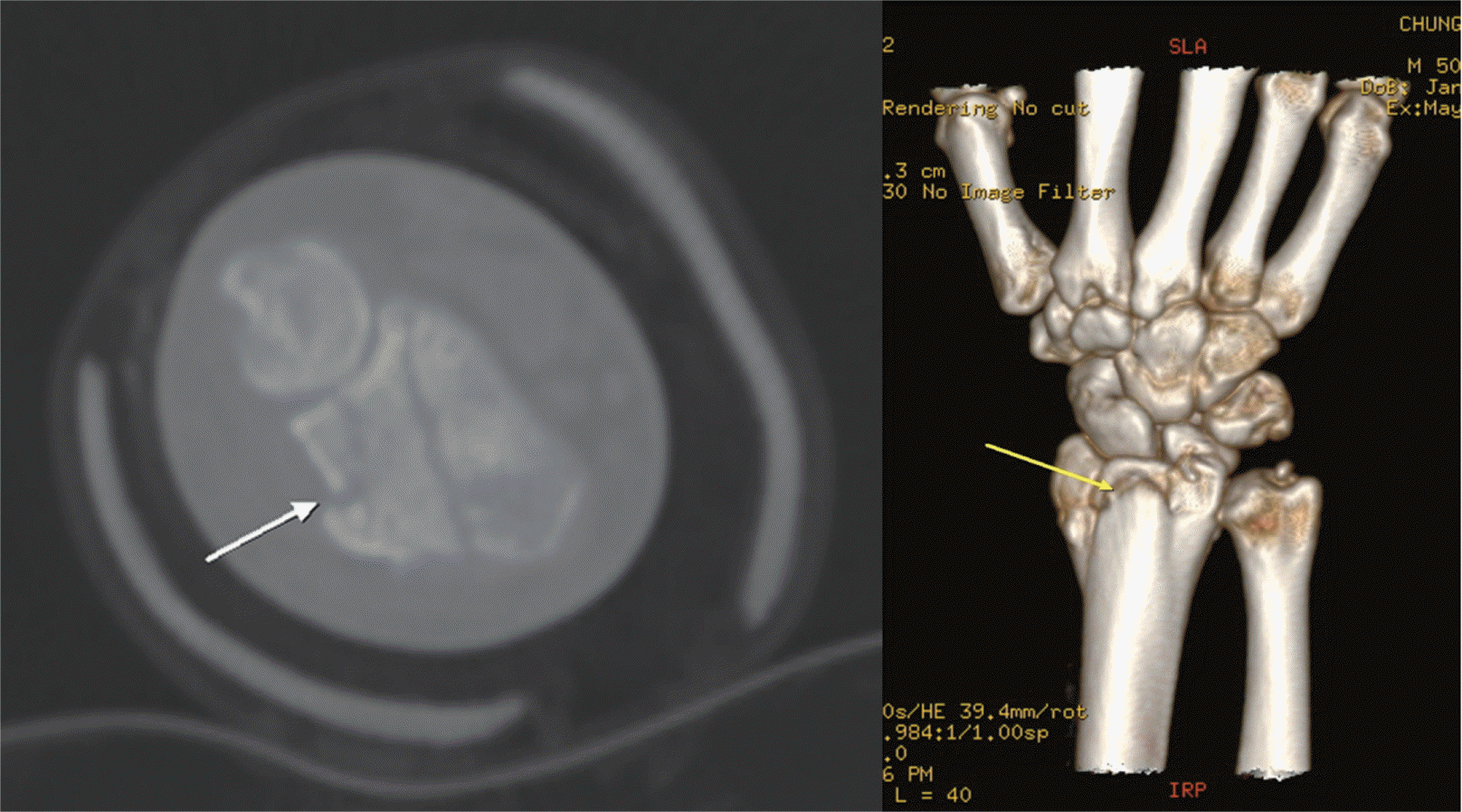
Fig. 2.
Preoperative and postoperative plain radiography showed that the fracture gap of the Lister’s tubercle persisted.
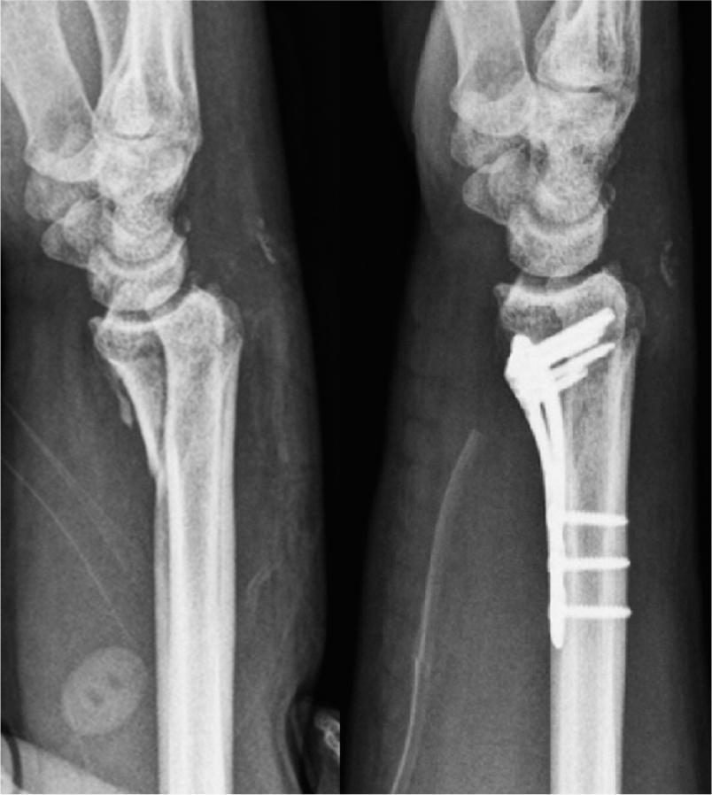
Fig. 3.
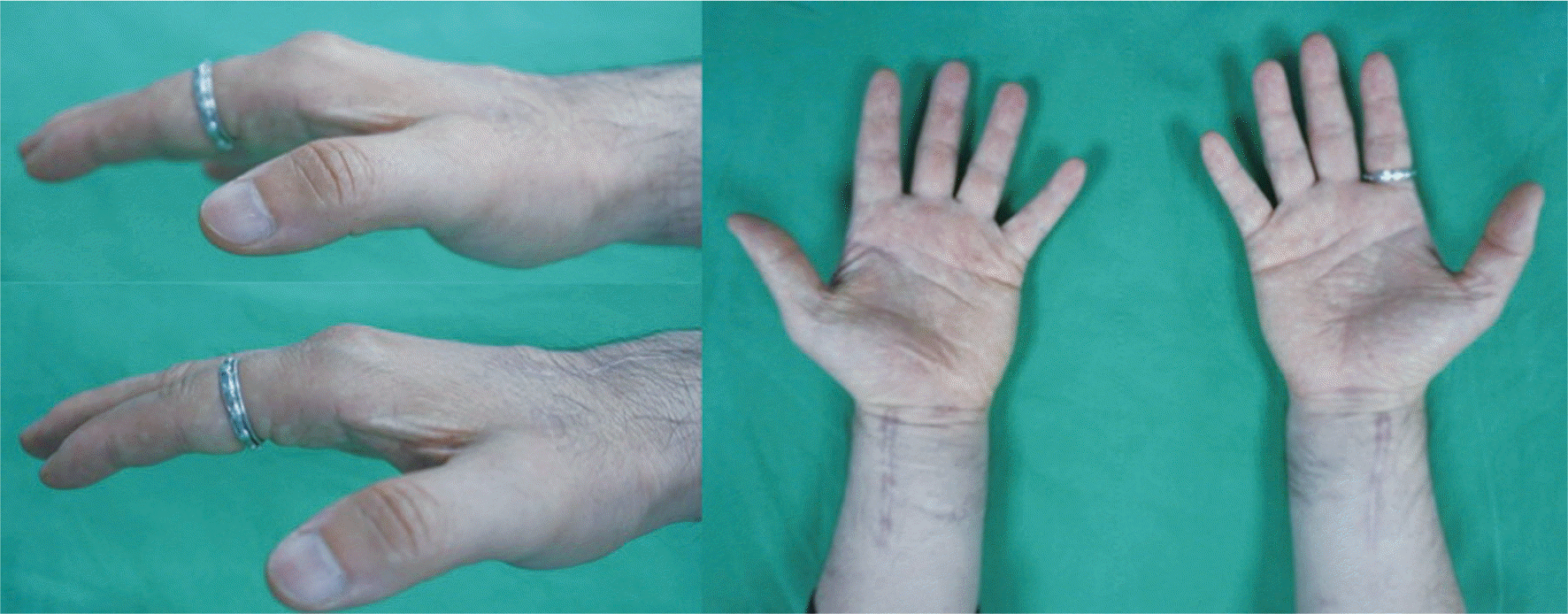
In the physical examination, limitations in thumb interphalangeal and index metacarpophalangeal joint extensions were observed when compared with those on the normal side.
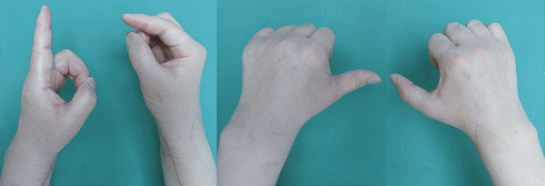
Fig. 4.
Axial image of ultrasonography showed complete disruptions of the extensor pollicis longus, (solid arrow) and fracture gap of Lister’s tubercle (vacant arrow).
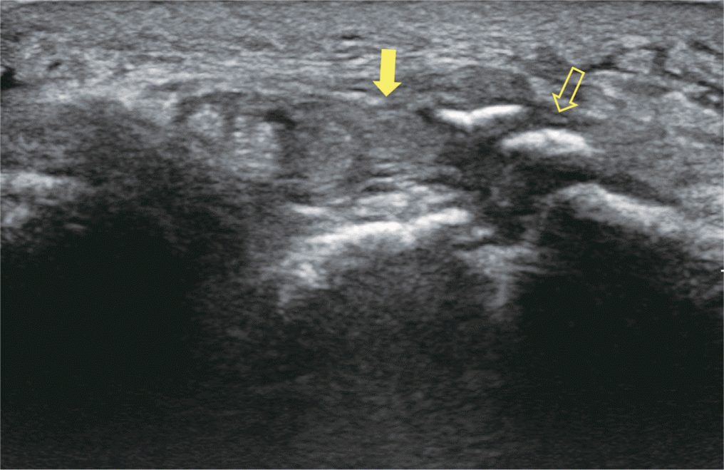
Fig. 5.
The ruptures of the extensor pollicis longus (arrowhead), extensor indicis proprius (solid arrow), and extensor digitorum communis II (vacant arrow). The asterisk shows a bony spur near Lister’s tubercle.
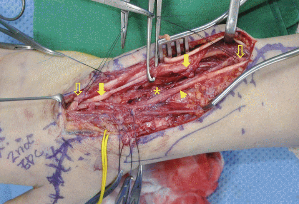
Fig. 6.
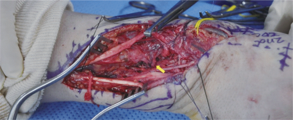
After removing the bony protrusion, the surrounding soft tissue was pulled to the ulnar side and sutured to the remaining tendon sheath and periosteum of the 3rd compartment to prevent further tendon tear (solid arrow).


 PDF
PDF ePub
ePub Citation
 Print
Print


 XML Download