

 PDF
PDF ePub
ePub Citation
 Print
Print


Abstract
Purpose
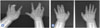
Non-preaxial polydactyly of the hand refers to axial polysyndactyly involving the 2nd, 3rd, or 4th finger and postaxial polydactyly involving the 5th finger. It has a much lower incidence and a higher genetic penetrance than preaxial type.
Methods
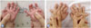
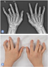
Medical records of the patients who had operation for their polydactyly between July 1997 and July 2015 were retrospectively reviewed. The clinical data of the patients were investigated regarding demographics, clinical findings of the involved digits, foot involvement, and genetic penetrance. Through postoperative follow-up based on physical and radiologic examinations, we assessed functional and aesthetic outcomes, postoperative complications, and reoperation rate.
Results
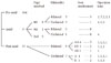
Twenty-four patients (17 males and 7 females) underwent surgery for non-preaxial polydactyly of the hand. There were 15 postaxial type polydactyly, and 9 axial type polysyndactyly. Thirteen patients had bilateral involvement (54.2%), while 5 patients (20.8%) were right-sided and 6 patients (25%) were left-sided. In the axial type, 4th finger was the most frequently involved in 8 patients, followed by the 3rd finger in 1 patient. Thirteen patients (54.2%) had concurrent congenital foot anomalies. One patient (4.2%) had a family history of congenital hand anomaly. Patients with axial type polysyndactyly had poorer postoperative outcome than those with postaxial type, regarding reoperation rate.
Figures and Tables
References
1. Finley WH, Gustavson KH, Hall TM, Hurst DC, Barganier CM, Wiedmeyer JA. Birth defects surveillance: Jefferson County, Alabama, and Uppsala County, Sweden. South Med J. 1994; 87:440–445.

2. Kim D, Park SK, Kim DC, Oh SJ, Yoo KY. Nationwide estimation for incidence at birth of congenital polydactyly and syndactyly in Korean. J Korean Soc Plast Reconstr Surg. 2003; 30:24–32.
3. Flatt AE. A test of a classification of congenital anomalies of the upper extremity. Surg Clin North Am. 1970; 50:509–516.
4. Woolf CM, Myrianthopoulos NC. Polydactyly in American negroes and whites. Am J Hum Genet. 1973; 25:397–404.
5. Wood VE. Treatment of central polydactyly. Clin Orthop Relat Res. 1971; 74:196–205.
6. Tada K, Kurisaki E, Yonenobu K, Tsuyuguchi Y, Kawai H. Central polydactyly: a review of 12 cases and their surgical treatment. J Hand Surg Am. 1982; 7:460–465.
7. Temtamy SA, McKusick VA. The genetics of hand malformations. Birth Defects Orig Artic Ser. 1978; 14:i–xviii. 1–619.
8. Stelling F. The upper extremity. In : Ferguson AB, editor. Orthopaedic surgery in infancy and childhood. Baltimore, MD: Williams & Wilkins;1963. p. 304–308.
9. Turek SL. Orthopaedics: principles and their application. Philadelphia, PA: Lippincott;1967.
10. Watson BT, Hennrikus WL. Postaxial type-B polydactyly: prevalence and treatment. J Bone Joint Surg Am. 1997; 79:65–68.
11. Malik S, Ullah S, Afzal M, Lal K, Haque S. Clinical and descriptive genetic study of polydactyly: a Pakistani experience of 313 cases. Clin Genet. 2014; 85:482–486.
12. Xiang Y, Bian J, Wang Z, Xu Y, Fu Q. Clinical study of 459 polydactyly cases in China, 2010 to 2014. Congenit Anom (Kyoto). 2016; 56:226–232.
13. Cantu JM, del Castillo V, Cortes R, Urrusti J. Autosomal recessive postaxial polydactyly: report of a family. Birth Defects Orig Artic Ser. 1974; 10:19–22.
14. Miura T, Nakamura R, Imamura T. Polydactyly of the hands and feet. J Hand Surg Am. 1987; 12:474–476.
15. Akarsu AN, Stoilov I, Yilmaz E, Sayli BS, Sarfarazi M. Genomic structure of HOXD13 gene: a nine polyalanine duplication causes synpolydactyly in two unrelated families. Hum Mol Genet. 1996; 5:945–952.
16. Radhakrishna U, Blouin JL, Mehenni H, et al. Mapping one form of autosomal dominant postaxial polydactyly type A to chromosome 7p15-q11.23 by linkage analysis. Am J Hum Genet. 1997; 60:597–604.
17. Zhao H, Tian Y, Breedveld G, et al. Postaxial polydactyly type A/B (PAP-A/B) is linked to chromosome 19p13.1-13.2 in a Chinese kindred. Eur J Hum Genet. 2002; 10:162–166.
18. De Smet L. International Federation for Societies for Surgery of the Hand. Japanese Society for Surgery of the Hand. Classification for congenital anomalies of the hand: the IFSSH classification and the JSSH modification. Genet Couns. 2002; 13:331–338.
19. Graham TJ, Ress AM. Finger polydactyly. Hand Clin. 1998; 14:49–64.
20. Goodman FR. Limb malformations and the human HOX genes. Am J Med Genet. 2002; 112:256–265.
21. Horsnell K, Ali M, Malik S, et al. Clinical phenotype associated with homozygosity for a HOXD13 7-residue polyalanine tract expansion. Eur J Med Genet. 2006; 49:396–401.
22. Wang C, Huang X, Tan W. A new skill for treating unclassified thumb polydactyly: ablation via a periosteal incision. Aesthetic Plast Surg. 2012; 36:928–933.
23. Johnson JM, Higgins TJ, Lemos D. Appearance of the delta phalanx (longitudinally bracketed epiphysis) with MR imaging. Pediatr Radiol. 2011; 41:394–396.
 XML Download